Brain structures Flashcards
Nervous system
is a system of networks of specialised cells (neurons) that connect different parts of the body to each other and the brain via electrochemical signals.
Neurons
are individual nerve cells that receive, transmit and process information
Central nervous system
is a major division of the nervous system consisting of all the nerves in the brain and spinal cord. Enables the brain to communicate with the rest of the body. Top half of spinal chord = upper body, lower body = lower half

Peripheral nervous system
is a major division of the nervous system consisting of all the nerves outside the CNS; transmits sensory information inwards to the CNS and transmits motor messages from the brain outwards to the rest of the body vie motor neurons from the CNS

Somatic nervous system
- division of the PNS - repsonsible for voluntary movement of skeletal muscles (striped) - motor neurons communicate from CNS to particular muscles - sensory neurons convey sensations (interact w. motor through interneurons)

Autonomic nervous system
- division of the PNS - responsible for communcation of info between CNS and non-skeletal muscles (smooth, visceral) as well as internal organs and glands - operated without voluntary control or consciousness awareness (can be controlled tho) - enables organism to have the cognitive resources to pay attention to other matters - through medulla
Sympathetic nervous system
- branch of the autonomic system - becomes active when an organism perceives itself to be in danger or stress - readies the body for action, such as running away (fight or flight) - dilates pupils - no effects on tear glands - weak stimulation of salivary glands - accelerates heart rate and constricts arterioles - dilates bronchi - inhibits stomach and pancreas - stimulates intenstines - relaxes bladders - stimulates ejaculation
Parasympathetic nervous system
- branch of the autonomic system - operates in circumstances when it is relatively calm - maintains automatic bodily functions and normal breathing - normal function = homeostasis - affects same organs and sympathetic, but different ways - pupils contricted - tear glands stimulates - salivary glands simulated - slowed heart rate, dilates arterioles - bronchi constricted - stomach, pancreas and intestines stimulate - contract bladders - stimulate erection
Cerebral cortex
- 3 mm layer of tissue that forms the outer layer and surface of the brain’s cerebellum - responsible for basic sensory and motor functions, as well as higher mental processes. - bulges (gyri) and valleys (sulci)
Corpus collosum
is a set of neural fibres that bridges the gap between the two hemispheres (left and right) of the brain
Central fissure
is a deep group that runs from the top of a hemisphere to the bottom to separate the front (anterior) from the rear (posterior).
Frontal lobe
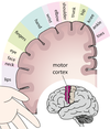
- largest of the lobes - initiates movement of the body (motor functions), language, association area controls planning, judgement, problem solving, aspecs of personality and emotions - Broca’s area is responsible for production of speech (part of association) - primary motor cortex at rear of each frontal lobe, adjacent to central fissure; responsible for movement of skeletal muscles - functions contralaterally (left side controlls right hand side of body) - toe part located at top, mouth at bottom

Parietal lobe
- enables peson to perceive your own body, and location of things in immediate environment - right pareital lobe enables person to perceive 3d shapes and designs - left pareital lobe is reading, writing and mental arithmetic + pointing out own body parts or things in a room - primary somatosensory cortex situated at front of each parietal lobe - responsible for processing sensation (pain, temp), functions contralaterally - if right damaged,can’t process information on left side

Occipital lobe
- vision - right side of each retina = right occipital lobe - information from centre of visual field is processed in both occipital lobe - different parts of primary visual cortex process different types of visual stimuli - if association cortex damaged, can’t recognise things by sight
Temporal lobe
- receives and processes auditory information and also the recognition of faces. - Contains Wernicke’s Area which is responsible for comprehending speech of others - Contains the Primary Auditory Cortex which receives auditory information. (upper part of temporal lobe) - damaged right temporal lobe = no recog of faces, songs,paintings - association areas important for processing of memory
Right hemisphere
- left side of body - analysis by touch - auditory cortex of left ear - spatial visualisation and analysis - some things are lateralised (only one side) eg language - left - music appreciation - art appreciation -fantasy - perception

Left hemisphere
- right side of body - speech centre - right hand touch - auditory cortex (right ear) - general interprative centre (language and maths) - logic

Association areas
- involved in integration of info between motor and sensory areas and higher-order mental processes - less specific function of neurons - damage = difficulty speaking, thinking, behaving normally

Location of lobes

Location of cortexes + lobes

Areas responsible for speech prod


