Basal Ganglia Anatomy and Function Flashcards
Frontal slide through the cerebrum showing both sides of the brain
White grey= grey matter; dark grey= white matter
Where are the basal ganglia located?
deep within the cerebrum (or telencephalon), the front most part of the brain
What are the basal ganglia?
Caudate
Putamen
Globus pallidus (internal and external)
Why is the substantia nigra black?
because it contains nuclei that contain black melanin
Close up of the basal ganglia. Where is the caudate nucleus located?
medial and along the lateral ventricles
IC= internal capsule
CC= corpus callosum
The basal ganglia are primarily involved in what?
movement control (somatic basal ganglia mostly)
motivation, reward, and affect (limbic basal ganglia)
The basal ganglia can be divided into what two categories of structure?
the somatic basal ganglia (dorsal) and the limbic basal ganglia (ventral)
What is the limbic basal ganglia composed of?
the nucleus accumbens, olfactory tubercles, and the ventral pallidum
Note that the somatic BG include the caudate, putamen, and globus pallidus
Left side view of the basal ganglia.
Note that the internal capsule seperates the caudate nucleus from the putamen in space
The tail of the caudate wraps behind and then ventral underneath the putamen to end as what structure?
amygdaloid nucleus
Note how the putamen and caudate nucleus are collectively called what?
the striatum
This image is a brain with a stain showing which nuclei make dopamine. Notice how the substantia nigra is the most dominant dopamine making nuclei
You can also see that the putamen, and the head and tail of the caudate are all brown. Why?
Because the substantia nigra ha axons that travel into the head of the caudate, the putamen, and tail of caudate, and form dopaminergic terminals there (the globus pallidus does not receive many dopamine terminals, so its pale)
The _____ ______ is the midbrain continuation of the internal capsule
the cerebral peduncle
Note the labels individual dopaminergic neurons of the substantia nigra. Where are they located?
in an upper part of the substantia nigra, called the pars compacta (because they are arranged in a compact layer)
The region beow the pars compact is called what?
the pars reticulata (a reticulum is a net-like meshwork of fibers) - note that this area contains few dopaminergic neurons. Rather, its neurons commonly very much resemble those in the globus pallidus in their chemistry, shape and function.
Thus, while the pars compacta and the pars reticulata together make up the substantia nigra, only the pars compacta really contains melanin
What neuron types make up the striatum?
A (aspiny) neurons and SN (spiny) neurons
In the striatum, 5% are A neurons
Describe A neurons
An aspiny neuron has dendrites that do not possess stubby protrusions on them (the stubby protrusions are called spines). About 95% of neurons in the striatum are SN neurons
T or F. SN neurons are usually smaller than A neurons
T.
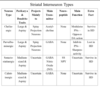
Closeup of a SN neuron. Again, A neurons are larger than SN neurons
Neurons that need to integrate info from diverse sources possess dendritic spines
One important point about spiny neurons is that they have what?
a long axon that leaves the striatum (by contrast, A neurons have axons that are short and dont leave the striatum)
Because of this, SN neurons in the striatum are called projection neurons, while the A neurons are called the local circuit neurons, or interneurons. Thus, it is spiny neurons that transmit decisions to other brain areas
All spiny neurons use what neurotransmitter?
GABA
What are the different types of projection (spiny) neurons based on?
-type of neuropeptide contained (neuropeptides are adjunct neurotransmitters that neurons often use, but they also can be neurochemical signatures for defining neuron subtypes)
Subtypes of spiny neurons in the striatum
1) One subtype of projection neuron in the striatum, in addition to making GABA, makes the opiod neuropeptide enkephalin (ENK).
2) The second subtype makes the neuropeptide substance P (these neurons project to the GPi)
The characteristics of the ENK neurons in the striatum is that they nearly all project where?
the GPe
Where are substance P and ENK found in the substantia nigra?
Substance P terminals are located in the upper shelf of the SN where dopaminergic neurons are located (the pars compacta, aka SNc) and in the region of the SN below the dopaminergic neurons (the pars reticulata, aka SNr). Thus, substance P neurons in the striatum include neurons projecting to the GPi, SNc, and SNR
By contrast, the ENK neurons in the striatum have only a hint of a projection to the substantia nigra, and that is only to the very most medial aprt of the SN
One important thing to know about substance P and ENK neurons in the striatum is that they are intermingled
In addition to containing substance P, these neurons also _______
a second neuropeptide, an opiod dynorphin
Notice how all striatal projection neurons possess a certain type of dopamine receptor. Those projecting to the GPe (ENK neurons) bind to what receptor?
D2
Notice how all striatal projection neurons possess a certain type of dopamine receptor. Those projecting to the GPi, SNr, and SNc (Substance P/Dynorphin neurons) bind to what receptor?
D1 receptors
NOTE: Remember that D1 and D2 receptors function differently
What do striato-GPe neurons (ENK) do?
inhibit conflicting movement with desired movement
What do striato-GPi neurons (SP/DYN) do?
promote/initiate limb movement
What do striato-Snr neurons (SP/DYN) do?
promote eye movement
What do striato-SNc neurons (SP/DYN) do?
regulate dopamine neurons
There are four types of interneurons in the striatum, distinguished by their neurochemistry, by the size of their cell body, and by which neuron in the striatum they project, and which parts of the brain they receive input from. What are they?
cholinergic
paravalbuminergic
somatostatinergic
calretinergic
Note that each interneuron type makes up about 1% of neurons in the striatum and together they make up about 5%
One important interneuron type in the striatum that hsa eben the target of therapies directed at basal ganglia disease is the ______
cholinergic- this interneuron has a big cell body
Colinergic interneurons in the striatum use what neurotransmitter?
ACh
Parvalbuminergic interneurons contian calcium binding protein and use what transmitter?
GABA (the neurons are larger than projection neurons, but smaller than the large cholinergic interneurons)
The somatostatinergic interneurons, containing somatostatin (a neuropeptide), uses what transmitter?
GABA (these neurons have cell bodies the same size as spiny neurons)
Calretinergic interneurons also use GABA as their neurotransmitter and contain _____
calretinin, a calcium binding protein
Which interneurons are impervious to Huntington disease?
somatostatin interneurons
Somatostatin interneurons also produce _____
Neuropeptide Y (NPY)
Note that as Huntington progresses, spiny neurons die. By grade 3, 60% of SNs are lost, and the striatum, GPe, and GPi are greatly shrunken as a result. Nonetheless, somatostatin neuron abundance is preserved in grade 3 and 4.
Note that ____ and ______ interneurons also survive well in Huntington disease
Cholinergic and calretinergic interneurons; but Parvalbuminergic interneurons do not survive well
What is the main function of cholinergic interneurons?
modulate PNs- opposes DA action
What is the main function of parvalbuminergic interneurons?
inhibit PNs- sharpen activity
role of somatostatinergic and calretinergic interneurons unknown
What is the major neuron type with the globus pallidus?
large A neurons, with long dendritic trees arranged in teh vertical axis of the GPe and GPi. Many striatal neurons send their terminal onto the dendrities of any given globus pallidus neuron and they integrate info from the many striatal neurons that project to it.
Both GPe and GPi neurons are ____-ergic and they send their axons out of the globus pallidus to their target area
GABA
Where does the GPe project to?
the subthalamic nucleus
Where does the GPi project to?
the motor thalamus
There are two nuclei making up the motor thalamus, namely:
the ventral anterior nucleus (VA)
ventral lateral nucleus (VL) (two parts- the C (caudalis) and O (oralis) parts)
The ______ ___ ______ _____ also projects to the motor thalamus
substantia nigra pars reticulata
Cortical projection targets of the motor thalamus. Where does the VA project to? VL?
VA: area 6, which is pre-motor cortex
VL: area 4, which is motor cortex
Note that this illustration makes the point that the basal ganglia send much of its output to motor-related cortex
What are the major inputs into the striatum?
the cortex
the thalamus
dopaminergic input from the substantia nigra pars compacta
What is the CM of the thalamus?
the centre median, part of the intralaminer thalamus (note that the intralaminar thalamus is the source of one of the major inouts to the striatum, but it is not currently well defined)
The major input to straitum that is clearly central to the function of the basal ganglia is what?
input from the cerebral cortex- almost all of the cerebral cortex projects to the striatum
The cortical neurons that project to the striatum are what?
layer 5 pyramidal neurons. Many of the neurons in layer 5 that project to the striatum also send motor commands to the spinal cord and pre-motor neurons of teh hindbrain
Note that dopaminergic terminals (from the pars compacta of the SN) for input into the basal ganglia are located where?
in the caudate and putamen, not the GPe or GPi
How does the cortical input transmit to the basal ganglia?
the cerebral cortex input ends on the tips of the spines of the spiny neurons, using glutamate as its neurotransmiiter.
How does the SN dopaminergic input transmit to the basal ganglia?
it ends on the sides of the spines of spiny neurons (this the dopaminergic input can modulate the corticla input to the spine ends)
Interplay of the striatal input to the globus pallidus and the subthalamic nucleus input. Explain
WHile the GPe projects to the subthalamic nucleus, the subthalamic nucleus mainly projects back to the GPi, by wrapping around the smooth dendrities of the GPi neurons, which also receive terminals from striatal substance P containing neurons
