Bacteria Flashcards
defensins
hydrophobic, cationic
pisitive - stick to neg charged membrane in bacteria and make pores
gram stain
- blue stain
- compexing agent - make stain into larger molecules
- extraction agent - pull out stain, but only works on gram negative!!
- red stain - only sticks on gram negative

gram positive cell membrane structure
peptido glycan cell wall - 1 really thick cell wall
single plasma membrane
many layers and extensive crosslinking in cell wall - gram stain cannot be washed out

Gram negative cell envelope structure
cytoplasmic membrane, very thin, non complex cell wall, outer cell membrane with LPS on the very outside
minimum number of layers and minimum cross linking - stain can be washed out

LPS
on outside of cell envelope in gram NEGATIVE - inflammatory and parrier
gram negative ONLY - for viability and some innate antibiotic resistance (i.e. PCN doesn’t wrk against a lot of gram negative because it has to get through LPS)
peptidoglycan sructure
N-acetylmuramic acid, N acetylglocusamine, pentapeptide ending in D-ala, D-ala
linked and then cross linked to make cell wall
disaccharaide w pentopeptide side chain, op together for cell wall and give the bacteria shape
GM + and -
osmotic integritity and shape - strength

Type 3 secretion system
only find in gram neg- 2 membranes
molecular syringe, inject proteins into cell - in eukaryotic cell
do bad things - paralyze or kill cell
evolutionarily related to flagella
LPS Lipid A
if purify and give to someone - massive immune respoinse!
fatty acids attach and anchor LPS into outer membrane
phosphorylated glucosamine disaccharide backbone

Core polysaccharides of LPS
branched polysaccharide of 9-12 sugars
if gram neg - need LPS to be vaible - core and lipid A

LPS O-specific antigen
repeating unit structure
long linear polysacchadie - variable, different repeating sugars
major serologic determinant
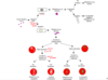
Transformation
bacterium takes up free DNA and adds into genome
takes up as single strand and releases soluble nts, then repairs to have 2nd strand
big receptor or small receptor

neisseria antigenic variation
transformation!
Neisseria species can vary their surface structures, including pilli and capsule. It is clear that natural transformation plays a role in this process, allowing Neisseria to share genes encoding variations of these structures.
PCN resistance in streptococcus pneumoniae
Transformation
penicillin resistance has become widespread amongst Streptococcus pneumoniae strains. In this case the penicillin resistance is due to altered penicillin-binding proteins (PBPs) which exhibit a low affinity for beta lactam antibiotics. Comparison of the nucleotide sequences encoding the PBPs in S. pneumoniae and S. mitis demonstrates that horizontal gene transfer has occurred between these two bacteria.
transduction
bacteriophages
viruses that attack bacteria and are specific for closely related bacterial species
virulent phage
virulent phages - always cause lysis and release of phage particles - clear plaques on bacterial lawns
temperate phage
may cause lysis OR may integrate stably into the bacterial host’s chromosome, generate turbind paques and persst as prophages
induced by DNA damage to excise and repliicae
all prophage genes are repressed except for a phage repressor gene
Temperate phages may establish a state of dormancy within the cell, often by integrating into the chromosomal DNA at a specific attachment site, by means of a phage-coded integrase enzyme. The dormant phage is known as a prophage and the state of dormancy is known as the lysogenic state

intermediate phages
replicate stably int he host cell and continually release progeny
no lysis
lysogenic state
phage is in a dormant state in bacteria
integrating chromosomal DNA at a specific site
dormant phage = prophage
lysogenic conversion
Certain temperate phages have incorporated bacterial genes that have nothing to do with the phage life cycle. When a bacterial cell is lysogenized by such a phage, any such incorporated gene is expressed and becomes a phenotypic trait of the bacterium. The best-known genes of this type are toxin genes, including genes for the diphtheria, tetanus, and scarlatiniform toxins
generalized transduction
occasionally encapsulate host DNA, which is transferred to any new host upon infection
have different chromosomal segments stuffed in pacteriophage head
any bacterial gene transferred
typically only bacteria genes - no viral genes
probably accidental consequence of phage multiplication - no proven clinical relevance

specialized transduction
always take same genetic info!
pecialized transduction is the process by which a restricted set of bacterial genes is transferred to another bacterium. The genes that get transferred (donor genes) depend on where the phage genome is located on the chromosome. Specialized transduction occurs when the prophage excises imprecisely from the chromosome so that bacterial genes lying adjacent to the prophage are included in the excised DNA. The excised DNA is then packaged into a new virus particle, which then delivers the DNA to a new bacterium, where the donor genes can be inserted into the recipient chromosome or remain in the cytoplasm, depending on the nature of the bacteriophage.

plasmids
non essential but hereditarily stable, self replicating
circular and supercoiled
medically important accessory functions
bacterial mating!
conjugation
pilus - gram 0 membrane fusion - single strand into 2nd bacteria - replicate into plasmid or integrate into chromosome
plasmids spread rapidly - toxins are plsmid encoded - abx resistance

transposons
discrete segments of DNA that encode recombination enzymes - transposases
move from one dnA location to another