B5.019 - Non-Neoplastic GI Pathology Histo COPY Flashcards
cell types in the esophagus and stomach
esophagus - squamous
stomach - columnar

normal esophagus

normal esophagus
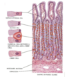
layers of normal esophagus


esophageal mucosa: stratified squamous epithelium with papillae
symptoms of esophageal disorders
dysphagia
odynophagia - pain upon swallowing
heartburn - retrosternal chest pain
hematemesis - vomiting of blood
melena - blood in stools
esophagitis
an inlammatory process of the esophagus cuased by biochemical acid reflux, infectious, inflammatory or chemical agents
symptoms of infectious esophagitis
patients usually present with odynophagia
more common in immunosuppressed and elderly
most common causes of infectious esophagitis
HSV and CMV - reactivation of latent virus in laryngeal or superior cervical nerves
Candida - normal flora, colonzation due to structure or obstruction
describe epidemiology of HSV and gross/micro
usually opportunistic/immunosuppressed paitients. Self limited in healthy
gross: shallow vesicles and ulcers
micro: viral inclusions present and mulitnucleated squamous cells at margin of ulcer with thickened nuclear membrane and ground glass inclusions that fill nuclei

punched out ulcers from HSV infection

shallow ulcer with granulation tissue and superficial necrosis (L) and squamous mucosa (R) seen in HSV

high power of rim/edge of ulcer demonstrating pahtognomic cytologic featurs of HSV
red arrow - multinucleation, nuclear molding and
yellow arrow - nuclear margination
in squamous epithelium
HSV
what are the 3 Ms and what are they associated with
Multinucleation
Margination
Molding
HSV
describe the epidemiology of CMV and gross/micro appearance
immunocompromised patients
gross: punched out mucosal ulcers similar to herpes
micro: virus present in endothelium and enlarged stroma cells at ulcer base; inclusions are intranuclear surround by clear halo, often with coarse intracytoplasmic granules
owl eye inclusions

punched out ulcers seen in CMV or HSV

granulatino tissue in bed of ulcer (infecting endothelial and stromal cells) with nuclear and cytoplasmic inclusions
CMV

CMV in gastric pyloric glands with classic Owl eye nuclear inclusions
what is the most common cause of infectious esophagitis
candida
describe candida esophagitis
associated with antibiotc use in non immunocompromised
usually due to candida albicans
fungal invasion a requirement for dx since its normal flora in GI tract
endoscopy findings of candida esophagitis
gray white pseudomembrane or plaques in mid to distal esophagus; mucosa is erythematous, edematous, ulcerated or friable.

candida esophagitis
top arrow - distal esophagus
middle arrow - white plaques
bottom arrow - erythematous mucosa

candida esophagitis
superficial squamous mucosa with neutrophils

candida esophagitis
Gomori methamine silver stain highlighting fungal hyphae
note: it has to be invaded otherwise it could be normal flora
non infectious causes of esophagitis
reflux
eosinophilic
pill esophagitis
toxins/chemicals
clinical symptoms and sequelae of reflux esophagitis
clinical - heartburn, regurgitation and chest pain
sequelae - bleeding, strictures and barrets esophagus
pathogenesis of reflux esophagitis
multifactorial, incompetent LES, hiatal hernia, increased gastric volume, obesity, alcohol, tobacco, CNS depressants, pregnancy
reflux esophagitis gross appearance and histo
gross - redness, erosions
histo - elongation of papillae, basal cell hyperplasia, intraepithelial eosinophils and neutrophils

normal esophagus

reflux esophagus

normal esophagus

reflux esophagitis
Note the basal cell hyperplasia and papillary elongation. Maturation of the epithelium is decreased with more immature cells present above the normal 1-2 cell thickness. Also note the elongation of the lamina propria papilla extending to upper third of epithelium

numerous intraepithelial eosinophils
reflux esophagitis
what are symptoms of eosinophilic esophagitis and treatment
most atopic
symptoms - food impactions, dysphagia, GERD like in children
treatment - diatary restriction (six food elimination diet SFED: milk, egg, soy, wheat, peanuts/tree nuts, fish/shellfish, other)
steroid inhalation
gross and histo appearance of eosinophilic esophagitis
furrowed esophagus, trachealized esophagus (felinzation)
Histo - similar to reflux, papillary hyperplasia, basal hyperplasia, eosiophils; also superficial clustering, degranulation of eosinophils
note: the eosninophils are not confined to distal esophagus

trachealization or felinzation of esophagus seen in EoE

EoE
Some eosinophilic microabcsesses and mostly superficial location
what is barretts esophagus
probably complication of longstanding reflux
more common in middle aged white males
replacement of normal distal stratified squamous mucosa with intestinal type glandular mucosa
pathogenesis of barrets esophagus
reflex induces inflammation and mucosal injury
healing occurs by ingrowth of stem cells and re-epithelialization
cells differentiatin into abnormal inestinal mucosa that may be more injury resistant
gross and histo appearance of barretts esophagus
gross - irregular band dark pink, velvety mucosa extending upwards as tongues of mucosa, may be very patchy
histo - metaplastic columnar epithelium with goblet cells

barrets esophagus
endoscopically the gastroesophageal junction has tongues of velvety red tongues of metaplastic mucosa extending upward with adjacent pale squamous mucosa

barretts esophagus

higher power view of abundant intestinal metaplasia in esophagus
barrets esophagus
goblet cells - arrows
barrets esophagus sequelae
ulceration
bleeding
stricture
dysplasia
name the layers


name the cell types


body/fundus of stomach with pairetal and chief cells

antrum with mucous cells
what is gastritis and what are the types
inflammation of gastric mucosa caused by a variety of agents, chemical, infectious, autoimmune
acute - transient and self limiting, hemorrhagic, erosive
chronic - environmental - H. pylori, autoimmune
what is gastropathy
when inflammatory cells are absent or rare
diverse set of disorders marked by injury or dysfunction
NSAIDs, alcohol, bile, stress induced injury
what is acute gastritis
common mucosal acute inflammatory process
often transiet
can be accompanied by erosions, hemorrhage
severity ranges from mild to massive
usually resolves if stimulus is removed
gross and micro path of acute gastritis
gross - hemorrhage, erosions, edema
micro - inflammation, neutrophilic>chronic
focal or diffuse
superficial or full thickness
what is chronic gastritis
often asymptomatic, pain, nausea, vomiting
defined as a chronic inflammatory process which may lead to:
ulceration
atrophy
metaplasia –> dysplasia –> carcinoma
lymphoma (Helicobacter)
etiology of chronic gastritis
H pylori
autoimmune
chemical/reactive (NSAIDs, bile reflux, alcohol)
other - uremia, radiation
what is H. pylori
non invasive spirillar gram negative rod (spirochete) producing urease and other toxins
highly prevalent esp in developing countries
risk factors are socioeconomic
reservoir/transmission and gross appearance of H. pylori
humans/water
fecal-oral, oral oral, not really known
increased risk of carcinoma and lymphoma
gross - normal usually, may see erythema, nodularity, ulcers

H pylori gastritis

h pylori gastritis

h pylori gastritis
chronic HP associated gastritis histo
dense lamina propria lymphoplasmacytic infiltrate
+/- neutrophils infiltrating glands
HP may be found in mucus layer (NOT within cells)
antrum > body
what can HP gastritis lead to
atrophy, metaplasia, dysplasia

low power view of antral type mucosa with expansion of lamina propria by chronic inflammatory cells, including lymphoid aggregate and active inflammation.
H pylori gastritis
inset shows helicobacter pylori immunostain with organisms in mucin between cells NOT WITHIN CELLS

H pylori gastritis
chronic active gastritis with active inflammation with neutrophils in epithelium and expanded lamina propria with predominantly plasma cells
intraepithelial neutrophils and subepithelial plasma cells are characteristic of H pylori

h pylori gastritis
complications of h pylori gastritis
peptic ulcer disease
atrophic antral gastritis leading to intestinal metaplasia
adenocarcinoma
lymphoma
treatment of HP gastritis
triple therapy
proton pump inhibitor (omeprazole) and 2 antibiotics

atrophy with IM, complication of h pylori
clinical features of peptic ulcer disease
dyspepsia, epigastric pain, melena, hematemisis, anemia
pathogenesis of peptic ulcer disease
h pylori infection most common
hyperacidity - zollinger ellison syndrome
NSAIDs
where is peptic ulcer disease found and how is it treated
98% in antrum and duodenal bulb
lesser curvature of stomach
clean base and smooth edge
responsive to antibiotics
biopsy
what is AMAG
autoimmune metaplastic atrophic gastritis
describe AMAG
antibodies are present - antiparietal cell antibody
anti-intrinsic factor antibody
affects parital cells, decreased parietal cells and decreased acid production
leads to decreased B12 absorption
what is pernicious anemia
from loss of B12, a complication of AMAG
gross appearance, histo of AMAG
flattened/atrophic gastric mucosa
histo - biopsy of the body/fundus shows lack of parietal cells; normal cells replaced by mucous cells (looks like antrum) or intestinal metaplasia
lab tests for AMAG
high gastrin, anti parietal or anti intrinsic factor antibody; microcytic anemia at first but then changes to macrocytic anemia
treatment of AMAG
B12 supplementation (shots)

AMAG
atrophy with intestinal metaplasia (goblet cells)

shows IF anti parietal cell Ab in AMAG
compare and contrast HP and AMAG

what part of the GI system does celiacs affect
small bowel

small bowel
mucosa comprised of epithelium and lamina propria in a villous architecture. epithelium is columnar, single layer, with goblet cells
what is celiacs disease presenation, gross/micro appearance
malabsorption, diarrhea (light colored, foul smelling)
gross - cracked earth appearance
micro - blunting of villi, increased intraepithelial lymphocytes
treatment of celiacs
gluten free diet

celiac disease

celiac - left; blunted villi and increased intraepithelial lymphocytes
normal - right

normal colon
mucosal lining similar to small bowel but no villli formation

normal colon - equal parts epithelium and lamina propria, organized and regular test tume like crypts
what are ulcerative colitis and crohns disease
UC - confined to colon, distribution usually confluent and starts at left side/rectum
crohns - can involve entire GI, skip lesions, granulomas and deep ulcers with stricture are hallmarks
what test can you do to look for ulcerative colitis that sets it apart
pANCA - 60-80%+
presentation of inflammatory bowel disease
chronic diarrhea
crohns may have upper GI symptoms or present with bowel obstruction symptoms
endoscopy and histo of IBD
endoscopy - erythema and friability of mucosa; ulcerations
histo - chronic colitis; crypt architecture distortion
basal lamina propria lymphoplasmacytosis
active disease has neutrophils

chronic colitis - crypt distortion

crypt distortion typical of IBD
branching crypts, no longer test tube shaped
green arrow - basal lymphoplasmacytosis

chronic active colitis
crypt abscesses
UC prognosis/treatment
no cure
treatment - anti-inflammatory and immunosuppressive drugs; surgery if not responsive
crohns therapy and prognosis
no cure
treatment - anti inflammatory and immunosuppressive drugs, monoclonal TNA alpha ab
surgery for complications
increased risk of adenocarcinoma (UC as well)
microscopic colitis pathogenesis and presentation
presentation - chronic, watery diarrhea in middle aged to elderly patients
pathegenesis - incompletely understood, thought to be autoimmune
gross/endoscopy apperance, histo, treatment of microscopic colitis
gross - normal
histo - surface epithelium attenuation, lymphocytic, collagenous
treatment - symptom management, anti inflammatories

lymphocytic colitis (microscopic)

collagenous colitis (microscopic)
symptoms of ischemic colitis
acute onset, bloody diarrhea, abdominal pain, usually elderly
pathogenesis of ischemic colitis
atherosclerotic CVD
hypercoagulability
global ischemia
gross appearance of ischemic colitis
dark red/hyperemic abrupt transition to normal mucosa; watershed areas affected more often (splenic flexure, sigmoid colon) can have patchy psudomembranes
microscopic apprearance of ischemic colitis
attenuation of epithelium beginning at surface, can lead to crypt drop out (withered crypts)
necrosis if severe
hyalinization of lamina propria chronic
treatment of ischemic colitis
supportive
correct cause
may need surgical resection

ischemic colitis
normal colon on left, transitioning to crypt epithelial attenuation at arrow. Also note deep pink color of lamina propria - hyalinization

ischemic colitis
surface epithelial attenuation and pink hyalinization of lamina propria
differential for ischemic colitis
psudomembranous colitis - pathy pseudomembranes, hyanalized lamina propria and withered crypts favor ischemia
EHEC - right sided involvement and fibrin thrombi favor this