Axial Muscles Flashcards

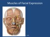
A. Levator labii superioris alaeque nasi
B. Levator labiisuperioris
C. Zygomaticus minor
D. Zygomaticus major
E. Rosorius
F. Platysma
G. Levator anguli oris
H. Orbicularis oris
I Mentalis
Epicranius (Frontal and occipital belly of occipitofrontalis, Galea aponeurotica)
A Corrugator supercilii
B Orbicularis oculi
C Procerus
D Nasalis
E Buccinator
F Depressor labii inferioris
G Depressor anguli oris

A. Auricularis anterior
B Temporoparietalis
C Auricularis Superior
D Auricularis posterior
E Occipital belly of occipitofrontalis
Depressor labii inferioris action
Depresses lower lip
Levator Labii Superioris Action
Elevates upper lip
Levator anguli oris action
Elevates corner of mouth
Mentalis action
Elevates and protrudes lower lip
Obicularis oris action
Compresses, purses lips
Risouris action
Draws corner of mouth to side
Depressor anguli oris action
Depresses corner of mouth
Zygomaticus action
Retracts and elevates corner of mouth
Zygomaticus minor action
Retracts and elevates upper lip
Corrugator supercilii action
eyebrow movement
Levator palpebrae superioris action
Elevates eyelid
Obicularis oculi action
Closes eye
Buccinator Origin and insertion
O: Alveolar process of maxilla and mandible and pterygomandibular raphe
I: blends into orbicularis oris fibers
CN 7
Pierced by parotid duct
Pterygomandibular raphe
Upper and Lower attachments
What attaches to it
Linear cord like connective tissue ligament
- Upper attachment
- Pterygoid hamulus
- Lower attachment
- Mandible, posterior to 3rd molar
- Point of attachment for buccinator and superior pharyngeal contrictor muscles

Lateral Pterygoid
Superior O and I
Superior
O: Greater sphenoid wing, lateral surface
I: TMJ articular disc and capsule
Lateral Pterygoid
Inferior O and I
Inferior
O: Lateral pterygoid lateral surface
I: Ramus and condylar
Lateral Pterygoid Action
Protrudes
Depresses
Lateral movement (deviates to injured side)
DOES NOT ELEVATE
Lateral pterygoid surrounding structures

- Maxillary artery runs either superficial or deep to it
- Surrounded by pterygoid venous plexus
- Buccal branch of CNV passes btw the 2 heads
Medial Pterygoid
Superficial O and I
O: Maxillary tuberosity
I: Medial surface of ramus and angle
Medial Pterygoid
Deep O and I
O: Lateral pterygoid medial surface
Medial pterygoid lateral surface
Pterygoid fossa
I: Medial surface of ramus and angle
Medial pterygoid actions
Elevates! Slight protusion and lateral movement
Medial Pterygoid Contacts 5 structures
Clical significance
- Contacts
- Parotid gland
- Inferior alveolar artery
- Lingual nerve
- Inferior alveolar nerve
- Chorda tympani nerve
- Deepest muscle of mastication
- If needle is inserted below the mandibular foramen can be pierced during mandibular block
Masseter
Superficial O and I
O: Anterior 2/3 of zygomatic arch
I: Angle and ramus lateral surface
Masseter
Deep O and I
O: Posterior 1/3 of zygomatic arch
I: Coronoid and ramus lateral surface
Masseter action
Elvate! Some protrude and lateral
What 3 structures pass the Masseter
- Passing superficial to masseter
- Parotid duct
- Tranverse facial a.
- Branches of CNVII (7)
Temporalis portions
- Anterior
- Vertical fibers
- Middle
- Oblique fibers
- Posterior
- Horizontal fibers
Temporalis
O and I
O: Temporal fossa, inferior temporal line, Temporal fascia
I:Anterior border of ramus
Temporalis action
Elevates mandible into resting position
Retract and lateral movement
What are 4 other muscles innervated by CN V3
Mylohyoid
Tensor tympani
Tensor veli palatini
Anterior digastric
Sternocleidomastoid
O and I
- O: Manubrium and clavicle
- I: Mastoid region of the skull
Sternocleidomastoid action
Nerves
Bilateral, flex neck
Unilateral, rotation
Face turns to contralateral side
Head tilts ipsilateral side
N: CN XI and C2-3
Oblique Cervical Muscles
Anterior Scalene
Middle Scalene
Posterior Scalene
Scalenes
Attach
Action
Innervates
Attach: TP of C2-C7 and first 2 ribs
Action: Elevate ribs or flex neck
Innervated: by C4-C6
Scalene, structures associated
Subclavian artery and brachial plexus pass between the anterior and middle scalene
Subclavian vein and phrenic nerve pass anteriorly to the anterior scalene
Suprahyoid Muscles
Digastric
Stylohyoid
Mylohyoid
Geniohyoid
Digastric
Anterior and posterior
- Anterior belly
- Origin
- Mandible
- Insertion
- Hyoid
- Origin
- Posterior
- Origin
- Mastoid Notch
- Insertion
- Hyoid
- Origin
Digastric Action
Elevate hyoid
Depresses/Retracts mandible during swallowing
Digastric Innervation
Anterior: CN V3 (Mylohyoid branch)
Posterior: CN VII (Digastric branch)
Stylohyoid
O
I
N
A
Origin: Styloid process
Inserion: Hyoid
N:CN VII
A: Elvate hyoid during swallowing
Mylohyoid
O
I
N
A
O: Mylohyoid line of mandible
I: Hyoid via fibrous raphe
N: CN V3 (Mylohyoid branch)
A: Tightens and elevates oral floor
Depresses, retracts, side to side mandible
Geniohyoid
O
I
N
A
O: Inferior mental spines (genial tubercle)
I: Hyoid
N: C1 via CN XII
A: draws hyoid forward
depresses/retracts mandible
What makes up the floor of the oral cavity
- Mylohyoid muscles
- Muscular diaphragm
- Geniohyoid muscles
- cord like muscles
- Superior to mylohyoid
- Tongue
- Superior to geniohyoid
