Auditory function and the vestibular system Flashcards
Define pitch
Perception of frequency.
Define frequency.
Speed of vibration. Wave cycles per second (Hz).
Define intensity.
Power of the sound. Determined by amount of energy delivered per second (joules/second passing through 1m2).
Diagram outling gross anatomy of the ear.
Explain the divisions of the external, middle and internal ear.
External - part of year attached to lateral aspect of the head.
Middle - Cavity in petrous part of temporal bone. Separated from inner by tympanic membrane.
Internal ear - series of cavities within petrous part of the bone, with the internal acoustic meatus medially to allow passage of vestibulocochlear nerve
What is the external acoustic meatus?
cavity that extends from deepest part of concha –> tympanic membrane.
What lines the external acoustic meatus?
walled by cartilage and bone; lined by skin with hair and modified swear glands
What does the tympanic membrane separate?
external acoustic meatus from middle ear (malleus bone) (sits at an angle)
What lines the tympanic membrane on either side?
skin on outside and mucous membrane on inside
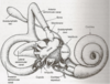
Gross illustration of middle and inner ear.
Label middle ear structures present.
Explain the gross anatomy of the middle ear.
air-filled space in temporal bone lined by mucous membrane.
Consists of tympanic cavity adjacent to the membrane and epitympanic recess superiorly
Where do the bones of the middle ear reside?
in the epitympanic recess.
What is the role of the middle ear?
transmits vibrations of the tympanic membrane across cavity to the internal ear via three interconnected but moveable bones
What are the bones of the middle ear and their connections?
Malleus: connected to membrane
Incus: connected to malleus via synovial joint
Stapes: connected to incus via synovial joint and attached to lateral wall of internal ear at the oval window
What is the function of the bones of the middle ear?
transmit vibrations by matching impedance and reducing loss of energy (frequency at which impedance of system is minimal is the resonant frequency)
What muscles are located in the middle ear and what is their role?
tensor tympanic and strapedius muscles.
Control tension of tympanic membrane.
What is the function of the inner ear?
cochlear duct facilitates hearing.
semi-circular ducts, utricle and saccule facilitate balance.
What innervates the balance organs of the inner ear?
Where does it exit?
Vestibocochlear nerve (VIII)
via internal acoustic meatus.
What are the two gross components of the internal ear?
bony labyrinth (bony cavities)
membranous labyrinth (membranous ducts/sacs)
Where is the internal ear located?
petrous part of the temporal bone.
Explain the mechanism of sound transmission.
vibration of tympanic moves malleus and incus laterally, pushing the staples medially onto the oval window –> a wave in fluid filled cochlea.
Wave moves through scala vestibuli –> bulging of secondary tympanic membrane –> basilar membrane vibration + stimulation of spiral organ receptor cells.
Wave flows round scala tympani and exits at round window.
Diagram to show direction of cochlear wave movement.
How does the motion of the stapes generate vibration of the basilar membrane?
medial movement of stapes –> pressure difference between liquid filled chambers in the cochlea generates a wave of vibration.
What is the role of the basilar membrane?
elastic structure with heterogenous mechanical properties.
vibrates at different positions along length in response to different frequencies.
breaks complex sounds down by distributing energy of each component along length.
What are the components of the organ of corti?
Outer and inner hair cells.
Deiter cells (supporting cells)
Basilar and tectorial membranes.
DIagram of organ of corti.
What are the components?
CT = tunnel of Corti.
SC = Deiter cells (supporting cells)
How do hair cells generate an electric signal?
motion of basilar membrane deflects hair bundle –> bending of stereocilia –> tallest stereocilium.
Internal voltage of cell changed –> electric signal generated (i.e. mechanotranduction)
How are stereocilia connected? What is the role of this connection?
filamentous linkages called tip links.
Tip links share location with ion channels allowing for disruption by loud noises.
How does mechanotransduction lead to generation of electrical impulses?
tip links stretching –> openubng of ion channels + influx of ions –> AP generation.
Why is amplification of sounds necessary?
energy lost in damping effects of cochlea liquids.
How is sound amplified?
Active process.
Live BMs vibrate far more in response to its resonant frequency, and at selective frequencies, than the low-level and broad response of a dead BM
Low intensity stimulation is amplified 100-fold, but larger stimuli are amplified less
Compare the roles of inner and outer hair cells.
Inner: provide sensory tranduction. Account for 95% of afferent projections.
Outer: receive most efferent projections. activation of efferent fibres leads to enhance selectivity and sensitivity for specific frequencies;
How do outer hair cells modulate sensitivity and selectivity for specific sound frequencies?
cell body of efferent fibres shortens and elongates when internal voltage is changed due to prestin (protein) reorientation
Where do IHCs synapse with sensory neurones? How is NT release modulated here?
in the cochlear ganglion.
Constant NT release at rest. Rate adjusted in response to change in presynaptic voltage (due to mechanotransduction gating).
Where does the cochlear nerve transmit information to?
cochlear nucleus.
(each axon of nerve responsive to a single frequency)
Explain how cochlear tonotopic mapping arises.
ganglion cells respond best to resonant frequency of basilar membrane in same area.
Neurones in cochlear nuclei of brainstem arranged so that low frequencies are carried ventrally and high frequencies dorsally.
How do cochlear nuclei in the medulla help determine sound origin location?
receives glossopharyngeal input from ears and interpret visual cues.
High frequency sounds can produce constructive/destructive interference depending on site of origin.
This allows ears to detect sound location via the asymmetry of waves on each side.
What is the role of T-stellate cells?
encode sound frequency and intensity of narrowband stimuli
What is the role of Bushy cells?
produce more sharp but less temporally precise versions of cochlear nerve fibres to allow encoding of relative time of arrival of inputs to two ears
What is the gross role of the superior olivary complex in the Pons?
receive glossopharyngeal input + compare the bilateral activity of cochlear nuclei.
What is the role of the medial superior olive?
computes interaural time difference with bushy cell information about timing of inputs
What is the role of the lateral superior olive?
detects difference in intensity between ears because neurones are excited by sounds arising from ear in same side and inhibited by opposite sounds - interaural level difference computed to localise sounds in horizontal plane
What are the outputs of the superior olivary complex?
Send feedback to hair cells to balance responses from both ears and reduce sensitivity of the cochlear.
MSO: to IHCs bilaterally
LSO: to OHCs ipsilaterally.
What is the role of the inferior colliculus?
Convergence of ascending pathways (only central nucleus is tonotopically organised).
Neurones become responsive to complex sounds.
Signal integration, frequency recognition and pitch discrimination.
What is the role of the superior colliculus?
Merging of auditory and visual maps.
Neurones respond to stimuli with specific sound direction.
Auditory map generated here is essential for reflex head and eye response to auditory stimuli.
Where is the primary auditory cortex located?
superior bank of temporal lobe.
Diagram to show gross auditory pathway
Define conductive deafness.
ear not capable of transmitting vibration of sound onto cochlear
What can cause conductive deafness?
Fluid accumulation in the middle ear
Perforated tympanic membrane
Abnormal bone growth obstructing the ear canal
Barotrauma
Define sensorineural deafness.
hearing loss due to a problem in the sensory apparatus of the inner ear or vestibulocochlear nerve
What can cause sensorineural deafness?
Loud noise exposure
Meniere’s disease - excess of fluid in cochlea
Genetic mutations in Organ of Corti
Aminoglycoside antibiotic toxicity in hair cells
Congenital diseases
Ageing
Give examples of how brain dysfunction might lead to hearing loss.
Demyelination due to inflammation or viral causes.
Blast injuries causing disruption in balance between inhibition and excitation.
What are the roles of the vestibular system?
Perception of movement in space and tilt with respect to gravity
Provide reflexive balance reactions to unstable gait
Stabilise the eyes on fixed targets to preserve visual acuity during movement
Assist BP/HR control during tilting
Provide a reference for absolute motion in space
What is the gross function of the vestibular system?
control balance, orientation and sensation of movement.
Diagram showing key structures of the vestibular sytem.
What are the gross structures of the vestibular system?
Semicircular canals: anterior / posterior / lateral; connected to utricle at both ends, with an ampulla at one end
Utricle: centre of the system, connected to canals and saccule
Saccule: connects the utricle and the cochlea
What is the function of the otolith organs?
allow sensation of linear acceleration in 3D space
(utricle + saccule)
How do the utricle and saccule allow for the sensation of linear acceleration?
hair cells are stimulated by deflection of forces of inertial resistance to acceleration.
Plane of utricle horizontal with verticle hair cells sensitive to lateral + A/P movement.
Plane of saccule verticle with horizontal hair cells sensitive to verticle + A/P movement
What is the role of the kinetic labyrinth?
allow sensation of rotational acceleration.
What are the angles of the semicircular canals?
Anterior canal: 45deg anterior
Posterior canal: 45deg posterior
Lateral canal: lie horizontally
How do hair cells allow for the sensation of rotational acceleration?
hair cells exist in the ampulla of the canals, and each has a tonic firing rate - when still, tonuses of each side cancel out; rotation acceleration leads to preferential stimulation of the canals in that side (e.g. Turning right stimulates right canals), and then stopping leads to stimulation of the opposite side; pattern from all canals computed to yield rotation in 3D space
Explain cupula movement
fluid movement form semicircular canal toward utricle is excitation; fluid movement from utricle to canal leads to inhibition
Explain how hair cell firing rate is modulated.
transduction - tilting/movement of head –> movement of hair cells.
Deviation –> kinocilium –> depolarisation –> increased firing frequency (vice versa).
What is the difference between type 1 and type 2 hair cells?
Type 1: greater in number. Direct afferents, indirect efferents, rounded shape.
Type 2: fewer in number, direct afferents and efferents, columnar in shape.
How is the vestibular nerve divided?
into inferior and superior nerves.
Both pass through internal auditory meatus in vestibulocochlear nerve leading to the vestibular nuclei in the brainstem.
Explain the vestibular blood supply.
basilar artery forms anterior cerebellar artery to supply cochlea and vestibular systems.
What vestibular nuclei are the static and kinetic labyrinths connect to?
Static labyrinth: connected to lateral and inferior nuclei
Kinetic labyrinth: connected to medial and superior nuclei
What is the purpose of the vestibulo-ocular reflex?
keep imaged fixed while head is moving.
Explain the vestibulo-ocular reflex (3 stages).
- Vestibular nucleus on side in direction of turn sends excitatory impulse –> contralateral abducens nucleus. opposite inhibits the other nucleus
- Nucleus abducens on side opposite to rotation uses the abducens nerve to contract lateral rectus + an interneuron to contract medial rectus
- Nucleus abducens on side of rotation inhibits abducens nerve and oculomotor CN III, so lateral rectus of that eye and medial rectus of the other are inhibited
Result: eyes move in opposite direction with same velocity. Eye + head movement cancel each other out.
Diagram of vestibulo-ocular reflex.
Explain the connections of the vestibulo-spinal reflexes in the lateral vestibulo-spinal tract.
(ipsilateral motor neurones –> limb muscles)
vestibular nerve synapses in lateral vestibular nucleus, fibres then descend down cord in LV tract to synapse in ventral horn at correct level.
Explain the connections of the vestibulo-spinal reflexes in the lateral vestibulo-spinal tract.
(bilateral motor neurones to neck and back muscles).
Vestibular nerve synapses in medial vestibular nucleus. Motor bundle then splits - some contralateral, most ipsilateral. Both travel down MV tract and synapse in ventral horn at correct spinal level.
Define vertigo.
illusion of movement - eyes moving (traditionally with nystagmus)
Define oscillopsia.
visual field oscillates as a result of ocular instability after damage to the oculomotor system; world will appear to bounce or lag behind head movement
What conditions might be red herrings for an acute vertigo diagnosis?
Presyncope with postural blood pressure
Pulmonary embolism with arterial saturation
Cardiac arrhythmia with an ECG
What can cause acute vertigo?
Benign paroxysmal positional vertigo (BPPV)
Vestibular neuritis - bell’s palsy CN VIII
Migrainous vertigo
Cerebellar stroke
Meniere’s (rare)
What are the red flags for acute vertigo?
Headache: posterior circulation stroke
Gait ataxia: only non-vertiginous sign of cerebellar stroke
Hyperacute onset: suggests vascular origin
Vertigo and hearing loss: urgent ENT problem
Prolonged Sx: floor of 4th ventricle problem
Define benign paroxymal vertigo.
repeated, brief (<1 minute) periods of vertigo with spinning sensation on moving the head - presents with nystagmus
What are some features of an acute vestibular migraine?
recurrent, with Hx migraines and acute vertigo (even without prominent headache).
What are some features of vertigo caused by a cerebellar stroke?
thunderclap onset vertigo, with inability to walk or sit and headache
What are the effects of unilateral vestibular ataxia?
one-sided vestibular disorder leads to tendency for head/body to lean and fall to the lesioned side - becoming pronounced when difficult to balance
What are the concequences of bilateral vestibular ataxia?
both-sided vestibular disorder leads to mild gait ataxia, worse at speed and when on rough ground
Compare symptoms of bilateral and unilateral oscillopsia
Unilateral: head swing to good side will cause eyes to remain fixed on target, whereas swinging to bad side causes eyes to move with head, and saccades are executed to correct
Bilateral: head swing to either side will cause eyes to move with head, and saccades are needed to correct
What additional consequences might arise with damage to the vestibular system (besides vertigo)?
Homeostasis: vestibular system provides input to HR and RR, so dysfunction may lead to hypotensive episodes - leading to presyncope
N&V: vestibular nuclei project to autonomic brainstem/hypothalamus structures that can lead to N&V