Anatomy review, Stress testing, Pacemakers Exam #1 Flashcards

1
Q
A
2
Q

Purpose of a stress test: What is it that’s being stressed?

A

compare resting baseline to post-exercise Cardiovascular system, usually cardiac profusion

3
Q

Name five Methods/Types of stress tests:

A

Harvard step test Treadmill Bicycle or arm ergometry Toe raises Walking a prescribed distance Medications Active plantar flexes

4
Q

Why so many different protocols for stress testing?

A

We need accommodate our patients’ varying physical conditions.

5
Q

Indications for Stress Testing

A

Angina Suspected CAD Detection of exercise-induced arrhythmias Evaluation of cardiac function Vascular lab: evaluate LE arterial disease Evaluation of therapy Sports medicine

6
Q

Which method of stress testing is most common?

A

Tredmill (TM)

7
Q

What is a stress test?

A

Exercise used to induce ischemia Exercise is increased until a “target” heart rate is achieved Stressing the patient is a provocative measure;we want to provoke symptoms if they are there(angina, claudication, etc.)to disclose disease.It must be done with caution.

8
Q

What is MVO2?

A

Myocardial oxygen demand (MVO2) Oxygen is demanded by the heart during systole Oxygen is supplied to the heart during diastole If the supply of oxygen does not meet MVO2 , ischemia will result

9
Q

inotropic state:

A

to do with strength of myocardial contraction

10
Q

Stress induced ischemia will (may) reveal the presence of

A

CAD

11
Q

At rest, myocardial oxygen demand is:

A

low

12
Q

With exertion and increased heart activity, myocardial oxygen demand is:

A

high

13
Q

What is CAD?

A

Coronary Artery Disease

14
Q

Small problem with TM testing: How severe must plaque be to cause M.I.?

A

It’s looking as if plaques don’t have to be hemodynamically significant to cause M.I.According to at least one recent study, many or most M.I.s result from plaques less than 50%that thrombose and cause acute occlusion.

15
Q

Define ischemia.

A

lack of O2

16
Q

In the cardiac cycle, when is O2 demand created, and when is it satisfied?

A

the demand is created in systole and is satisfied in diastole because that is when the heart is profused

17
Q

What causes it CAD?

A

Build up of plaque Narrowing of coronary artery lumen Reduces blood flow to myocardium Increases probability of blood clot formation

18
Q

What is the usual mechanism of M.I.?

A

Plaque surface eroding and thrombosing at the site. mechanism of tissue getting ischemic and dying. plaque ruptures, thrombosis and causes a sudden total occlusion.

19
Q

What implication does that have for the utility of stress testing?

A

it means that if the plaque is not hemodynamically significant then it will not show up on a stress test.

20
Q

Ischemia

A

Insufficient supply of oxygen to the tissue

21
Q

TPA

A

desolves clots

22
Q

How is cardiac ischemia detected during stress testing?

A

ST segment changes ST depression of 2mm ST elevation of 1 mm ST slope T-wave inversion

23
Q

What is the J point?

A

Where QRS endsand ST segment begins.Sometimes difficult to spot.

24
Q

Basic Concept of Stress Testing

A

Increase MVO2 and watch for indication of ischemia Ischemia indicates that the demand for oxygen exceeds the coronary system’s ability to supply oxygen Detects the presence of CAD
25
ST Segment Changes
ST depression of 2mm or greater ST segment ≥ 1 mm with Horizontal slope Downslope
26
AHA Criteria for Ischemia
Upsloping ST segments carries a 30% to 40% false positive Horizontal slopes have a near zero false positive Downsloping ST segments less than 10% false positive
27
Typical Patient Presentation
Patient complains of chest pain (angina pectoris) on exertion Stable angina Unstable angina Resting ECG reveals no ischemia or infarction Patient referred for cardiac stress test
28
Stress test to R/O CAD
Use exercise to induce myocardial ischemia in the presence of CAD Must increase MVO2 to stress the coronary circulation’s ability to deliver blood (oxygen) Under stress the narrow lumen may not deliver the required amount of oxygen (May be combined with nuc med test: thallium treadmill)
29
Sensitivity
Sensitivity: A test’s ability to detect the presence of disease when the disease is actually present (i.e., to call abnormal when it’s really abnormal) Avg. 65%
30
Specificity
Specificity: A test’s ability to call absence of disease when it is actually absent (i.e., to call normal when it’s really normal) Avg. 85%
31
Stress Testing: False Positives
Left bundle-branch block Pre-excitation syndromes -Such as WPW Mitral valve prolapse Digitalis (see next slide) Diuretics Some psychotropic drugs
32
Stress Testing:False Negatives
Right bundle-branch block Left-axis deviation Previous myocardial infarction Some psychotropic drugs False Negative or False Positive: about 25%
33
The Stress Test may be terminated if:
A positive result is obtained The target rate is achieved w/o a positive indicator The test is too risky to continue
34
Stress Test Data Accumulated
Duration of exercise Heart rate Blood pressure S-T segment changes Arrhythmias Anginal pain and/or claudication Dyspnea Oxygen consumption (calculate or device)
35
Actual Test Performance
Obtain consent Instruct the patient Bathroom! Patient prep Baseline recordings Stress test Recovery period
36
Obtaining Consent
Preset, hospital approved consent form Patient signs, you witness Reluctant patient… Call doctor if unable to obtain
37
Patient Instructions
Lay out the plan to the patient Allow patient to change clothing (if needed) Show the patient how to get on/off treadmill Make sure the patient knows to inform you if angina appears Have the patient use the bathroom! (Did we mention that already?)
38
What to watch for when ECG Monitoring
Detection of cardiac ischemia Detection of arrhythmias Detection of axis changes with exercise Heart rate Exercise targets
39
Application of ECG leads
Patient prep Use alcohol wipes Application of leads Precordial leads Keep limb leads level with each other RL, LL leads level RA, LA leads level Apply blood pressure cuff with tape to stabilize it
40
What to watch for when Patient Monitoring
Blood Pressure ECG (12-lead) Pulse oxymetry for O2 sat Visual
41
What is Blood pressure made up of?
Systolic BP Peak pressure produced by cardiac contraction Diastolic BP Lowest pressure produced during cardiac relaxation Mean Arterial Pressure (MAP) “Average” blood pressure during both systole/diastole—throughout cardiac cycle
42
What is Pulse Oxymetry and how do you obtain it?
Uses infrared light Measures % oxygen saturation of arterial blood Probe usually placed on the finger Not used in all labs Provides information on respiratory function (i.e., how’s the O2?
43
What are you Visually monitoring on the patient?
Skin color Diaphoresis (sweating) SOB (short of breath) (accessory breathing muscles) Patient in distress, etc. Talk to the patient, get responses: “How are you doing? Is this fun or what?”
44
What are normal Exercise changes?
Increased SV produces increased cardiac output (CO) Cardiac Output = HR x SV Increased CO produces increased BP BP = CO x SVR (i.e., ∆P = Q x R)
45
What should be the Final Result of a normal Exercise stress test?
Increased blood pressure Increased MVO2 Increased coronary blood flow required
46
What are some different Exercise Mechanisms for stress testing?
Treadmill Exercise bike Step test Other exercise (e.g., walk up and down hallway) Drug-induced stress Dobutamine- or other drugs that are more current
47
What do you control during a Treadmill stress test?
Control rate of pt. walking (mph) Control the slope of incline (%) Easier for elderly patients than some other modes of stress
48
What are the Baseline Recordings that need to be taken for a stress test?
Resting ECG Patient supine/sitting Resting BP Determine max HR Determine target rate
49
what should the patients target HR be at for as long as possible?
Set a maximum predicted heart rate (220 – age) Target rate of 85% of maximum rate Encourage the patient to exercise at 85% of max for as long as possible Few patients complete an entire test
50
How do you calculate the patients target HR?
Common formula: 220 bpm – patient’s age Example: 60 year old pt. would have a maximum heart rate of 220 – 60 = 160 bpm Target Rate 85% of 160 85 % of 160 = 136 bpm
51
What is Bruce Protocol?
Series of 3-min stages of increasingly difficult exercise Stages Warm-up period 1 1.7 mph @ 10% grade 2 2.5 mph @ 12% grade 3 3.4 mph @ 14% grade 4 4.2 mph @ 16% grade 5 5.0 mph @ 18% grade (Trust me, this gets strenuous very quickly even if you’re young and healthy.)
52
What is the Testing Procedure for Bruce protocol?
Obtain the resting data (sitting or supine) Warm-up period Begin protocol Record ECG and BP at end of each stage Carefully monitor ECG for ST segments Monitor for arrhythmias
53
Test termination: reasons to stop
Positive for CAD: 2mm ST segment depression Any significant ST segment elevation Maximum heart rate reached (85%) Pain (angina; sometimes claudication prevents cardiac testing) Dyspnea (SOB) Patient finishes all stages w/o reaching Max HR Arrhythmias develop Blood pressure does not increase or even drops w/exercise–may indicate aortic insufficiency Significant drop in O2 sat Patient just wants to quit
54
What kind of Arrhythmias would cause for termination of the stress test?
Runs of V tach develop Frequent PVCs Coupled or paired PVCs 2nd or 3rd degree HB develops
55
When does the stress test end?
Positive finding or Patient reaches target heart rate or Simply insists on stopping
56
What happens during the Recovery Period?
Exercise has been terminated Often the most critical time; watch pt. carefully Monitor patient continuously Usually lasts 12 min Take BP every 3 min Should be decreasing to resting level Record final resting ECG
57
What is Evaluated after the stress test?
A series of ECGs have been recorded Trends can be displayed on a “trend sheet” ST segments analyzed Much of this is done by the computer these days, though certainly the report is read by a cardiologist
58
If Patient Positive, what happens?
May be informed by M.D. May be directly admitted Probably scheduled for cardiac cath Treatment initiated as indicated
59
Why does the Vascular lab stress testing?
Evaluate lower-extremity arterial perfusion Can use treadmill: not Bruce protocol, but steady rate and incline for perhaps 5 minutes Can do toe-raises (active pedal plantarflexion) Or just walk up and down hallway
60
What is the order of the Vascular lab stress test procedure?
Get resting ankle/brachial index (and waveforms) Carry out exercise Immediately get post-exercise ankle and arm pressures Compare to resting Increased flow through stenosis (why?) exacerbates the loss of energy; this is manifested as a drop in pressure
61
What is the indication of disease in the Vascular lab?
There will be no drop in normal patient Significant drop (\>10-20%) points to LE arterial stenosis (somewhere—doesn’t localize disease) 20% drop is mildly abnormal 50% drop is severely abnormal What if resting ABI is \< 0.40? Exercise won’t be useful (patient is already maximally vasodilated…)
62
Why pacemakers?
To augment and/or correct problems with the heart’s conductive system Too fast Too slow Heart block Correct for fib (atrial or ventricular) Stabilize cardiac rhythm temporarily during therapy
63
What are Pacemakers?
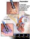
Electronic devices that stimulate myocardium to contract Pacing and sensing Defibrillation?
64
Pacemaker function
Correct slow heart rates Artificial conduction system Act as a“safety net” for certain arrhythmias
65
What two different types of generators are there?
External Generator outside the body Implantable (permanent) Generator implanted inside the body
66
What does the Generator do?
Provides the stimulation Monitors heart activity and decides whether to stimulate
67
What does the Leads do?
Carry the stimulation from the generator to the myocardium Endocardial Epicardial External Transvenous
68
Implantable Generator (pic on back) External Generator (pic on front)
Implantable: Implanted in the patient, Programmable through patient skin (magnetic field changes…) Contains: Battery, Computer chip, Lead attachment head, “Reed switch”: magnetic External: Lies outside the body, Connects to wires emerging from the patient, Programmable, Type of pacing, Adjustable, Output (m/a), Rate
69
Reed Switch
Inside an implantable pacemaker “on and off” switch Turned off by placing a magnet over the pacemaker Pacemaker back “on” once magnet is removed
70
How do Pacemaker Leads work?
Connect the Generator to myocardium Allow for two way communication: Epicardial: Outer surface of the heart Endocardial: Inner myocardium Right atrium Right ventricle
71
What are the two different kinds of fixation of the Pacemaker leads?
Fixation The method of attaching the leads Active Fixation “screw in” mechanism Passive Fixation Hooks or “tines” Attaches in the myocardium (trabeculae)
72
What is Transvenous Pacing?
Pacing catheter Introduced by percutaneous technique Placed in the RV Connects to external generator
73
What are some Indications for Pacing ?
Slow heart rates: Brady arrhythmias 3rd degree AV block S/P cardiac surgery Asystole: Pacemaker dependent
74
What does the Pacemaker provide?
Provides a stimulus voltage to myocardial tissue when needed Can provide a “safety net” Can temporarily shut off in the presence of intrinsic activity Both pacing and sensing
75
What are the two Pacemaker Functions?
Pacing Stimulating the myocardium to depolarize Sensing The pacemaker’s ability to sense intrinsic beats and rates
76
What is pacing and what is it dependent upon?
Capture: The ability of the pacemaker to depolarize the myocardium Dependent upon: The pacemaker’s voltage production The condition of the myocardium The resistance offered by the pacing lead
77
What is Capture?
ECG: Capture occurs when a pacing spike is followed by a QRS ( or p wave if atrial paced) Failure to capture: Occurs when a pacing spike is NOT followed by a QRS (or p wave).
78
When does Capture occur?
Each “spike” is the voltage produced by the pacemaker Capture occurs when a QRS (or p wave) follows the spike
79
What is Sensing?
The ability of a pacer to detect intrinsic activity There are two different kinds of sensing: Under-sensing Over-Sensing
80
what is Under-sensing
The pacer does not detect intrinsic beats
81
what is Over-sensing?
The pacer detects non-existent intrinsic activity
82
What are the two different Pacemaker Types?
Single Chamber Paces and senses one chamber Either atrium or ventricle Dual Chamber Paces and senses two chambers Both atrium and ventricle
83
What are the limitations and advantages of a Single chamber pacer and what is it used for?
Most common type used for temporary pacing Limitations Cannot synchronize atria/ventricles Advantages Simple, low cost
84
What is the Dual Chamber Pacer do?
Can synchronize atrium/ventricle Allows use of P waves to trigger ventricles Paces Atrium Ventricle Senses Atrium Ventricle
85
When is a Dual Chamber pacer used?
A-V sequential pacemaker Paces the atria and ventricles in sequence Most commonly used today Effective in 3rd degree HB, others.
86
Heart Rhythm Society
Used to be NASPE: North American Society of Pacing and Electrophysiology They’ve established standard coding for the various types and functions of pacemakers.
87
Pacemaker Codes
3-5 Letters are used Denotes the pacer program Often found in the patient’s chart Patients rarely know their own settings
88
PSR(sorta like “paser”)
P for chamber paced S for chamber sensed R for response to sensing
89
First Letter of Pacer code.
Indicates the chamber that is paced Atrial: A Ventricle: V If both are paced: D
90
Second Letter
Indicates the Chamber that is sensed Atrial: A Ventricle: V Both: D
91
Third letter
Indicates what the pacer does when it senses an intrinsic beat The pacer’s response Inhibits : I Does nothing! Triggers: T Paces Does both: D
92
VVI Pacer Code(common program for elderly patients)
Paces: Ventricle Senses: Ventricle Response: Inhibits
93
VAT Pacer code
Paces: Ventricles Senses: Atria Response: Trigger (i.e., pace the ventricle if no atrial depolarization)
94
DDD Pacer Code (“D” for dual)
Paces: Atria and ventricles Senses: Atria and ventricles Response: May trigger or inhibit
95
AAI pacer code
What gets paced? What gets sensed? What is the response if the pacer senses intrinsic activity?
96
Pacemaker Evaluation
Capture Is the pacemaker properly capturing? Sense Is the pacemaker sensing properly? Under-sensing Over-sensing
97
How do you know if the pacemaker's Capture working properly or not? Picture is of proper capture on front Capture failure on back
If each pacer spike is followed by a QRS or p wave, then the pacer is properly capturing. If just one pacer spike fails to produce a QRS or p wave, we say the pacer is NOT CAPTURING!
98
What are some reasons for Failure of Temporary pacing capture?
Lead dislodged Reposition Inadequate voltage output Increase output
99
How do you know if the pacemakers Sensing is working properly or not?
A pacemaker should stop pacing once the heart generates its own intrinsic beats Intrinsic beats are more effective than paced beats (and use less battery!)
100
Sensing is kind of like a fence… If the fence is too high, then the pacemaker doesn’t get to see some of the legitimate intrinsic activity; it would pace too much. e.g., R waves are hidden… If the fence is too low, the pacemaker gets noise that it wrongly interprets as intrinsic activity; it might pace too much or it might pace too little (depending on what it’s sensing).
In either case, the pacemaker isn’t getting good information about what the heart’s actual electrical activity is.It’s important to find the good threshold of sensing— to set the fence at just the right height.
101
Under-sensing
If a pacemaker does not change the distance of its pacing spikes in the presence of intrinsic activity, the pacer is UNDERSENSING!
102
Over-sensing
If the pacemaker changes its spike-to- spike distance without any intrinsic activity, the pacer is OVER-SENSING!
103
Regional terms
104
Quadrants
105
anatomical landmarks and ribs
106
107
108
109
110
111
112
113
Decubitus, as in left-lateral decubitus position
114
What is a stress test?
Provocative measure to disclose disease
115
What is it that's being stressed?
Cardiovascular system, usually cardiac perfusion
116
117
118
119
120
121
122
123
Decubitus, as in left-lateral decubitus position
124
anatomical landmarks and ribs
125
Quadrants
126
Regional terms