Anatomy Flashcards
Key aspects of anatomy and applied knowledge of anatomy. (39 cards)
Anatomy:
What are the 3 positional terminologies of anatomy?
- Anatomical Position: Standing, feet, and palms facing towards the front
- Supine: Lying on the back
- Prone: Lying on your abdomen.
Anatomy:
What are the Directional Terminologies?
- Anterior (to the front) and Posterior (to the back)
- Lateral (toward the side) and medial (toward the centre)
- Inferior (below) and Superior (above)
- Proximal (closer to the centre) and Distal (further from the centre)
- Abduct (away from the midline) and Adduct (towards the midline)
Anatomy:
What movements can a joint preform?
- Flexion: folding a joint or moving in an anterior direction.
- Extension: posterior movement or straightening of a joint.
- Abduction: moving away from the midline.
- Adduction: moving toward the midline.
- Medial or internal rotation: Rotating towards the centre or inwards.
- Lateral or external rotation: rotating or turning towards the side or outwards
- Dorsi Flexion: flexing the foot
- Plantar Flexion: pointing the foot (towards the plants)
Anatomy:
What is hyperextension?
Exessive movement in the direction of extension. The agonist is working shortened and the antagonist is lengthened or weak.
Anatomy:
What is Circumduction?
A combination of flexion, extension, adduction, and abduction. Like making circles.
Anatomy:
What are the movement categories of muscles?
- Agonist: Primary mover or movement producing muscle.
- Antagonist: Opposite worker to the agonist.
- Synergist: Assists lager agonists.
Anatomy:
What are the 3 planes of motion?
- Sagittal Plane: divides the body equally into left and right or flexion and extensions.
- Coronal Plane: divides the body into front and back or abduction and adduction.
- Transverse Plane: divides the body into upper and lower or rotations.
Anatomy:
What are the different sections that make up the spinal column, how many vertebrae do they consist of and what is their orientation?
24 vertebrae in total (like the hours in a day)
- Cervical: 7 vertebrae the uppermost part. Ther Upper cervical vertebrae are in flexion and lower cervical are in extension.
- Thoracic: 12 vertebrae. Mostly in flexion, T11 to T12 starts extension. 12 ribs attach from T1 to T12.
- Lumbar: 5 vertebrae. All in extension.
- Sacral: 5 vertebrae. Joins the pelvis to the spine at the Sacrio-iliac joint.
- Coccyx: 4 vertebrae.
Anatomy:
What are the 3 most important ligaments in the spine?
1.The Ligamentum Flavum:
forms a cover over the dura mater: a layer of tissue that protects the spinal cord. This ligament connects under the facet joints to create a small curtain over the posterior openings between the vertebrae.
2.The Anterior Longitudinal Ligament:
attaches to the front (anterior) of each vertebra. This ligament runs up and down the spine (vertical or longitudinal).
3.The Posterior Longitudinal Ligament:
runs up and down behind (posterior) the spine and inside the spinal canal.

Anatomy:
What are the structures of the scapular or shoulder blade and what is its function?
Structures:
- The inferior and superior angle.
- The Medial and Superior border
Function:
It is a gliding joint and glides across the back of the rib cage.
It has various muscles attaching to it that facilitate shoulder stability.

Anatomy:
What is the Gleno-Humeral Joint?
Otherwise known as the shoulder joint.
The head fits into the glenohumeral cavity.
It is a ball in socket joint which is shallow allowing for large range of motion yet the least stability.
The Glenohumeral joint works together with the scapulae for shoulder stability.

Anatomy:
What is the olecranon?
This is the anatomical word for the elbow. Also known as the Decranon.
Anatomy:
What bones are fused together to make the pelvis and which other bones form apart of the pelvis?
The Ilium, Ischium, and Pubis are fused to form the pelvis.
The other bones are the Sacrum and Coccyx also form apart of the pelvis.
The pelvis attaches to the spine at the Sacro-Iliac Joint (SIJ)

Anatomy:
What is the ASIS?
The Anterior Superior Iliac Spine.
This is the hip bone on the anterior side of the body.
It is a refrence point when refering to neutral pelvis.

Anatomy:
What is the Pubic Symphysis?
Referred to as the pubic bone. It is a cartilaginous joint and an important reference to the neutral pelvis.

Anatomy:
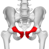
What is the Acetabulum?
This is the hip socket where the head of the femur firts deep into the cup-shaped acetabulum forming the ball and socket hip joint.
Anatomy:
Where is the Tibia situated and what condition is it prone to?
The Tibia is the medial shin bone with ends in the medial malleolus (ankle bone). It is much thicker than the Fibula.
This bone is prone to tibial torsion where the personal externally rotate the feet/ knee instead of from the hip.

Anatomy:
Where is the Fibula located?
It is the lateral shin bone and starts at the base of the knee and ends in the lateral malleolus (ankle bone)

Anatomy:
What affects the alignment of the Patella?
The Patella is a sesamoid bone or floating bone.
It is affected by the balance in strength between the medial and lateral quadriceps, as well as the tibia and fibula’s positioning.
Anatomy:
What is the difference between a posterior pelvic tilt and an anterior pelvic tilt?
Posterior Pelvic Tilt:
The PS (pubic symphysis) is higher than the ASIS (Anterior Superior Illiac Spine). Known as a tuck.
Anterior Pelvic Tilt:
The ASIS is higher than the PS. Known as a arch.
Anatomy:
What groups of muscles around the pelvis affect the stability of the pelvis and what are the muscles in each of these groups?
- Abdominals: (more for posterior pelvic tilt)
- Internal obliques
- Rectus Abdominus
- External Obliques - Back Extensors: (more for anterior pelvic tilt)
- Multifidus
- Erector Spinae (Spinalis, Longissimus, and Iliocostalis)
- Quadratus Lumborum - Hip Flexors: (more for anterior pelvic tilt)
- Illiopsoas (Illiacus and Psoas) - Hip Extensors:
- Gluteus Maximus
- Semi-tendinosis, Semi-membranosus and Bicep Femoris - Hip Abductors:
- Gluteus Minimus
- Gluteus Medius
- Tensor Fascia Latae - Hip Adductors:
- Gracilis
- Adductors (Longus, Magnus and Brevis)
- Pectineus
Anatomy:
Which muscles are shortened and which muscles are lengthened in an anterior tilt?
Shortened:
- Hip flexors
- Back extensors
Lengthened:
- Hip extensors
- Abdominals
Anatomy:
Which muscles are lengthened and which muscles are shortened in a posterior tilt?
Shortened:
- Hip extensors
- Abdominals
Lengthened:
- Hip flexors
- Back extensors
Anatomy:
Which muscles are shortened and which muscles are lengthened in a lateral pelvic tilt?
Shortened:
- Same side as the higher hip Quadratus Lamborium
- Same side as the higher hip oblique
- Opposite side glute med
- Same side as higher hip Abductors
Lengthened:
- Opposite Oblique
- Opposite Quadratus Lamborium
- Same side as the higher hip glute
- Opposite adductor


