Anatomy Flashcards
The hip joint
ASIS - anterior superior iliac spine

Knee bones

Ankle bones

Inguinal ring borders

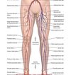
Saphenofemoral junction
2.5cm below and lateral to the pubic tubercle
Quadricep Muscles
Rectus femoris
vastus lateralis
vastes medialis
vastus intermedius
hamstrings
biceps femoris
semitendinosus
semimembrinosus
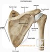
shoudler joint

Calf
Gastrocnemius - lateral epicondyle
subclavian steal
narrow proximal subclavian artery (proximal to where vertebral artery leaves)
decreased pressure distally
:. subclavian artery takes blood from contralateral vertebral artery (via circle of willis and back down the vertebral artery)
can steal from internal mammary - CABG
lehriche
aortoiliac occlusion
absent femorals
buttock claudication
erectile dysfunction
gunstock deformity
malunion of a spuracondylar fracture
wedge osteotomy of lateral humerus
mitral regurgitation
jet width 0.6cm+
regurgitant volume more than 60 ml
aortic stenosis
pressure gradiant >40
valve area <1cm^2
6 Ps
acute limb ischaemia
pulseless
painful
Pallor
perishingly cold
paraesthesia
paralysis
Still’s
juvenila idiopathic arthritis
salmon coloured rash comes and goes
mig infuinal point scar
Navy
nerve
artery
vein
y fronts - lateral to medial
Posterior cruciate ligament anatomy
lateral edge of medial femoral condyle
tibial plateau
anterior cruciate ligament
posterior to anterior
lateral to medial
superior to inferior
attachments -
- notch of distal femur (lateral femoral condyle)
- tibial plateau
Acromegaly management
ocreotide - somatostatin analogue
pegvisomant - GH antagonist
examine
hands
face
visual fields - bitemporal hemianopia
Graves
carbimazole
propilthyouracil
radioiodine
Dercum’s
adiposis dolorosa
multiple painful benign lipomas
in obese
ICD
implanatable cardioverter defribrillator
Shoulders

Diabetic Retinopathy
Background
- Venodilation
- Microaneurysms (red dots)
- Hard exudates
Pre-proliferative
- Soft exudates (cotton wool spots)
Proliferative
- New vessels
Hypertensive Retinopathy
Grade 1
Arteriolar narrowing + silver wiring
Grade 2
AV nipping
Grade 3
Flame shaped haemorrhages + cotton wool spots
Grade 4
Papilloedema
Carpal Tunnel Syndrome
numbness, tingling, burning thumb and fingers (esp index and middle fingers and radial half of ring finger)
thenar eminence wasting
Goal of surgery - divide transverse carpal ligament and distal aspect of volar ante brachial fascia
Optic Atrophy
0.3+ cup to disc ratio of optic nerve
Local
- Optic Neuritis
- Advanced Glaucoma
- Ischaemia-Retinal artery occlusion (GCA)
- SOL
Systemic
- B12/Folate
- Alcohol
Horner’s Syndrome Causes
lesion of sympathetic - hypothalamus, preganglionic tract, post ganglionic tract
CNS - MS, stroke, SOL - syrinx
Pancoast’s Tumour
Carotid artery aneurysm
Trauma - Carotid endarterectomy, central line
Migraine
Ptosis differentials
Horner’s
CN3 palsy
Guillain Barre
MG, Lambert Eaton
Myotonic Dystrophy
Blue sclera
Ehler’s Danlos
Osteogenesis Imperfecta (poor bone formation due to lack of type I collagen)
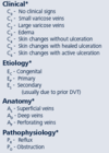
CEAP Classification

Femoral head blood supply
Deep femoral nerve
circumflex arteries
retinacular vessels
Median Nerve Injury
inability to abduct and oppose thumb - paralysis of the thenar muscles ape-hand deformity
Sensory loss - thumb, index finger, long finger, radial aspect ring finger
Weakness in forearm pronation and wrist and finger flexion
Activities of daily living such as brushing teeth, tying shoes, making phone calls, turning door knobs and writing, may become difficult with a median nerve injury.


