Anatomical Areas Flashcards
Clinical link: Carpal tunnel syndrome
Mechanism and symptoms
Mechanism: Caused by entrapment or irritation of the median nerve within the carpal tunnel. Such as through thickening of the flexor retinaculum
Symptoms: Paresthesia in the median nerve distribution, wasting of the thenar muscles, reduced strength of grip, reduced thumb abduction; numbness in the thumb, middle and index fingers (not palm due to cutaneous branch).
Clinical link: Blood supply of the hand - Allen’s Test
- Used to test the perfusion capability of the ulnar artery to the hand
- The both arteries and occluded. Pallour seen
- The ulnar artery is released.
- If perfusion is adequate then the hand will return to normal colour (flushing) in 5-15 seconds - positive
- If negative the radial artery should not be punctured for an arterial blood sample
Palmar spaces and synoival sheaths: Describe the palmar spaces and synovial flexor sheaths of the hand.
Clinical relevance?
Infections may ‘jump’ between these spaces. Particularly important is the midpalmar space
☆Tenosynovial lining of flexor sheaths

Elbow joint: Dislocation and annular joint subluxation
Dislocation: Commonly occurs in the posterior direction
Annular ligament: Surrounds the radius
- Function: Allows sliding of the ulnar on the radius and pivoting of the radius bone during supination and pronation
- Can be subluxed due to excessive force
Fractures of the forearm: Describe Colle’s, Smith’s and Bennett’s fractures
Colle’s: Sees dorsal displacement of the distal fragment of the radius. Shortening and dorsal angulation seen
- FOOSH
Smith’s: Palmar displacement of the distal fragment of the radius. Shortening and volar angulation.
- Fall onto flexed wrist, direct blow to dorsal forearm
Bennett’s: Fracture of the carpometacarpal joint
- Forced abduction of the thumb
Lower limb: Lateral compartment of the leg
Innervation: Superficial fibular nerve (L4-S3)
Muscles: Fibularis longus and brevis
Action: Eversion of the foot
Clinical relevance: Nerve injury may occur in neck of fibular fracture, sciatic nerve compression will also affect the compartment.
Inversion injuries can damage the muscle tendons and cause an avulsion injury of the 5th metatarsal
☆ The superficial fibular nerve and small saphenous vein pass posteriorly to the lateral malleolus
- Great saphenous vein passes anteriorly to the medial malleolus
Scaphoid: Consequence of fractures and region for palpation
Fracture: Avascular necrosis may result as blood supply is provided in a distal to proximal fashion. A fracture in the middle of the scaphoid may disrupt this and avascularise the proximal portion
Palpation: The scaphoid bone can be palpated within the anatomical snuffbox
Lower limb: Posterior compartment of the thigh
Innervation: Sciatic nerve (L4 - S3)
Muscles: Hamstring muscles (semimembranous, semitendinous and biceps femoris)
Action: Hip extension, knee flexion
Clinical relevance: The sciatic nerve may be impinged through vertebral disc herniation, at the sciatic notch, by piriformis
Ischial tuberosity avulsion fracture
Upper limb: Dorsal scapular spaces
Quadrangular space: Axillary nerve
Triangular interval: Profunda brachii artery and radial nerve

Pectoral girdle: Deltoid and pectoral muscles

Pectoralis major: Medial and lateral pectoral nerve
- Adducts, flexes and medially rotates the arm
Pectoralis minor: Medial pectoral nerve
Deltoid: Axillary nerve
- Abducts the arm (beyond the first 15 degrees of supraspinatus)
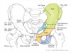
Lower limb: Outline the landmarks of the pelvis

☆ The greater sciatic foramen is formed by the sacrospinous and sacrotuberous ligaments
☆ Piriformis muscle divides the greater sciatic foramen into an upper and lower region.
Upper sciatic foramen: Superior gluteal nerve, artery and vein
Lower sciatic foramen: All other vessels pass through this foramen
Intervertebral foramen: Formation, patholgy
Formation: From the inferior vertebral notch of the superior vertebrae and the superior notch of the inferior vertebrae (of the pedicles)
Pathology: Narrowing of the intervertebral foramen
- Arthritis
- Spondylolisthesis
- Osteophyte growth (OA)
Clinical link: Medial humeral epicondyle fracture and tunnel through which the associated nerve runs
- Can damage the ulnar nerve
- Cubital tunnel of the elbow
- Runs through Guyons Canal at the wrist
Gluteal muscles
Gluteus maximus
Innervation: Inferior gluteal nerve (sacral plexus)
Action: Extends the thigh and lateral rotation
☆ Only active when force is required
Gluteus minimus and medius
Innervation: Superior gluteal nerve (sacral plexus)
Action: Abducts and medially rotates the thigh
Lower limb: Sensory innervation

- Consider that the sensory innervation of the leg is suppled mostly by branches of the sciatic nerve

Stability of the hip joint: Name the ligaments which contribute to the stabilty of the hip joint
- Iliofemoral
- Pubofermoral
- Ischiofemoral
-
Ligamentum teres
- Of the femoral head. Inserts into the fovea capitus. Important hip stabiliser.
- Transmits a nutrient artery to the femoral head in infants
Elbow joint: Name the ligaments of the elbow joint
- Radial collateral ligament
- Ulnar collaterol ligament
- Annular ligament
Clinical link: Lumbar puncture - positon and surface marking
Position: Patient to lay on their side with their knees raised to their chect
Surface marking: The posterior iliac crest. Intersects the L4 spinous process
Structures passed through: Skin, superficial fascia, supraspinous ligament, intraspinous ligament, ligamentum flavum, epidural space, dura mater, arachnoid mater, subarachnoid space

Pectoral girdle: Muscles of the anterior aspect

Suprascapular foramen: The suprascpaular nerve passes through this space
Subscapularis: Upper and lower subscapular nerves
Latissimus dorsi: Thoracodorsal nerve
Long and short heads of biceps brachi: Musculocutanous nerve
Bicipital aponeurosis: Seen at the sharp medial margin. Covers the brachial artery and the median nerve
Upper limb: Posterior compartment of the arm
Innervation: Radial nerve
Muscles: Triceps brachii
Actions: Extension at the elbow joint
Clinical relevance: Radial nerve may be injured at the lateral epicondyle, tight watch/handcuffs, mid-shaft humeral fractures (radial groove), falling asleep with hand over the back of the chair, upper limb dislocation - becomes stretched (commonly anterior)
The radiocarpal joint: Label the carpal bones

Scaphoid, lunate, triquestrum, pisiform (sesamoid bone), hamate, capitate, trapezoid, trapezium
Outline the blood supply to the upper limb
- include areas of ‘transition’

Lower limb: Medial compartment of the thigh
Innervation: Obturator nerve
Muscles: Adductor longus and brevis
Actions: Adducts the thigh
Clinical relevance: The obturator nerve may be damaged in birthing mothers and in sports (e.g. fencing)
Quadratus lumborum: Action and innervation
Action: Extension and lateral flexion of the vertebral column
Innervation: Anterior rami of spinal nerves T12-L4
Erector Spinae Muscles: Action and innervation
Action:
Unilateral contraction: Lateral flexion
Bilateral contraction: Supports the vertebral column and increases intra-abdominal pressure
Innervation: Dorsal rami of spinal nerves
Clinical link: Surgical neck and mid-shaft humerus fracture.
Surgical neck:
- Can damage the axillary nerve.
- Leads to loss of innervation to the deltoid muscle and teres minor
- Weakened abduction and lateral rotation
Mid-shaft humerus fracture:
- Can damage the radial nerve
- Leads to loss of innervation of the triceps branchii
- Loss of extension of the elbow joint
- Loss of innervation of the dorsal forearm
- Sensory loss to dorsal forearm and hand
Spinal nerves: Outline the 2 branches derived from spinal nerves
Spinal nerves (emerging from ventral rami) →
Large anterior rami: Innervates most skeletal muscles and skin
Small posterior rami: Innervates the intrinsic muscles of the back and narrow strip of associated skin
Intervertbral disc herniation: Most common ‘direction’ and region
Posterior: Most common due to tapering of the posterior longitudinal ligament of the spine, weaker/less organised, flexion is much more common, nucleus becomes more posterior moving down the spine
Region: Most commonly occurs in the lumbar region of the spine. This is due to the curvature of the spine and the large amount of pressure placed upon this region
Muscles of the forearm: Outline the functions of the FDPs and FPS
Flex the digits at the IPJs, can flex the MCPJs
Upper limb: anterior compartment of the forearm
Innervation: Median nerve (C5-T1)
- Except medial half of FDP and FCU which are innervated by the ulnar nerve
Muscles: FDS, FPL, pronator teres
Actions: Flexion of the wrist and digits, pronation
Clinical relevance: The median nerve may be damaged at the cubital fossa, brachial plexus lesions (upper or lower), supracondylar fracture of the humerus, damage at the wrist (e.g. self-harm)
Intervertebral discs: Anatomy
IV disc bodies: Increase in size from rostral to caudal. Flexibility is sacrificed to allow stabilty. The amount of force apparent on the dics increases caudally. (shapes)
Central nucleus pulposus: Derived from the notochord. Keeps the annulus fibrosus under constant tension. Contains proteoglycans with GAG side chains, type II collagen. Compression resistant.
Annulus fibrosus: Layers of oliquely organised fibrocartilage. Less well organised posteriorly.
Venous drainage of the upper limb: Name the deep and superficial vessels
Superficial: Basilic and cephalic veins
Deep: Radial, ulnar and branchial veins
Hip joint: Outline the relation of the hip joint to our centre of gravity when standing
The hip joint lies anterior to the centre of gravity.
The knee joint is posterior to the centre of gravity.
Allows us to ‘lock’ joints, reducing the muscular energy required for standing.
The axilla: Describe the boundaries of the axilla
Inlet: Lateral margin of rib I
Anterior wall: Pectoralis major and minor muscles
Medial wall: Upper thoracic wall, serratus anterior muscle
Posterior wall: Subscapularis, teres major and latissimus dorsi
Lateral wall: Intertubecular sulcus
Floor: Skin of the armpit
Outline the blood supply to the lower limb
Common iliac → Internal and external iliac
Internal iliac → Superior and inferior gluteal, obturator
External iliac → Femoral artery → *Branch: Profunda Femoris→ medial and lateral circumflex *→ Popliteal artery → anterior and posterior tibial
Anterior tibial → dorsalis pedis
Posterior tibial → Fibular, medial and lateral plantar

Hip joint: Label the hip joint and describe the stabilty of the joint

The pelvis is formed by the ilium, ischium and pubic bones.
The acetabulum of the pelvis and the head of the femur form the hip joint.
Acetabulum: Fossa, notch and labrum
- Large and cup shaped
- Allows weight to be transferred to the femur
The joint is very stable, to accomodate the large amount of force placed upon it. However, this is at the expense of movement.
The acetabulum is large whilst the head of the femur is small.
The axilla: Describe the contents of the axilla
AXillary artery and vein
Infraclavicular portion of the brachial plexus
Lypmh nodes
Long thoracic nerve
Adipose tissue
Elbow joint: Epicondylitis
Lateral epicondylitis: (tennis elbow) Inflammation at the common extensor origin
Medial epicodylitis: (golfer’s elbow) Inflammation at the common flexor origin
Forearm: Label the key anatomical regions of the forearm

Nerve: Radial
- Loss leads to wrist drop
- Can be injured at the lateral epicondyle and also by tight wrist watches/handcuffs
The interosseus membrane
- Provides attachment for muscles
- Fibres orientated such that forces are transmitted from the hand to the humerous
- Allow attachment of the radius and ulnar without restriction pro- and supination
Subclavian vein: Relation to the subclavian artery and its approximate surface markings
- Both cross the first rib
The subclavian vein crosses the first rib in the midclavicular line, extending to the medial clavicular edge.
Spinal nerves:
- At which vertebral level does the spinal cord end?
- Define the cauda equina
Spinal cord:
- Ends at L1 in adults and L2/L3 in children
- Conus medullaris
Cauda equina: Formed from the ventral and dorsal rootlets of spinal nerves
Clinical link: The cauda equina may be compressed in vertebral disc prolapse.
☆ Red flag symptoms: Loss of anal tone, faecal and urine incontinence, paraesthesia in the saddle region
Gleno-humeral joint: Decribe the anatomy of the gleno-humeral joint
- A synovial ball and socket joint
- Formed from the head of the humerus inserting into the glenoid cavity, which has a surrounding glenoid labrum
- The large head and small cavity cause the joint to be relatively unstable and therefore requires stabilisation by surrounding ligaments and muscles
- The tendon of the long head of biceps is found near the joint
- Blood supply: Medial and lateral circumflex arteries of the profunda brachii artery

Facet/Zygapophyseal Joints: Formation, innervation, orientation and function
Formation: Formed by the anterior and superior articular processes of each vertebrae
Innervation: Dorsal rami of spinal nerves
Orientation: Move from a horizontal to a vertical position moving from superiorly to inferiorly.
- This ↓ flexibility but ↑ stability
Function: Guide and constrain motion of the vertebrae, faciliate load transmission
Intrinsic muscles of the hand: Outline the actions of the dorsal and palmar interossei and the lumbricals
Dorsal interossei: Abduction
Palmar interossei: Adduction
Lumbricals: Flexion of the MCPs and extension of the IP joints. Crucial for movement of the fingers
Vertebral bodies: Name the features of the vertebral bodies

Transverse processes
Spinous process
Pedicle
Lamina
Neural arch
Body

Palmar aponeurosis: Location and function
- A sheet of fascia with a central thickening covering the palmar surface of the hand
- Function: Protection of the underlying tendons and forms the fibrous digital sheaths
The carpal tunnel: Outline the contents of the carpal tunnel
- A bony canal formed by the carpal bones
Roof: Flexor retinaculum
Contents: Median nerve, FDP, FDS and FPL tendons
Dermatomes: Label the dermatomes of the upper limb

Upper limb: Anterior compartment of the arm
Innervation: Musculocutaneous (C5-C7)
Muscles: Biceps brachii
Actions: Flexion at the elbow joint
Clinical relevance: The nerve may be damaged through stab wounds to upper arm or upper brachial plexus lesions
Pectoral girdle: Muscles of the back

Trapezius: Accessory nerve
- Movement of the scapula
Latissimus dorsi: Thoracodorsal nerve
- Extends, adducts and medially rotates the humerus
Levator scapulae: Dorsal scapular nerve
- Elevates scapula
Rhomboid minor and major: Dorsal scapular nerve
- Adducts and elevates the scapula
Intrinsic muscles of the hand: Outline the innervation of the intrinsic muscles of the hand
LOAF muscles (including thenar eminence) supplied by the median nerve (recurrent branch):
- Lateral 2 lumbricals
- Opposens pollicis
- Abductor pollicis brevis
- Flexor pollicis brevis
All other intrinsic muscles, including the hypothenar eminence of the hand are supplied by the ulnar nerve.
- Enters the hand via Guyon’s canal
Clinical link: Outline the boundaries for IM injection of the gluteal muscles
- Vertical line from the posterior iliac crest to the ischial tuberosity
- Horizontal line from the midpoint of this line
- The left upper quadrant is the ‘safe’ area
Clinical relevance: Failure to correctly recognise this site may lead to injury to the sciatic nerve
☆ REMEMBER ☆ The sciatic nerve ultimately innervates the leg and provides sensory innervation to the majority of the area

Pronation and supination: Name the muscles which perform pronation and supination of the wrist
Pronation: Pronator teres and pronator quadratus (C7-C8)
- Located in the anterior compartment
- Pronator teres innervated by median nerve, quadratus by interroseous nerve
Supination: Supinator and biceps brachii (C5-C6)
- Biceps brachii innervated by musculocutaneous
- Supinator innervated by the radial nerve
Pectoral girdle: Muscles of the posterior aspect

Supraspinatus: Suprascapular nerve
Infraspinatus: Suprascapular nerve
Teres minor: Axillary nerve
Teres major: Inferior subscapular nerve
Long head of triceps brachii
Triangular space: Circumflex scapular artery
Quadrangular space: Axillary nerve, posterior circumflex arterty and vein
Triangular interval: Radial nerveand profunda branchii artery
Muscles of the forearm: Pronation and supination muscles, which movement is strongest
Supination is the strongest movement
Pronation: Pronator teres and quadratus (C7-C8)
Supination: Biceps brachii and supinator (C5-C6)
Outline the borders and contents of the tarsal tunnel
Borders: Passes posteriorly to the medial malleolus, held in place by fibrous band (flexor retinaculum)
Contents: Tibialis anterior, flexor digitorum longus, flexor digitorum brevis, posterior tibial artery, tibial nerve, posterior tibial vein

Spinal ligaments: Name the ligaments of the spinal column

Anterior and posterior longitudinal ligaments: Support the vertebrae
Ligamentum flavum: Resists separation of laminae
Inter- and supraspinatous ligaments: Limit flexion
Ligamentum nuchae: Supports the head, resists flexion, returns the head to anatomical postion
- Attaches to the skull, extending down to the spinous process of cervical vertebrae VII

Lower limb: Posterior compartment of the leg
Innervation: Tibial nerve (branch of sciatic nerve) (L4-S3)
Muscles: Gastrocnemius, soleus, calcaneal tendon
Actions: Plantarflexion, flexion of toes, inversion of the foot
Clinical relevance:
- Injury to the medial malleolus may sever the tibial nerve
- The compartment may be affected by sciatic nerve damage
- The calcaneal tendon may be injured by forceful plantarflexion
Clinical link: Pathology of the subacromian space
- The subacromian space lies between the acromian and the head of the humerus
- The supraspinatus tendon passes through the space
- The tendon or bursa within the space may become inflammed causing impingement and subsequent pain
-
Outcome:
- Overhead activities become painful
- Inabilty to actively abduct the GHJ
- Supraspinatus tendonitis generates a painful arc
- Thickening of the coracoacromial ligament can also caused impingement of the tendon
Elbow joint: Decribe the basic anatomy of the elbow joint
- A synovial hinge joint
- Formed by articulation of the radial head on the capitulum of the humerus and articular of the ulnar head on the trochlea of the trochlea

Osteology: Identify the key features of the humerus

Anatomical and surgical necks
Greater and less tubercles
Bicipital groove: The tendon of the long head of biceps passes through this groove. The tenosynovium enclosing the tendon may become inflammed - tenosynovitis.
Medial and lateral epicondyles
Olecranon fossa
Lateral epicondyle
Clinical link: Shoulder (GHJ) dislocation
Most likely to occur in the anterior direction
Appearance: Externally rotated and abducted
The axillary nerve is at risk in GHJ dislocation. Leads to loss of abduction and sensory loss over the regimental badge area.
☆Remember the axillary nerve passes through the quadrangular space and round the back of the humerus
The radial nerve may also be damaged
Outline the borders and contents of the cubital fossa
Borders: Pronator teres medially, brachioradialis laterally and between the lateral and medial epicondyles superiorly
Contents: Median nerve, Brachial artery, Tendon of biceps brachii, Radial nerve

Muscles of the forearm: Outline the function of the carpi muscles
Carpi = wrist
Extensor carpi = extends and abducts the wrist
Flexor carpi = flexes and adducts the wrist
Lower limb: Knee Joint
Formation, ligaments, menisci, bursa, clinical revelvance
Synovial modified hinge joint
Formed by: The distal head of the femus articulating with the proximal head of the tibia and the patella (sesamoid bone)
Ligaments:
- Anterior cruciate ligament: Extends from the tibia (anterior part of the intercondylar area) to the lateral (wall of the intercondylar fossa of the) femur. Resists anterior displacement.
- Posterior cruciate ligament: Extends from the tibia (posterior intercondylar area) to the medial (wall of the intercondylar fossa of the) femur. Resists posterior displacement.
- Lateral collaterol ligament: Lateral femur epicondyle to the lateral surface of the fibular head. Resists varus forces.
- Medial collaterol ligament: Medial femur epicondyle to the medial surface of the tibial head. Resists valgus forces.
Menisci: Fibrocartilage, improve joint congruence, stabilty and provide proprioceptive feedback
- Medial meniscus: ATTACHED to the joint capsule and medial collaterol ligament.
Clinical relevance: This leaves the medial meniscus more vulnerable to damage.
Unhappy triad of knee injury: Medial meniscus, ACL and medial collateral ligament
Bursa: Fluid filled sacs that act to reduce friction between surfaces.
Clinical relevance: A large joint effusion would typically be seen in the suprapatella pouch
Lower limb: Anterior compartment of the thigh
Innervation: Femoral nerve (L2 -L4)
☆ Psoas innervated by anterior rami of L1-L3
- Injury within the femoral triangle, tumour
- Consequence: Reduced hip flexion and knee extension, fuctional problems climbing stairs
Muscles: Quadriceps (vastus medius), sartorius, rectus femoris
Actions: Generally hip flexion (rectus femoris, psoas major and ilacus), knee extension
Clinical relevance: Femoral stretch test (damage to the femoral nerve will cause reduced contraction of the quadriceps femoris)
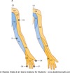
Venous drainage of the upper limb: Describe the route of the main venous vessels
Basilic vein: Travels from the dorsal venous network up the arm, on the medial/ulnar side.
Cephalic vein: Travels from the dorsal venous network up the arm, on the lateral/radial side.
Median cubital veins: Links the basilic and cephalic veins at the cubital fossa. Ideal for venepuncture
Brachial veins: Deep paired veins which run alongside the brachial artery
Intracapsular fracture of the femoral head: Outline the blood supply to the femoral head and the consequences of this event
Blood supply: Medial and lateral circumflex branches of the femoral artery, branch of the obturator and from the superior gluteal artery
The blood supply of the femoral head enters the capsule of the joint from below the joint (distal to proximal) and hence can be severed in fractures of the femur. This can then leave the area without a blood supply, causing avascular necrosis.
☆ Intracapsular fracture
Clinical link: Loss of serratus anterior innervation
Innervation, function, observation and clinical test
Innervation: Long thoracic nerve (C5-C7)
Function: Protraction and lateral rotation of the scapular
Observation: Winging of the scapular (as the scapular is elevated away from the thoracic wall)
Clinical test: Press-up against the wall
Hip joint: Proximal femur

- Fovea
- Head of the femur
- Neck of femur
- Greater trochanter
- Lesser trochanter
- Gluteal tuberosity
The radio-carpal joint: Describe the articulation seen at the joint
- A synocial ellipsoid joint
- The radius articulates with the carpal bones
- The TFCC is seen between the ulnar bone and the carpals
- Ulnar and radial styloid processes
Muscles of the forearm: State the general function for muscles originating at the epicondyles
Lateral epicondyles: Extensors
- Tennis elbow
Medial epicondyles: Flexors
- Golfer’s elbow
Upper limb: Posterior compartment of the forearm
Innervation: Radial nerve
Muscles: EPL, EPB, EDL, supinator
Actions: Extension of the wrist and digits, supination
Clinical relevance: The radial nerve may be damage in mid-humerus fractures (winds around the spiral groove), at the lateral epicondyle, supra-epicondyle fractures, tight watch/handcuffs, falling asleep on arm, pressure of crutches
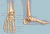
Lower limb: Intrinsic muscles of the foot
☆ Plantar surface is separated in 4 layers. The muscles support the digits during standing and gait. They also act as dynamic arch support.
Plantar aponeurosis: Deep fascia of the foot
Innervation: Branches of the tibial nerve
The axilla: Name the 5 lymph node groups of the axilla. Which areas which drain to these nodes and when may this be interrupted?
- Central
- Humeral
- Apical
- Pectoral
- Subscapular
All upper limb and lateral breast lymphatics drain into the axillary nodes.
Drainage may be interrupted following mastectomy or surgical node clearance (for breast cancer).
Meninges: Outline the layers of the meninges
Pia
Subarachnoid space: End at L3 vertebral level
Arachnoid
Subdural space: Only seen in pathology
Dura
Scapula: Name the vessels which contribute to the arterial anatomoses of the scapula. Outline the importance of this
- Suprascapular artery
- Transverse cervical
- Circumflex scapula (of the axillary artery)
- Thoracodorsal (of the axillary artery)
- Posterior intercostal
The anastomoses ensure that blood supply will not be prevented in the event of an occlusion to one of the vessels
The extensor hood: Outline the formation and function of the extensor hood
Formation: Extensor tendons expand at the MCPJs, forming an extensor hood
Function: As the lumbricals and interossei contract they pull on the extensor hood. MCPJs flex and IPJs extend.
Lower limb: Ankle joints
- Movements and ligaments
Movement: Plantar and dorsiflexion
- Myotomes: Plantarflexion (S1-S2)
- Plantarflexion is unstable due to the narrow area of talus in contact with the distal tibular. Mortise when more stable
Ligaments:
Medially: Anterior and posterior tibiotalar, tibionavicular and tibiocalcaneal, plantar calcaneonavicular ligament (spring)
Laterally: Anterior and posterior talofibular, calcaneofibular ligaments
Glenohumeral joint: Stabilizing muscles and ligaments
Muscles:
Biceps brachii: A humeral head depressor
Rotator Cuff Muscles SITS
- Supraspinatus - suprascapular nerve
- Infraspinatus - suprascapular nerve
- Teres minor - axillary nerve
- Subscapularis - upper and lower subscapular nerves
Ligaments: Coracohumeral ligament, glenohumeral ligaments
Glenoid labrum: Deepens the glenoid cavity
Outline the borders and contents of the femoral triangle
Borders: Adductor longus medially, sartorius laterally, inguinal ligament superiorly, fasica lata roof
Contents: Femoral nerve, femoral artery, femoral vein, empty space and lymph nodes
☆ Femoral artery and femoral vein are in the femoral sheath
☆Femoral artery travels down the femoral canal, gives off its profunda femoris branch and passes through the adductor hiatus to enter the posterior compartment of the thigh
Clinical link: Locating the femoral pulse. Found half way between ASIS and the pubic symphysis

Clinical link: Intervertebral disc pathology
Changes with age, herniation and extrusion
Changes with age: With age the nucelus pulposus becomes dessicated, becoming more fibrotic and less gel like
Herniation/prolapse: The cartilaginous disc protrudes between the vertbrae
Extrusion: The nucleus pulposus pushes out of the annulus fibrosus beyond the confines of the disc
☆ IV disc herniation can cause compression of spinal nerves leading to neurological issues
Lower limb: Anterior compartment of the leg
Innervation: Deep fibular nerve (from common fibular nerve) (L4-S3)
Muscles: Extensor digitorum longus, tibialis anterior
Actions: Extensor of toes (dorsiflexion) and inversion compartment
Clinical relevance: Damaged in neck of fibular fracture, leads to foot drop/high stepping gate (due to unopposed plantarflexion), sciatic nerve injury may also affect the compartment
Outline the borders and contents of the anatomical snuffbox
Borders: EPL laterally, APL and APB medially
Contents: Cephalic vein, radial artery and cutanous branch of radial artery, scaphoid, trapezium

Muscles of the forearm: Brachioradialis - location and action
Located within the posterior compartment of the forearm
Acts to flex the wrist
