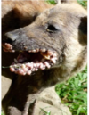
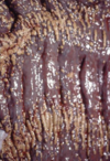
Alimentary Flashcards

1
Q

What are the predominant alimentary diseases for:

Small animals:

Ruminants and pigs:

Horses:

A

Small animals: alimentary neoplasia

Ruminants and pigs: infectious diseases

Horses: intestinal displacements/colic

2
Q

What is the most common portal of entry of pathogenic agents into the alimentary system?

A

Ingestion

3
Q

Cause of death of a calf was determined to be aspiration pneumonia due to incomplete fusion of the palatine shelves. What is a likely cause of this abnormality?

A

Ingestion of teratogenic plants by the mother during gestation (Veratrum californicum, lupines, poison hemlock)

*Palatoschisis can be caused by exposure to Griseofulvin in queens and mares, and steroids in primates

4
Q

What disorder seen in this sheep can result in difficulty with prehension and mastication of feed?

A

Brachygnathia

5
Q

What disorder is seen in this horse?

A

Prognathia

6
Q

Step mouth is characterized by abnormal wear of the teeth, and is most common in _______.
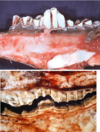
A

Herbivores

7
Q

Periodontal disease involves damage to enamel, gums, and periodontal ligaments, and resorption of alveolar bone. What is the mechanism?

A

Acids and enzymes produced by resident bacterial films

8
Q

This 48 year old Chimpanzee has lost tooth structure as a result of chewing. What is this called?

A

Dental attrition

9
Q

Enamel hypoplasia, seen here in a dog and a calf, can be caused by which diseases in each species?

A

Dog: Canine Distemper Virus (death of cells that make enamel)

Calf: BVDV infection while in utero

10
Q

This histology slide taken from a cow shows radiating clubs of eosinophilic material caused by colonies of Actinobacillus lignieresii. What gross lesion would you expect to see in the live cow?

A

Wooden tongue

11
Q

A necropsy shows white lesions in the esophagus, and you determine that the causative agent is Candida albicans. What likely caused thrush in this animal?
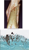
A

Long term treatment with antibiotics

12
Q

A 10 year old cat comes in with lesions on the ventral surface of his tongue. You determine that the cat has uremic glossitis. What may be causing these lesions?

A

Renal disease

MDx: ulcerative and necrotizing glossitis

13
Q

A FeLV positive cat presents with severe inflammation of the gums. Histology shows lymphoplasmacytic gingivitis, so you diagnose stomatitis. What is the preferred treatment?

A

Partial or full-mouth extraction of the teeth and debridement of the associated soft and hard tissues.

14
Q

A cat presents to your clinic for drooling, weight loss, and suspected oral pain. The cat is FIV positive and the owner thinks this illness may be related. You sedate the cat for a full oral exam and discover these lesions in her throat. What is the disease?

A

Feline chronic gingivo-stomatitis (FCGS)

15
Q

Chronic ulcerative (lymphoplasmacytic) paradental stomatitis is most common in what age group of dogs?

A

Older

16
Q

Vesicular lesions in the oral cavity of small animals can be caused by what immune-mediated diseases?

A

Bullous pemphigoid

Pemphigus Vulgaris

*Calicivirus can cause oral lesions in cats

17
Q

A cat infected with calicivirus presents to your clinic with vesicular glossitis. What would you expect to see on histology?

A

Ballooning degeneration of keratinocytes (intracellular edema)

18
Q

A farmer calls you out to his farm to inspect some cows that have lesions on their tongues and dental pads. The cows are drooling and some are lame. The farmer said the illness has been spreading rapidly throughout his herd, but no cows have died from it. How is this disease transmitted?

A

Ingestion or inhalation

*This is Foot and Mouth disease. Viremia follows infection and lesions develop in areas subject to mechanical injury.

19
Q

Qualified veterinarians perform a necropsy on a young pig who died while under FMD quarantine. This pig didn’t have any vesicles, but “tiger heart” is noted on the report. What was the cause of death?

A

Heart failure due to a malignant form of FMD that causes myocardial necrosis

20
Q

A farmer decides to euthanize his elderly horse who hasn’t been doing well, and has been off feed for awhile. After euthanasia, you notice these lesions in the oral cavity of the animal. What vesicular diseases are on your differential?

A

Vesicular Stomatitis ONLY in horses!

21
Q

You diagnose Vesicular Exanthema in a number of pigs on a farm. What virus are they infected with?

A

Calicivirus

22
Q

The erosive, ulcerative stomatitis seen in these cattle can be caused by which viruses?

A

BVDV

Herpesvirus (Malignant Catarrhal Fever)

23
Q

A few immunocompromised individuals in a herd of cattle have papules on their nares, muzzle, and oral cavity. A closer look reveals coin-shaped ulcers in their mouths. You suspect that they are infected with a parapoxvirus. What disease are these cows suffering from?

A

Bovine papular stomatitis

24
Q

A local farmer calls you out to check on some of his lambs who have stopped grazing and are losing weight. You note pustular dermatitis around the mouth and coronary bands of a 4 month old lamb, and a skin scrape reveals infection with Dermatopilus congolensis. What is likely the primary pathogen involved?

A

Parapoxvirus

*Orf - contagious viral pustular dermatitis.

ZOONOSIS

25
Q

You perform histology on a calf with ulcerative and necrotizing inflammation of the oral cavity and find filamentous bacteria on the slide. You believe this is a case of Calf Diphtheria. What is the causative agent?

A

Fusobacterium necrophorum

26
Q

A client brings her cat in to have a firm, red swelling on her lip examined. On oral exam you are able to visualize ulcers on the palate. You perform a biopsy of the lesion and see “flame figures” on the histology. What is your diagnosis?

A

Feline eosinophilic granuloma

* “Rodent/labial” ulcer

27
Q

What is the generic term for tumor-like masses on the gingiva?

A

Epulis

28
Q

A reactive lesion composed of granulation tissue, characterized by a bright red or blue mass on the gums of dogs:

A

Pyogenic granuloma

29
Q

A reactive lesion in dogs and cats that presents as a red, smooth, or pedunculated, gingival mass.

A

Peripheral giant cell granuloma

30
Q

A Boxer presents with these hard, nodular lesions on his gingiva. What is the best treatment option?

A

Surgical removal

*This is fibrous hyperplasia. It may be generalized/diffuse or focal.

31
Q

On necropsy of an old dog, you note a tumor of the peridontal ligament-type stroma. If the mass appears benign, what kind of epulis could it be?

A

Fibromatous or ossifying

*Acanthomatous ameloblastomas are locally aggressive and often recur after excision.

32
Q

A client brings his 9 month old dog in to have multiple wart-like growths on his mouth examined. You determine that the dog has Canine Oral Papillomatosis. What is the preferred treatment and what precautions should the owner take with the other young dogs at home?

A

Lesions will regress spontaneously and the dog will become immune. No treatment is necessary.

The owner should know that canine oral papillomatosis is caused by CPV-1 and CPV-13, and is transmissable to young and immunosupressed animals.

33
Q

What is this lesion of thick, keratinized, stratified squamous epithelium with a pedunculated core?

A

Oral papilloma

34
Q

What are predisposing factors for this oral neoplasia?

A

Smaller breeds

Oral pigmentation

*This is oral melanoma. 90% of these neoplasias in dogs are malignant.

35
Q

What is the most common oral malignancy in dogs?

A

Oral melanoma

36
Q

What is the most common oral malignancy in cats?

A

Squamous cell carcinoma

37
Q

Upon necropsy of a Quarter Horse gelding with marked emaciation, you discover this fibrosarcoma on the palate and maxilla. Is this malignant or benign?

A

Malignant, but don’t tend to metastasize

38
Q

Define the following terms:

Sialolith

Sialocele

Sialoadenitis

A

Sialolith: calcareous concretion or calculus in the salivary ducts or glands

Sialocele: salivary mucocele (accumulation of salivary secretions in cavities not lined by secretory epithelium.

Sialoadenitis: inflammation of the salivary glands

39
Q

What is the cause of this anomaly in canines?

A

Persistent right aortic arch

*This is congenital megaesophagus, evident from the fact that only the oral portion of the esophagus is affected.

40
Q

A dog presents for coughing and nasal discharge. The dog has a BCS of 1/5 and it dehydrated. Xrays reveal a uniformly enlarged esophagus. What could be causing esophageal ectasia in this patient?

A

Idiopathic or Myasthenia Gravis

*Since the esophagus is uniformly dilated, we know this is acquired megaesophagus

41
Q

A dog dies days after an exploratory surgery to try to determine the cause of an illness. On necropsy, you find these lesions in the esophagus. Is this a significant finding?

A

No. This is reflux esophagitis, a sequel to surgery involving general anaesthesia. It may also follow chronic gastric regurgitation or vomition for any reason.

42
Q

A cow dies after infection with BVDV. Necropsy shows these multifocal to coalescing lesions in the esophagus. What is the MDx?

A

Erosive-ulcerative esophagitis

43
Q

Necropsy on a dog reveals granulomatous esophagitis with some formation of osteosarcomas. What is the causative agent?

A

Spirocerca lupi

44
Q

During a routine necropsy on a cow, you find squiggly, blood-filled tracks and a small hematoma in the esophageal mucosa. You believe this was caused by the nematode parasite Gongylonema pulchrum. What clinical implications would this infection have had for the cow?

A

No clinical consequence in ruminants

45
Q

You arrive on a farm to examine a cow whose left paralumbar fossa is markedly distended. You rule out secondary ruminal tympany because there is no esophageal obstruction preventing eructation. What is the cause of bloat in this cow?

A

Primary ruminal tympany caused by a new diet promoting the formation of a stable foam.

46
Q

Traumatic reticuloperitonitis can lead to vagus indigestion and ruminal atony. This can be a cause for what clinical presentation?

A

Bloat.

47
Q

Grain overload can lead to lactic acidosis in cows, and is responsible for what mechanism of injury in the rumen and reticulum?

A

Chemical rumenitis/reticulitis

48
Q

Infection with Fusobacterium necrophorum causes coalescing areas of necrosis on the rumenal mucosa. What is the main complication with this type of rumenitis?

A

Liver abscesses

49
Q

Necropsy on a cow reveals “stellate ulcers” or ruminal scars. What would have been the cause of these lesions and what implication would this have had for the living cow?

A

Necrobacillary rumenitis (F. necrophorum)

These scars leave less functional rumenal surface

50
Q

You are performing a necropsy on a 6 month old heifer with a history of grain overload. Examination of the rumen reveals submucosal venular thrombosis. What type of overgrowth likely caused this type of rumenitis?

A

Fungus

*Mycotic rumenitis involves the blood vessels because fungi (aspergillus, rhizopus, etc.) need O2 to live.

51
Q

A client brings her Rhodesian Ridgeback on emergency. She says the dog was fine this morning, ate breakfast, and then played in the yard with the kids. Shortly after, he became restless and hasn’t been acting himself. The dog is retching and drooling on presentation, and his abdomen looks distended. Considering the breed and history, what is your first differential?

A

Gastric dilation-volvulus

52
Q

You are performing a necropsy on a German Shepherd who died suddenly at home. You find that the vessels of the stomach are engorged, and the lungs, intestines, and spleen are swollen and red. You suspect GDV. What is the mechanism causing the gross changes to the organs?

A

Venous infarction of the stomach, restricting vascular outflow, and causing distension and congestion.

53
Q

A farmer calls you out to look at one of his post-parturient dairy cows who has been off feed, hasn’t been producing feces, and seems to be in pain. Thoracic auscultation indicates tachycardia, and abdominal auscultatinon and percussion of the right side produces a high pitched ping. You suspect that this is a case of displaced abomasum. Considering the clinical signs, what side is the abomasum displaced to and what complication can follow?

A

Right side abomasal displacement

20% of these cases result in abomasal volvulus

54
Q

What is the most common cause of gastric rupture in equines?

A

intestinal obstruction (ileus)

55
Q

While on a horse farm, you notice many of these fruits growing along the fence. The farm owner says that she doesn’t know what they are, but some of the horses will occasionally eat them. Enlighten her.

A

These are persimmons and they can cause gastric impaction and rupture by forming a mass in the stomach.

56
Q

While helping a student perform a necropsy on a pig. you find the stomach with these lesions. The student knows gastric ulcers in dogs can be caused by high levels of histamine as a result of mast cell tumors, but is unfamiliar with the mechanism in pigs. What factors may have caused ulcers in the gastric mucosa of this animal?

A

Fed highly gorund rations

High wheat or low protein feed

High copper levels

High dietary unsaturated fatty acids

Histamine and stress

57
Q

Necropsy on a horse with a history of lameness reveals ulceration to the non-glandular stomach. What could have caused these lesions?

A

NSAIDs

*Contstriction of vessels leading to ischemia and tissue death. NSAIDs also inhibit protective prostaglandins.

58
Q

Aspiration of this tumor on a dog’s chest reveals numerous mast cells. What is a potential complication of these tumors in dogs?

A

Gastric ulceration

*Mast cells release histamine into the blood, which binds receptors on the parietal cells of the stomach, increasing HCl secretion.

59
Q

What section of the equine large colon is subject to ulceration with NSAID use?

A

Right dorsal colon

60
Q

Gastric mucosa of a pig, showing thrombosis and hemorrhage (gastric venous infarction). This condition is secondary to endotoxemia or bacterial sepsis. What is the likely etiology in pigs? All species?

A

Pigs: Erysipelas, Swine Dysentery (Brachyspira hyodysenteriae), Glasser’s Disease (Haemophilus parasuis)

All species: Salmonellosis and E. coli septicemia

61
Q

Necropsy on a 2 year old Shih-tzu reveals mineralization of the gastric glands (uremic gastritis) secondary to renal disease. What other lesions would you expect to see in this dog?

A

Stomatitis

Left Vintricular Mural Myocarditis

Pneumonitis

Thrombosis

62
Q

A farmer requests a necropsy on a few of his lambs that died suddenly after their first time on the pasture following a harsh winter. PM exam shows an enlarged, red, edematous abomasum with emphysema in the walls. What is the responsible agent?

A

Clostridium septicum

*Braxy (bradsot). C septicum has 4 toxins that cause severe tissue damage, increase capillary permeability, and cause myonecrosis, facilitating the spread of infection.

MDx: necrotic and hemorrhagic abomatisis

63
Q

What is this histo slide from a dog showing?

A

Mycotic vasculitis. Some hyphae invade vessel to obtain oxygen the fungus needs to live.

64
Q

What is the cause of parasitic gastritis in this horse?

A

Gasterophilus intestinalis in the stomach

Gasterophilus nasalis in the small intestine

65
Q

Abomasum of a cow with proliferative abomasitis, causing moroccan leather appearance. What parasite is causing these changes?

A

Ostertagia spp

66
Q

Necropsy on a horse reveals granulomatous gastritis with pouches of small worms near the margot plicatus. Where else do you expect to find lesions from these parasites?

A

On the face (summer sores)

*Draschia megastoma is a stomach worm. Accidental placement of the parasite by transmitting flies near mucous membranes of the face causes summer sores.

67
Q

A farmer requests a necropsy on a sheep who died after developing anemia and bottle jaw. You find these barber-pole type worms in the abomasum. What is the parasite and how can the farmer assess the rest of the herd for treatment?

A

Haemonchus contortus

The farmer should use the FAMACHA card to assess anemia in the other sheep to decide whether or not they require antihelminthics.

68
Q

What is the most common gastric tumor in horses?

A

Squamous cell carcinoma

69
Q

On a necropsy of a horse with gastric squamous cell carcinoma, you find the abdominal cavity like this. What is the likely cause of these lesions?

A

Cancer cells leak out of the stomach into the peritoneum.

*Peritoneal carcinomatosis

70
Q

What type of gastric neoplasia is seen in this horse?

A

Gastric lymphosarcoma

71
Q

Atresia coli is the most common segmental anomaly of the intestine of domestic animals, and may be caused by caused by ischemia of a segment of the intestine during fetal development. In what species might it be genetically linked?

A

Cows - Holsteins

72
Q

What is a predisposing factor for calves born with atresia coli?

A

Paplation of the cow before 42 days gestation.

73
Q

What is the likely cause of megacolon in this 7 week old pig?

A

Atresia ani

74
Q

You are called to perform a necropsy on a white American Paint foal that died days after birth, and find the large colon to be severely underdeveloped. The farmer is worried the feed he bought from a new supplier is the cause, but you assure him this has nothing to do with what the horses are eating. What caused the death of this foal?

A

Lethal White Syndrome (congenital colonic agangliosis)

*Autosomal recessive disorder most prevalent in Paints. Absence of PNS ganglia in the wall of the intestines leads to underdevelopment and intestinal immotility.

75
Q

You remove an intestinal obstruction made of concentric lamellae of struvite (magnesium ammonium phosphate) deposited around a foreign body. What is it?

A

Enterolith

76
Q

These objects may be found in the forestomachs and abomasum of cattle. What are the called if they’re made of hair? Of plant material?

A

Trichobezoars

Phytobezoars

77
Q

Rectal strictures such as these can be caused by penetrating wounds or foreign bodies. What is an important cause of rectal stricture in pigs?

A

Chronic salmonellosis

*Salmonella typhimurium

78
Q

Entrapment of loops of the intestine within the epiploic foramen is what type of heria, which remains inside the abdonimal cavity?

A

Internal hernia (rare)

79
Q

Displacement of loops of the intestine outside the abdominal cavity, in which the contents are inside of a pouch (hernial sac) and protruding through a hernial ring, is what type of hernia?

A

External hernia

80
Q

You arrive at a farm to perform a necropsy on a horse in which the cecum is protruding through the abdominal wall, and is not covered by parietal peritoneum or skin. What is this called?

A

Eventration

81
Q

PM exam on a horse shows that a loop of intestine is edematous, congested, and hemorrhagic. Some areas of the intestinal wall are green-black, indicating gangrene. You suspect the animal died from septic peritonitis, even though the bowel wall has not been perforated. What may have caused this condition?

A

Incarceration/Entrapment

Strangulation (by pedunculated mass)

Torsion (rotation on long axis)

Volvulus (rotation on mesenteric axis)

Intussusception

*This is venous infarction (obstruction of efferent veins), and is the most common cause of intestinal ischemia.

82
Q

Umbilical herniation in small animals is a predisposing factor for what condition?

A

Cryptorchidism

83
Q

What is the cause of venous infarction in this horse?

A

Entrapment (internal hernia - small intestine through epiploic foramen)

84
Q

What is the cause of venous infarction in this horse?

A

Intestinal strangulation by pedunculated lipomas

85
Q

A horse presents to the ER with severe colic that was unresponsive to palliative treatment. The owner elects euthanasia. On PM exam, you find this in the abdominal cavity. What was the reason for colic in this horse?

A

Cecocolonic intussusception causing venous infarct (edema > congestion > hemorrhage > necrosis)

86
Q

How can inflammation of the intestines lead to circulatory failure?

A

Loss of mucosal integrity can lead to endotoxic shock. Endotoxins have a detrimental effect cardiovascular function.

87
Q

Ulcerative colitis in cows can be caused by what virus?

A

BVDV - pestivirus

*Persistently infected animals may develop a more severe form of the disease (mucosal) when infected with a CP strain, which has a high mortality. This form causes necrosis of the peyer’s patches.

88
Q

You are performing a necropsy on a cow who died after an illness involving mucoid discharge from the nose. You note lesions on the oral mucosa and take samples from the digestive, urinary, and respiratory tracts for histology. You suspect infection with Malignant Catarrhal Fever (a gamma herpesvirus). What do you expect to see on histology?

A

Fibrino-necrotizing vasculitis and lymphocytic perivascular cuffing

89
Q

Catarrhal inflammation is found most commonly in tissues with abundant goblet cells and mucus glands. What disorders in the gastrointestinal system and respiratory system involve catarrhal inflammation?

A

GI: chronic allergic and autoimmune diseases

Resp: chronic asthma

90
Q

On histology of tissues with catarrhal inflammation, which cells are hyperplastic?

A

Epithelial cells of mucus glands and goblet cells

*Will also see CT fibers separated by mucins.

91
Q

Malignant Catarrhal Fever targets the lymphoid organs, epithelial tissues (respiratory, GI, and urinary), and vasculature. What pathogenesis does this cause in the animal?

A

Lymphoproliferation

Vasculitis

Erosive to ulcerative mucosal lesions

92
Q

You get a call about multiple piglet deaths on a farm in the area. Necropsy on one of the piglets shows very watery intestinal contents and histo of the small intestine shows severe villous atrophy and fusion. What disease do you suspect is causing this?

A

Transmissable Gastroenteritis (TGE)

*Coronavirus

93
Q

What are the 3 main causes of neonatal diarrhea in calves?

A

Coronavirus

Rotavirus

Cryptosporidium

94
Q

These infections damage surface enterocytes, with variable degrees of villous atrophy, and can cause diarrhea in young animals of any species. Piglets are commonly subclinical.

A

Rotavirus infections

95
Q

Necropsy on a 16 week old puppy who died after acute episodes of bloody, mucousy diarrhea and lethargy shows segmental necrosis and hemorrhage in the small intestine. Histology shows damage to the intestinal crypts. What is the causative agent?

A

Canine Parvovirus 2

96
Q

Necropsy on a cat shows segmental fibrino-hemorrhagic enteritis. Histology shows damage to the intestinal crypts. What is the causative agent?

A

Feline Panleukopenia (parvovirus)

*May not see gross lesions. Histo is mandatory in cats!

97
Q

Necrotizing enteritis, similar to what is seen in panleukopenia, can be seen in older cats infected with what virus?

A

FeLV

98
Q

A 2 year old cat is brought to your clinic for necropsy after dying suddenly at home. The owner states that she recently purchased the cat from a cattery in the area. PM exam of the abdominal cavity shows effusion and fibrinous to granulomatous serositis and phlebitis. What infection was this cat suffering from?

A

Feline Infectious Peritonitis

(Feline Coronavirus 1)

99
Q

Although prevalence of FCoV infection is high, what percent of these cases will end up developing FIP?

A

5%

100
Q

In FCoV infection, strong immune response will terminate viral replication and disease.

A weak cell-mediated response will result in viral persistence in monocytes and macrophages, and the cat will be infected with the ________ form of the disease.

Lack of cell-mediated immune response will lead to viremia and Ab deposited in vessel walls. This leads to fibronecrotic peritonitis and vasculitis in the ______ form of the disease.

A

Dry, non-effusive form

Wet, effusive form

101
Q

A farmer calls you out to look at some of his young pigs who were just weaned a couple weeks ago and have been thriving until recently. You find the pigs to have swollen faces, and necropsy on one of the affected shows edema of the spiral colon. What do you recommend as treatment for the others?

A

Withhold food and then gradually add back.

*This is Edema Dz (enterotoxic colibacillosis). The STEC verotoxin causes endothelial injury in arterioles, resulting in fluid loss and edema. Young pigs on high protein rations are susceptible due to overgrowth of E. coli.

102
Q

In addition to edema disease, infection with verotoxic E. coli may cause what CNS pathology?

A

focal, bilaterally symmetrical encephalomalacia

(Cerebrospinal angiopathy of swine)

103
Q

You are called out to perform a necropsy on a sheep who was found dead this morning. A few others on the farm are having bloody diarrhea. You see intestinal lesions, and on examination of the brain, you note focal symmetrical encephalomalacia (FSE). What is the causative agent?

A

Clostridium perfringens type D

104
Q

What species are commonly affected by Clostridium perfringens type C?

A

piglets

foals

105
Q

Hitsology from a chicken shows necrotic enteritis with gram positive bacilli lining the villi. What is the likely agent?

A

Clostridium perfringens type A

106
Q

T/F: As with other bacteria, Salmonella in small amounts is a non-significant finding in an intestinal culture.

A

FALSE. Any amount of salmonella is a significant finding in an intestinal culture.

*ALL Salmonella species are pathogenic

107
Q

You are performing a necropsy on a horse who died days after being shipped to a new barn. The intestines are ulcerated and necrotic on PM exam, with a very foul-smelling, septic tank odor, and within the lumen you find mucus, fibrin, and some blood. What is the suspected pathogen?

A

Salmonella

*Stress is important in onset of disease!

108
Q

You are performing a necropsy on a horse who died days after being shipped to a new barn. The intestines are ulcerated and necrotic on PM exam, with a very foul-smelling, septic tank odor, and within the lumen you find mucus, fibrin, and some blood. You open the thoracic cavity to find multifocal, embolic lesions within the lungs. Could these lesions be related to the intestinal disease?

A

Yes. Mycotic Embolic Pneumonia is a sequel of Salmonellosis. Fungal infections are rarely primary diseases.

109
Q

You are called out to examine a pig who died after what the farmer believed was an episode of constipation. He said the pig was no longer defecating and its abdomen looked distended. Button ulcers in the intestine indicate chronic Salmonellosis. What is the reason for the fecal retention and distended abdomen?

A

Chronic salmonellosis causes button ulcers and rectal strictures. Rectal strictures lead to fecal retention, megacolon, and abdominal distention.

110
Q

Porcine Proliferative Enteropathy (PPE) causes 3 syndromes; porcine intestinal adenomatosis, necrotic enteritis, and proliferative hemorrhagic enteropathy. What is the responsible pathogen?

A

Lawsonia intracellularis

111
Q

Ileum of a pig infected with Lawsonia intracellularis. What is the morphological diagnosis?

A

Necrotic enteritis

112
Q

Terminal ileum of a pig infected with Lawsonia intracellularis. What is the morphological diagnosis?

A

Proliferative hemorrhagic enteritis

113
Q

You are performing a necropsy on a 12 week old pig who died after a history of mucousy, bloody diarrhea. The large intestine is inflammed and hemorrhagic, as pictured. What is the causative agent?

A

Brachyspira hyodysenteriae

*Swine Dysentery (spirochetal colitis)

114
Q

Section of large intestine from a foal infected with Rhodococcus equi. What is the MDx?

A

Pyogranulomatous colitis

115
Q

Necropsy on a 4 month old foal shows pyogranulomatous enteric lesions which are associated with prominent lymphadenitis. What is the pathogen causing these lesions and where else in the body would you expect to find pathology?

A

Rhodococcus equi

Expect to also find lesions in the lungs

116
Q

Necropsy on a 6 week old foal shows supperative and granulomatous lesions in the cranioventral portions of the lungs. How does the responsible agent cause chronic disease in foals?

A

Survives within the cytoplasm of macrophages.

*Rhodococcus equi

117
Q

You arrive on a farm to perform a necropsy on a 4 year old cow who is emaciated and had been having intermittent diarrhea for the past couple weeks. The farmer states that the cow was still eating normally, despite the loss of condition, and that he was given antibiotics by another veterinarian, but they didn’t seem to help. You open the ileocecal junction to find a granulomatous enteritis, as pictured. What is the etiology and disease name?

A

Mycobacterium avium subsp paratuberculosis

Johne’s Disease

118
Q

Lesions from Johne’s disease are caused by proliferation of what cell in the ileum, cecum, and proximal colon?

A

Macrophages

*Mycobacterium avium subsp paratuberculosis lives inside macrophages, making it difficult to treat

119
Q

What’s happening on this histo slide from the ileum of a cow with Johne’s disease?

A

Blunt, atrophic villi

Hyperplastic crypts

Inflammatory infiltrate in LP and submucosa

120
Q

How does Johne’s disease affect the mesenteric lymphatic system?

A

Causes granulomatous lymphangitis with lymphangiectasia (dilation of the lymph vessels)

121
Q

A 3 year old Boxer presents with a history of weight loss despite normal appetite, and chronic diarrhea. Previous treatments have not worked and the owner elects for colonoscopy and biopsy. On the scope you see a diffusely thickened and ulcerated mucosa. Histology of the colon shows loss of glandular structure and cellular infiltration. What bacterium is associated with this disease?

A

Intramucosal colonization by specific strains of E. coli

*This is Granulomatous colitis in Boxer dogs

122
Q

What species of hookworm infect dogs and cats, cause anemia and hypoproteinemia, and can be passed in the colostrum?

A

Ancylostoma caninum

Uncinaria stenocephala

123
Q

Anoplocephala perfoliata infection in a horse. What is the MDx?

A

Catarrhal enteritis

124
Q

What areas of the GI tract do Trichuris spp parasitize?

A

Cecum and colon

125
Q

What parasite of pigs infects the small intestine as an adult, but migrates through the liver as a larva, leaving fibrous CT tracts and giving the appearance of “milk spotted” liver?

A

Ascaris suum

126
Q

Histology on a calf who died with profuse, watery diarrhea shows numerous protozoal organisms attached to the apical surface of enterocytes. What is the agent?

A

Cryptosporidium

127
Q

Coccidial enteritis. What’s the MDx?

A

Proliferative enteritis

128
Q

Coccidial enteritis can cause hemorrhagic diarrhea. What additional problems does it cause in rabbits?

A

Liver lesions and bile duct pathology. Leads to liver failure.

129
Q

What is this tumor derived from MALT in a pig?

A

Lymphosarcoma.

130
Q

In what species are intestinal tumors most common?

A

Dogs and cats

131
Q

Most primary intestinal tumors are (carcinomas/sarcomas)?

A

carcinomas

132
Q

T/F: Most lymphosarcomas in the intestine are a manifestation of multicentric lymphosarcoma.

A

TRUE

133
Q

What is the most common neoplasm in cats, with the “alimentary form” having the highest incidence in this species?

A

Lymphosarcoma

134
Q

This disease causes annular thickening of the intestine, give a “napkin ring” appearance.

A

Intestinal carcinoma

135
Q

Gastric carcinomas (glands) are most common in _____, while intestinal and colonic adenocarcinomas are more common in _____.

A

Dogs; Cats