Actin in non-skeletal muscles Flashcards
(10 cards)
Cytokinesis
- Actin/myosin functions to form the contractile ring that separates the daughter cells during mitosis
- Contractile ring is similar to a sarcomere but it is unstable
- As it contracts, it depolymerises (gets smaller and smaller) until it disappears

Smooth muscle
- Myosin phosphorylation is a major regulator of smooth muscle contraction
- Smooth muscle contraction is involuntary (unlike skeleton; not triggered by nerves)
- Triggered by extracellular signals
- Slower and more persistent contraction than skeletal muscles (no transient calcium)
- Phosphorylation of the regulatory light chain (LC) from the myosin thick filament allows conformational changes and contraction

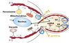
Vesicle transport
- Budding yeasts require the movement of DNA and organelles into the new bud when replicating
- Uses myosin 5-bound vesicles that are carried along actin filaments
- Myosin 5 is inactive in the absence of cargo
Cytoplasmic streaming
- Myosin generates cytoplasmic streaming: flow inside the cytoplasm, speeding up diffusion
- Key in plant cells
- Non-moving cortical actin goes all around the cell
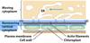
How do cells move?
- Cell movement depends on the actin-myosin cytoskeleton
- Stress fibres of actin and myosin are observed in the cell; similar to sarcomere
- They contact transmembrane proteins (integrins) at both ends which connect it to the extracellular matrix
- Integrins provide traction for movement
What is chemotaxis?
Chemotaxis: movement of a cell towards a molecule or in response to something (requires chemotactic receptors)
The receptors are located on the whole surface of the cell
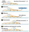
Describe the 4 steps of cell movement
- The cell is bound using focal adhesions and receives a chemotactic signal
- The focal adhesions are created by the integrins
- In between every focal adhesion are stress fibres
- Extension occurs as actin polymerises
- Forms filopodia (thin) and lamellipodia (large) structures
- These structures have integrins on it which are looking for a place to stick and form a new focal adhesion and additional stress fibre
- Adhesion and translocation occurs via actin-myosin contraction
- De-adhesin and endocytic recycling occurs
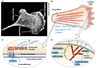
Describe the role of the proteins Cdc42, Rac, and Rho
Rho-GTP: regulates formin (unbranched actin regulation)
Cdc42/Rac: regulated Arp2/3 (branched actin regulation along with WASp and WAVE)
All of these are GTPase and must be in the GTP form
What does ‘dominant’ mean?
Describe dominant Rho, Cdc42, and Rac
- Dominant-active Rho shows it is responsible for unbranched stress fibre formation
- Dominant-active Cdc42 shows it forms small filopodia
- Dominant-active Rac shows it forms lamellipodia
Note that all of these work together (if one or more is missing, a “wound” cannot be filled)

Describe the coordination of cell migration
During this process, cell polarity causes microtubules to bring vesicles that are carrying G-actin, profilin, Arp2/3 and other resources to the front of the cell

