Abdominal Viscera and Collateral Circulation Flashcards
The two curvatures of the stomach and their derivation:
- lesser curvature (ventral side of foregut before rotation)
- greater curvature (dorsal side of foregut before rotation)
Lesser omentum attachment and derivation:
- attachment: lesser curvature of stomach
- derivation: ventral mesentery of stomach
The two parts of the lesser omentum:
- hepatoduodenal ligament
- hepatogastric ligament
Derivatives of the stomach dorsal mesentery:
- gastrosplenic ligament
- greater omentum
Label all:


Label:
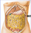
greater omentum
(arises from dorsal mesentery)
What structure is attached/fused to the posterior side of the greater omentum?
transverse colon
Label A and B:

- A = transverse mesocolon
- B = greater omentum
Greater Peritoneal Sac:
- All of the peritoneal cavity except for the lesser sac (omental bursa).
The greater peritoneal sac is subdivided into what two compartments?
- supracolic compartment
- above transverse colon and greater omentum (fused).
- infracolic compartment
- below transverse colon and greater omentum (fused).
The boundary between the supracolic compartment and infracolic compartment is:
- transverse mesocolon and greater omentum (which are fused together).

The three compartments of the peritoneal cavity:
- lesser sac
- supracolic (part of greater sac)
- infracolic (part of great sac)
What part of the greater omentum stretches from the greater curvature of the stomach to the level of the transverse colon?
gastrocolic ligament

Gastrocolic ligament:
- the part of the greater omentum above the transverse colon.
- a surgical entry into the lesser sac.
Label:


Contents of Supracolic compartment:
stomach, liver and spleen
Contents of Infracolic compartment:
small intestine, ascending colon, descending colon
The lesser sac communicates with greater sac via:
epiploic foramen
(foramen of Winslow)
The foregut gives rise to what organs?
PEG LSD
- Pancreas
- Esophagus
- Gallbladder
- Liver
- Stomach
- Duodenum (1, 2P parts)
The midgut gives rise to what organs?
JAILED CAT
- Jejunum
- Appendix
- ILEum
- Duodenum (2D, 3, 4, parts)
- Cecum
- Ascending colon
- Transverse colon (proximal 2/3)
The hindgut gives rise to what organs?
DESCENDING RATS
- DESCENDING colon
- Rectum
- Anal canal (upper part)
- Transverse colon (distal 1/3)
- Sigmoid colon
Celiac trunk is the blood supply to:
foregut and spleen
PEG LSD + spleen
Superior mesenteric artery is the blood supply to:
midgut + head of pancreas
JAILED CAT + head of pancreas
Inferior mesenteric artery is the blood supply to:
hindgut
DESCENDING RTS
The three branches of the celiac trunk:
- left gastric artery
- splenic artery
- common hepatic artery
Branches of the left gastric artery:
- esophageal artery
Branches of the splenic artery:
- short gastric artery
- left gastroomental artery
Branches of the common hepatic artery:
- proper hepatic artery
- gastroduodenal artery
Branches of the proper hepatic artery:
- left hepatic artery
- right hepatic artery
- right gastric artery
Blood supply to the gallbladder and the origin of this artery:
cystic artery
branch of right hepatic artery
First branch of the superior mesenteric artery:
inferior pancreaticoduodenal artery
Branches of the superior mesenteric artery:
- inferior pancreaticoduodenal artery
- middle colic artery
- right colic artery
- ileocolic artery
- jejunal arteries
- ileal arteries
Blood supply to the appendix and its derivation:
- appendicular artery
- from ileocolic artery, from SMA
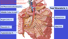
Label all:


Aorta ends (bifurcates) at what vertebral level?
L4
Inferior mesenteric artery branches off the aorta at what vertebral level?
L3
Branches of inferior mesenteric artery (3):
- left colic artery
- sigmoid artery
- superior rectal artery
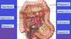
Label all:


What arteries branch and anastomose to form the Marginal Artery of Drummond (5)?
- Right colic a.
- Left colic a.
- Middle colic a.
- Ileocolic a.
- Sigmoid aa.
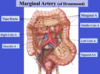
What arteries connect the Marginal Artery of Drummond to the colon?
- Vasa recta
- end arteries;
- do not anastomose with one another
What arteries have to be obstructed in order to create an area of ischemic bowel?
- vasa recta
- obstructing branches of SMA or IMA will not lead to ischemic bowel since they all anastomose to form the marginal artery of Drummond.
Where does diverticulitis occur in the colon?
- points where the vasa recta penetrate the colon wall
- “weak-points”
The two venous systems of the abdomen:
- inferior vena cava
- portal vein
What two veins come together to form the portal vein?
- splenic vein
- superior mesenteric vein
What vein drains into the splenic vein?
- inferior mesenteric vein
The IVC drains what organs of the abdomen?
- originally retroperitoneal organs
- (primary retroperitoneal organs)
The portal vein drains what organs of the abdomen?
- originally peritoneal organs
- (primary peritoneal and secondary retroperitoneal)
The spleen drains into what venous system of the abdomen?
portal venous system
Pathway of portal venous system drainage to the heart:
- splenic, superior mesenteric, inferior mesenteric veins
- hepatic portal vein
- venous sinusoids in liver
- hepatic veins
- inferior vena cava
- heart
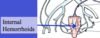
Portocaval anastomoses in the rectum:
- Superior rectal vein (portal) → middle rectal vein (caval) and inferior rectal vein (caval).
- in the wall of rectum.
Does the portal venous system have valves?
no; pressure gradient is based on pressure.
What will cause retrograde flow of blood in the portal venous system so that it drains in the IVC?
liver obstruction / portal hyperstension
What three veins of the rectum are susceptible to hemorrhoids in the setting of portal hypertension:
- superior rectal vein
- middle rectal vein
- inferior rectal vein
- due to anastomoses between portal and caval venous systems.
Portocaval anastomoses in the esophagus:
- Esophageal branch of left gastric vein (portal) to esophageal vein (caval) to azygos vein (caval).

Esophageal varices result from the anastomosis of what veins?
- Esophageal branch of left gastric vein (portal) to esophageal vein (caval) to azygos vein (caval).
Portocaval anastomoses in the umbilical region:
paraumbilical veins (portal) → epigastric veins (caval)

Caput medussae may arise from:
- Portocaval anastomoses in the umbilical region:
- paraumbilical veins (portal) to epigastric veins (caval).

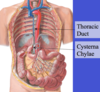
Lymphatic drainage of the abdomen:
- preaortic channels
- paraaortic channels
- cysterna chylae
- thoracic duct
Cysterna chylae is responsible for what lymphatic drainage:
all lymphatic drainage below the diaphragm
Label all:

Primary arterial supply to descending colon, which would have to be resected with the ascending colon in the setting of colon cancer:
- inferior mesenteric artery
- left colic artery
- LYMPH NODES FOLLOW VASCULATURE

Primary arterial supply to ascending colon, which would have to be resected with the descending colon in the setting of colon cancer:
- right colic artery
- middle colic artery
- ileocolic artery
- LYMPH NODES FOLLOW VASCULATURE

Draw the celiac trunk and its branches:

Draw the superior mesenteric artery and its branches:
Draw inferior mesenteric artery:

Draw marginal artery of drummond:
Draw IVC drainage in abdomen:
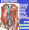
Draw portal vein drainage in abdomen:

