Abdominal Contents: Midgut & Hindgut Flashcards
Divisions of the gut tube
- What are the three divisions?
- What does each division share?
Divisions of the gut tube
- foregut
- midgut
- hindgut
Organs derived from the same division of the gut tube share:
- common blood supply
- route of venous drainage
- route of lymphatic drainage
- innvervation

Midgut
- Derivatives?
- Blood supply?
- Venous drainage?
- Lymphatic drainage?
Midgut
- Derivatives:
- distal duodenum
- jejunum
- ileum
- cecum
- appendix
- ascending colon
- proximal 2/3 of transverse colon
- Blood supply:
- SMA
- Venous drainage:
- SMV
- Lymphatic drainage:
- superior mesenteric lymph nodes

Hindgut
- Derivatives?
- Blood supply?
- Venous drainage?
- Lymphatic drainage?
Hindgut
- Derivatives:
- distal 1/3 of transverse colon
- descending colon
- sigmoid colon
- rectum
- Blood supply:
- IMA
- Venous drainage:
- IMV
- Lymphatic drainage:
- inferior mesenteric lymph nodes

Small intestine
- What are the three parts?
- What are the parts of the duodenum and where are they derived from? Quadrant of duodenum?
Small intestine
- Duodenum
- RUQ
- Proximal half
- 1st and 2nd parts are foregut
- Distal half
- 3rd and 4th parts are midgut
- Jejunum
- Ileum

Small intestine
Distal duodenum
Horizontal (3rd) part
- What kind of retroperitoneal organ?
- What kind of vessels pass by and where?
Small intestine
Distal duodenum
Horizontal (3rd) part
- Secondarily retroperitoneal
- Superior mesenteric vessels emerge superior and pass anterior to this part

Small intestine
Distal duodenum
Ascending (4th) part
- What kind of retroperitoneal organ?
- What is the acute angle and what does it mark?
- What is this structure supported by and what is it tethered to?
Small intestine
Distal duodenum
Ascending (4th) part
- Secondarily retroperitoneal
- Duodenojejunal flexure - acute angle that marks the duodenojujenal junction where it transitions from secondarily retroperitoneal duodenum to intraperitoneal jejunum
- Suspensory muscle of the duodenum (ligament of Treitz) - this supports the flexure, tethering it to the diaphragm

Small intestine
Jejunum
- Type of retroperitoneal organ?
- Location?
- What structures are prominent in jejunum?
Small intestine
Jejunum
- Intraperitoneal (robust mesentary)
- Central position in abdomen
- Plicae circulares - very prominent in jejunum

Small intestine
Ileum
- Type of retroperitoneal organ?
- Location?
- What structures are present and absent?
- Where does the ileum end?
Small intestine
Ileum
- Intraperitoneal (robut mesentary)
- Central position in abdomen
- Plicae circulares are sparse proximally and absent distally
- Lymphoid nodules (Peyer’s patches) are present in ileum
- Ileum ends at ileocecal junction

Small intestine
Ileal (Meckel’s) diverticulum of the ileum
- What is it?
- What is it a remnant of?
- Occurence?
- Clinical significance?
Small intestine
Ileal (Meckel’s) diverticulum of the ileum
- Blind pouch on the antimesenteric side of ileum
- Remnant of the yolk stalk (embryonic connection between the yolk sac and developing gut)
- 1-2% occurrence
- Congenital
- Typically 50 cm from ileocecal junction
- Inflammation of an ileal diverticulum can produce pain similar to appendicitis

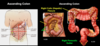
Large intestine
- What are the parts?
- Where does the midgut / hindgut transition lie?
Large intestine
- Includes:
- cecum
- appendix
- colon
- ascending colon
- transverse colon
- descending colon
- sigmoid colon
- rectum
- Includes both midgut and hindgut derivatives
- Midgut / hindgut transition lies at the junction of the proximal 2/3 and distal 1/3 of the transverse colon

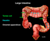
What are the features of the large intestine?
Features of the large intestine
- Teniae coli
- Three longitudinal bands of smooth muscle
- Teniae = ribbon in Latin
- Haustra
- Pouches between the teniae coli
- Omental (epiploic) appendices
- Fat
Large intestine
Cecum
- What kind of organ?
- Quadrant?
- Looks like a?
- What is the valve associated?
Large intestine
Cecum
- Intraperitoneal
- RLQ
- Round pouch
- Ileocecal valve - between the ileum and cecum, closes when cecum is distended

Large intestine
Appendix
- Type of organ?
- Quadrant?
- Location?
- Looks like?
- What converges there?
- What does root of appendix lie? Clinical significance of this?
Large intestine
Appendix
- Intraperitoneal (mesoappendix mesentery)
- RLQ
- Typically retrocecal (behind cecum) but this can vary
- Worm like appendage of the cecum
- Teniae coli converge at appendix
-
McBurney’s Point
- Root of appendix
- 1/3 of the way from the ASIS to the umbilicus
- Location of appendectomy incision

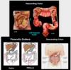
Large intestine
Ascending colon
- Type of organ?
- Quadrant?
- What is the fixture and gutter? Location?
Large intestine
Ascending colon
- Secondarily retroperitoneal
- Although it has mobile mesentery in 25% of people, so it can be intraperitoneal
- RLQ and RUQ
- Extends superiorly along right side of abdominal cavity between the cecum and the right colic (hepatic) flexure
- Right paracolic gutter is lateral to the ascending colon and fluid can accumulate there
Large intestine
Transverse colon
- Type of organ?
- Quadrant?
- Extends horizontally between what two flexures?
- Which flexure sits higher and why?
Large intestine
Transverse colon
- Intraperitoneal (transverse mesocolon mesentery)
- RUQ and LUQ
- Extends horizontally between the right colic (hepatic) flexure and the left colic (splenic) flexure
- Left colic flexure sits higher due to liver being on the right

Large intestine
Descending colon
- Type of organ?
- Quadrant?
- Extends inferiorly along what side of abdominal cavity and between what?
- What is directly lateral? Clinical significance of these structures?
Large intestine
Descending colon
- Secondarily retroperitoneal
- Although there is mobile mesentery in 33% of people, so it can be intraperitoneal
- LUQ and LLQ
- Extends inferiorly along the left side of abdominal cavity between the left colic flexure and sigmoid colon
-
Left paracolic gutter
- Lateral to descending colon
- Paracolic gutters are conduits for the spread of fluid within the peritoneal cavity
- If patient has abdominal infection and lays down, infection can spread to parietal pleura
- Patient with abdominal infections should stay sitting up
Large intestine
Sigmoid colon
- Type of organ?
- Quadrant?
- Extends between what?
- What shape does it form?
Large intestine
Sigmoid colon
- Intraperitoneal (sigmoid mesocolon mesentery)
- LLQ
- Extends from descending colon to rectum
- S shaped but variable in position

Large intestine
Rectum
- Type of organ?
- Continues proximally with? Distally with?
- Lacks what structures?
- Where do the rectum and anal canal meet?
- Location and function of rectal ampulla?
Large intestine
Rectum
- Primarily retroperitoneal
- Continous proximally with sigmoid colon and distally with anal canal
- Lacks:
- teniae coli
- haustra
- omental appendices
- Not a straight tube, has lateral flexures
-
Anorectal flexure
- The abrupt angle with the rectum and anal canal meet
- Maintained by the puborectalis muscle
-
Rectal ampulla
- Lies superior to the anorectal flexure
- Expands to store fecal matter

Midgut - SMA
- Arises from abdominal aorta at what level?
- Posterior to what?
- Passes anterior to what?
- What are the branches of the SMA?
Midgut - SMA
- Arises from the abdominal aorta at L1 vertebrae
- Posterior to the pancreas
- Passes anterior to the horizontal (3rd) part of the duodenum
- Branches of SMA:
- Anterior and posterior inferior pancreaticoduodenal arteries
- Jejunal and ileal arteries
- Colic arteries

Midgut - SMA
Anterior and posterior inferior pancreaticoduodenal arteries
Anastomose with what?
Midgut - SMA
Anterior and posterior inferior pancreaticoduodenal arteries
Anastomose with anterior and posterior superior pancreaticoduodenal arteries from the celiac trunk

Midgut - SMA
Jejunal and ileal arteries
- What pattern do these arteries form?
- How do you characterize jejunal vs ileal?
Midgut - SMA
Jejunal and ileal arteries
- Form anastomosing arcades that terminate in straight arteries (vasa recta)
- Jejunum arteries
- Few arcades
- Long straight arteries
- Ileum arteries
- Multiple arcades
- Short straight arteries

Midgut - SMA
Colic arteries
- Contribute to what artery?
- What are the three colic arteries?
- What is the branch off artery?
Midgut - SMA
Colic arteries
- Contribute to the marginal artery (an anastomosing loop that parallels the colon)
- Colic arteries
- Middle colic artery
- Right colic artery
- Ileocolic artery
- Appendicular artery (in mesoappendix)

Hindgut - IMA
- Arises from the abdominal aorta at what vertebrae?
- What are the three branches of the IMA?
- What do the branches contribute to?
Hindgut - IMA
- Arises from the abdominal aorta at L3
- Branches of IMA:
- Left colic artery (contributes to marginal artery)
- Sigmoid arteries (contribute to marginal artery)
- Superior rectal artery (terminal branch of IMA)

Portal system in the midgut and hindgut
- What vein goes to midgut and hindgut?
- What do the tributaries of the veins parallel?
- Where does it form or empty to?
Portal system in the midgut and hindgut
Midgut - SMV
- Tributaries parallel branches of the SMA
- Joins with the splenic vein to form the portal vein
Hindgut - IMV
- Tributaries parallel branches of IMA
- Empties into the splenic vein posterior to the body of the pancreas


