49-50 peptic ulcer & complications Flashcards
What is peptic ulcer
presence of ulcerative lesions in the stomach or lining of the duodenum(mc).
Ulcer = erosion/ break in the skin or mucous membrane
peptic ulcer = erosion in the mucouse membranes up to but not beyond the muscualris mucosa
3 layers of the the gastric mucosae
epithelial layer - glands that absorbs and secretes mucous
laimna propria= blood and lymph vess - vessels supply the gastric and duodenal mucosa with bi carbonate to nuetralise HCL
Muscularis mucosa- smooth muscle to brks down food
- the stomach mucosa is thick> duodenum d/2 increased contact w/ acidic content
anatomy and physiology of the stomach & duodenum relative to ulcers
- Cardia- foveolar cells that secrete mucus
-
Fundus & body-
- parietal cells -> HCL brks down food and convert pepsinogen into pepsin
- chief cells-> Pepsinongen ( a zymogen of pepsin)
- Pyloric antrum- G cells
- direct G cell mech-> secrete gastrin**- stim **parietal cells** to secrete **HCL
- indirect G cell mech -> stim enterochromafffin cells to release HIstamine -> acts on parietal cells -> HCL
- Pyloric sphincter- closes during eating allowing stomach to fuly digest food
- Duodenum - G cells & Brunner glands -> secrete mucous rich in bicarbonate- neutralise acidic content from the stomach
- Pancreas - G cells (accessory gland) and secretin -> stim mucosal cells to produce mucus
Function & location of G cells
location of G cells- Antrum, Duodenum, Pancreas
Physio of g cells is direct and indirect
- direct G cell mech-> secrete gastrin- stim parietal cells to secrete HCL
- indirecct G cell mech -> stim enterochromafffin cells to release HIstamine -> acts on parietal cells -> HCL
cells found in the fundus and body of the stomach
what do they do
parietal cells
+ by aCH, histamne, Gastrin. - by Prostaglandins and Somatostatin
- secrete HCL-> brks down food and convert pepsinogen into pepsin (stiim by gastrin and histamine)
- Secrete Intrinsic factor for binding and absorbtion vitamin B12.
chief cells
+ by ach, gastrin, secretin, Vasoactive Intestinal Polypeptide
- secrete Pepsinongen-> brks down protein after activated into the digestive enzyme pepsin when it comes in contac tHCL produced by gastric parietal cells. …
location and function of brunners glands
located in the duodenum
Brunner glands -> secrete mucous rich in bicarbonate- neutralise acidic content from the stomach which prevents the duodenum from being digested
Physiological role of Prostaglandins of the E type (PGE) in the stomach and duodenum
Stimulation of mucosal peptic mucosal protective factors
- stim mucus secretion in the epithelial layer
- stim production of bicarbonates in the duodenum
Vasodilation of adjacent blood vessels increasing local blood supply
- increases the supply of bicarbonates
- increases the growth of the epithelial layer
Classification of Peptic ulcers
Gastric ulcer: an ulcerative lesion in the stomach lining; typically manifests along the lesser curvature and the gastric antrum
Duodenal ulcer: an ulcerative lesion located in the duodenum, typically in the first part (i.e., the duodenal bulb)
Erosive gastritis: acute mucosal inflammation of the stomach that does not extend beyond the muscularis mucosae
RF of peptic ulcer
- Chronic gastritis caused by H. pylori, a curved, flagellated gram-negative rod
- Duodenal ulcers: up to 90% are due to H. pylori infection
- Gastric ulcers: up to 80% are due to H. pylori infection
- Chronic gastritis of other etiology
- Long-term use of NSAIDs: risk increases 5-fold - inhibit Cox 1&2 reduces Pg
- Long-term use of NSAIDs plus glucocorticoids: risk increases 10 to 15-fold!
- SSRIs -
- Smoking- decreased bf, increased irritation
- alcohol consumption - irritates mucosal lining
- Patients with blood type O have a higher risk for duodenal ulcers
- Age > 65 years
- Stress- actvates SNS, reduced bf to stomach
- Rare hypersecretory states:
- hyperparathyroidism, Zollinger-Ellison syndrome (gastrinoma), MEN,
- Genetics and family history
Pathophysiology of Peptic ulcer
reduction in protective factors
↓ protective prostaglandins - NSAIDS!!!!
↓ Clotting - SSRI, Cirrhosis
↓ blood flow- Curlings ulcer, Cushings ulcer, Smoking, Dieulafoy’s lesion
↓ Secretin -chronic Pancreatitis
↓ Cell restitution and Epithelial renewal
Increase in harmful factors
- ↑ acid secretion- H.pylori!!!, Zollinger ellison syndrome, Stress,
- irritation of the stomach lining -Alcohol, smoking, spicy foods, Acidic foods
- increased contact w/ gastric acid- GOO, Dumping syndrome, Hypercalcemia
why is duodenal ulcer usually on the duodenal bulb
typical ulcer location results from the effect of the gastric acid on the duodenal mucosa, which is highest in the duodenal bulb.
assoc rf
- blood type O
- 90% are due to H. pylori infection
causes of Gastric ulcer
H.pylori & NSaid syngeristic effect
Blood type A
Women
what is an An atypicalpeptic ulcer location suspicious for
carcinoma!
How does h.pylori cause Peptic ulcer
causes EMAG - Environmental Atrophic Gastritis
H.pylori is a gram negative
colonization by H. pylori → decreased production of mucins → increased production of gastric acids → increased penetration of gastric mucosa → inflammation primarily of the antrum→ ascending propagation → shift of the corpus-antrum border → in cases of chronification: atrophy of the gastric glands → hypochlorhydria (not achlorhydria) and epithelial metaplasia → increased risk of gastric cancers
why is H.pylori so viurlent in surviving in the acidic envirionment of the stomach
1-chemotaxis to avoid areas of low pH,
2- neutralizes the acid in its environment by producing large amounts of urease, which breaks down the urea present in the stomach to carbon dioxide and ammonia.
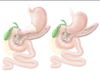
what is Zollinger Ellision Syndrome
gastrinoma (Zollinger-Ellison syndrome) is a gastrin-secreting neuroendocrine tumor that is most often localized to the duodenum and pancreas.
usually associated w/ Mx Endocrine Neoplasia
Gastrinomas release high levels of gastrin, which then increases the production of gastric acid.
dg = serum gastrin levels increase with the administration of secretin (positive secretin stimulation test).
rx = resection. PPi & Ocreotide
How do Nsaids cause peptic ulcers
Reversible inhibition (except aspirin) of the enzymes cyclooxygenase 1 and 2 (COX-1 and COX-2) → decreased prostaglandin synthesis
this causes reduction of PG mediated effects e.g.
- regulating pain receptor sensitivity,
- body temperature,
- renal blood flow,
- inflammatory processes.
- Bicarbonate synthesis & supply
- Epethlial mucus production
stress ulcers
Curling ulcer: patients with severe burns → ↓ plasma volume → ↓ gastric blood flow → hypoxic tissue injury of stomach surface epithelium → Acute weakening of the normal mucosal barrier & loss of Epithelial replacement
Cushing ulcer: In patients with incrreased ICP in brain injury, → Overwhelmingly increased vagal stimulation leads to an ACute increased production of stomach acid via acetylcholine release.
Dieulafoy’s lesion
(non cannon like malfoy)
rare disease where minor mucosal trauma can lead to major bleeding becauae of an abnormal submucosal artery.
Location: proximal stomach
Clinical presentation: signs of acute upper GI bleeding
Treatment: endoscopic hemostasis (injection therapy, hemoclips, etc.), excision of the susceptible mucosa
dx between duodenal ulcers and gastric ulcers
mc
causes
age
gender
onset after eating
relieving factors
excacerbating factors
type of bleeding presentation

common clinical features of gastric and duodenal ulcer
∼ 70% of patients with PUD are asymptomatic
Dyspepsia: postprandial heaviness, early satiety, and gnawing, aching or burning epigastric pain
Pain relief with antacids
Potential signs of internal bleeding (anemia, hematemesis, melena)
Stool sample positive for occult blood
General dg of peptic ulcer
-
≤ 60 years of age wit_hout alarm features_:
- Urea breath test for H. pylori
-
> 60 years of age or presence of 1+ alarm features:
- EsophagoGastroDuodenoscopy with biopsies
- rapid urease testing for H. pylori
If H.pylori and No histotry of Nsaid use
- serum gastrin level at baseline and after secretin stimulation test: high levels in gastrinoma (Zollinger-Ellison syndrome)
- Measure serum calcium and parathyroid hormone: high levels in primary hyperparathyroidism
Conservative TX of Peptic ulcer
DYSPEPSIA GENERAL MANAGEMENT
-
H. pylori positive → eradication therapy & medical acid suppression
- eradication therapy -> antibiotics
- acid suppresion PPI, H2blocker continue PPIs for 4–8 weeks → follow-u
- Mucosal protection: misoprostol (pg analong) Sucralfate (Aluminuim antacid)
- H. pylori negative → medical acid suppression only
Supportive rx
- Discontinue NSAIDs
- Restrict alcohol use/smoking/emotional stress
- Avoid eating before bedtime- reduces nocurnal acid levels
Surgical RX of peptic ulcers
With the advent of potent acid suppression in the form of PPIs, surgical intervention is rarely needed.
INDICATIONS
- fractory syndromes despite appropriate medical treatment
- If cancer is suspected
- Complications that cannot be treated endoscopically
OPERATIONS
- Bilroth 1
- BIlroth 2
- Vagotomy- division of vagal fibers decrease the production of gastric acid. It is rarely used today.