4. (Illustrated) Developmental problems and the child with special needs - Abnormal motor development (Cerebral Palsy) Flashcards
Cerebral Palsy
Definition of Cerebral Palsy
If the brain injury occurs after the age of 2 years, it is diagnosed as…
Cerebral Palsy is the most common…
CP is difficult to define, but the international group for
Surveillance of Cerebral Palsy in Europe defines it as
an umbrella term for a permanent disorder of movement
and/or posture and of motor function due to a
non-progressive abnormality in the developing brain.
The motor disorders of CP are often accompanied
by disturbances of cognition, communication, vision,
perception, sensation, behaviour, seizure disorder
and secondary musculoskeletal problems. Although
the causative lesion is non-progressive and damage
to the brain is static, clinical manifestations emerge
over time, reflecting the balance between normal
and abnormal cerebral maturation. Motor dysfunction
is usually evident early, often from birth. If the brain
injury occurs after the age of 2 years, it is diagnosed as
acquired brain injury.
CP is the most common cause of motor impairment
in children, affecting about 2 per 1000 live births.
Causes of Cerebral Palsy
About 80% of CP is antenatal due to…
Some of these problems are linked to…
Only about 10% of cases are thought to be due to…
Postnatal causes of Cerebral Palsy
About 80% of CP is antenatal in origin due to cerebrovascular
haemorrhage or ischaemia, cortical migration
disorders or structural maldevelopment of the brain
during gestation.
Some of these problems are linked
to gene deletions. Other antenatal causes are genetic
syndromes and congenital infection.
Only about 10% of cases are thought to be due to
hypoxic-ischaemic injury before or during delivery and
this proportion has remained relatively constant over
the last decade. About 10% are postnatal in origin.
Preterm infants are especially vulnerable to brain
damage from periventricular leukomalacia secondary
to ischaemia and/or severe intraventricular haemorrhage
and venous infarction. The improved survival of
extremely preterm infants has been accompanied by
an increase in survivors with CP, although the number
of such children is relatively small.
Postnatal causes are:
- meningitis/encephalitis/encephalopathy
- head trauma from accidental or non-accidental injury
- symptomatic hypoglycaemia
- hydrocephalus
- hyperbilirubinemia
MRI brain scans may assist in identifying the cause
of the CP, in directing further investigations and in supporting
explanations to the parents, but is not required
to make the diagnosis.
Clinical presentation of Cerebral Palsy
Early features of CP
Diagnosis is made by…
Categories of Cerebral Palsy
Many children who develop CP will have been identified
as being at risk in the neonatal period.
Early features of CP are:
• abnormal limb and/or trunk posture and tone in
infancy with delayed motor milestones (Fig. 4.3);
this may be accompanied by slowing of head
growth
• feeding difficulties, with oromotor incoordination,
slow feeding, gagging and vomiting
• abnormal gait once walking is achieved
• asymmetric hand function before 12 months
of age.
In CP, primitive reflexes, which facilitate the emergence
of normal patterns of movement and which need to
disappear for motor development to progress, may
persist and become obligatory (see Ch. 3).
The diagnosis is made by clinical examination, with
particular attention to assessment of posture and the
pattern of tone in the limbs and trunk, hand function
and gait.
CP is now categorized according to neurological
features as:
- spastic: bilateral, unilateral, not otherwise specified (90%)
- dyskinetic (6%)
- ataxic (4%)
- other.
The gross motor function level (functional ability) is
described using the Gross Motor Function Classification
System (Table 4.4).
In the past, the description was based on the parts of
the body affected (hemiplegia, quadriplegia, diplegia).
For children with high-risk factors for brain damage
such as significant prematurity or those with difficulties
around the time of birth, a formal standardized
assessment of general movements may identify at a
very young age those at greater risk of developing CP.
It is a specialized assessment usually performed by a
trained therapist or clinician.

Spastic cerebral palsy
Which neuronal pathway is affected/damaged?
How is limb tone affected?
Unilateral (hemiplegia):
- What parts of the body are involved?
- At what age does it often present?
- How is it presented?
- In some children, the condition is caused by…
Bilateral (quadriplegia):
- What parts of the body are involved?
- What is it more often associated with?
Bilateral (diplegia):
- What parts of the body are involved?
- What is it more associated with (Cause)?
- What may an MRI brain scan show?
In this type, there is damage to the upper motor neurone
(pyramidal or corticospinal tract) pathway.
Limb tone is
persistently increased (spasticity) with associated brisk
deep tendon reflexes and extensor plantar responses.
The tone in spasticity is velocity dependent, so the
faster the muscle is stretched the greater the resistance
it will have. This elicits a dynamic catch, which
is the hallmark of spasticity. The increased limb tone
may suddenly yield under pressure in a ‘clasp knife’
fashion. Limb involvement is increasingly described
as unilateral or bilateral to acknowledge asymmetrical
signs. Spasticity tends to present early and may even
be seen in the neonatal period. Sometimes there is
initial hypotonia, particularly of the head and trunk.
There are three main types of spastic CP:
• Unilateral (hemiplegia):
Unilateral involvement of
the arm and leg. The arm is usually affected more
than the leg, with the face spared.
Affected
children often present at 4–12 months of age with
fisting of the affected hand, a flexed arm, a
pronated forearm, asymmetric reaching, hand
function or toe pointing when lifting the child.
Subsequently, a tiptoe walk (toe–heel gait) on the
affected side may become evident.
Affected limbs
may initially be flaccid and hypotonic,
but increased tone soon emerges as the predominant
sign. The medical history may be normal, with an
unremarkable birth history and no evidence of
hypoxic-ischaemic encephalopathy giving rise to
the possibility of a prenatal cause, which is often
silent. In some children, the condition is caused by
neonatal stroke. More severe vascular insults may
cause a hemianopia (loss of half of visual field) of
the same side as the affected limbs
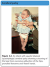
• Bilateral (quadriplegia)
All four limbs are affected,
often severely. The trunk is involved with a
tendency to opisthotonus (extensor posturing),
poor head control and low central tone (Fig. 4.4).
This more severe form of CP is often associated
with seizures, microcephaly and moderate or
severe intellectual impairment. There may have
been a history of perinatal hypoxic-ischaemic
encephalopathy
• Bilateral (diplegia)
All four limbs, but the legs are
affected to a much greater degree than the arms,
so that hand function may appear to be relatively
normal. Motor difficulties in the arms are most
apparent with functional use of the hands. Walking
is abnormal.
Diplegia is one of the patterns
associated with preterm birth due to
periventricular brain damage.
The MRI brain scan
may show periventricular leukomalacia.

Dyskinetic cerebral palsy
Definition of Dyskinesia
Description of Dyskinetic cerebral palsy (Features)
Affected children often present with…
Which neuronal pathway is affected/damaged?
Most common cause, in the past?
Most common cause, nowadays?
What will the MRI brain scan show?
Dyskinesia refers to movements that are involuntary,
uncontrolled, occasionally stereotyped and often more
evident with active movement or stress. Muscle tone is
variable and primitive motor reflex patterns predominate.
May be described as:
• chorea – irregular, sudden and brief non-repetitive
movements
• athetosis – slow writhing movements occurring
more distally such as fanning of the fingers
• dystonia – simultaneous contraction of agonist
and antagonist muscles of the trunk and proximal
muscles often giving a twisting appearance.
Intellect may be relatively unimpaired. Affected children
often present with floppiness, poor trunk control
and delayed motor development in infancy. Abnormal
movements may only appear towards the end of the
first year of life.
The signs are due to damage or dysfunction
in the basal ganglia or their associated pathways
(extra-pyramidal).
In the past, the most common cause
was hyperbilirubinemia (kernicterus) due to rhesus
disease of the newborn but it is now hypoxic-ischaemic
encephalopathy at term.
The MRI brain scan will often
show bilateral changes predominantly in the basal
ganglia.
Ataxic (hypotonic) cerebral palsy
Most common cause?
Early signs
Later signs
Most are genetically determined.
When due to acquired
brain injury (cerebellum or its connections), the signs
occur on the same side as the lesion but are usually
relatively symmetrical.
There is early trunk and limb
hypotonia, poor balance and delayed motor development.
Incoordinate movements, intention tremor and
an ataxic gait may be evident later.
The different types of CP are summarized in Fig. 4.5.

Management of Cerebral Palsy
Treatment for hypertonia in CP (4)
Parents should be given details of the diagnosis as early
as possible, but prognosis is difficult during infancy
until the severity and pattern of evolving signs and the
child’s developmental progress have become clearer
over several months or years of life.
Children with CP
are likely to have a wide range of associated medical,
psychological and social problems, making it essential
to adopt a multidisciplinary approach to assessment
and management, as described later in this chapter.
There are recently developed novel treatments
for treating hypertonia in CP such as botulinum toxin
injections to muscles, selective dorsal rhizotomy (a
proportion of the nerve roots in the spinal cord are
selectively cut to reduce spasticity), intrathecal baclofen
(a skeletal muscle relaxant) and deep brain stimulation
of the basal ganglia.


