3 - Cholesterol Flashcards

1
Q
  1. Where in the human body is cholesterol synthesized? Which tissues produce the most cholesterol?
A
  • Cholesterol is synthesized by virtually all tissues in humans.
  • Liver, intestine, adrenal cortex, and reproductive tissues, including ovaries, testes, and placenta, make the largest contributions to the body’s cholesterol pool.
2
Q

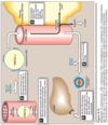
Major sources of liver cholesterol

A
3
Q
  1. Where in the cell is cholesterol manufactured?
A
  • Synthesis requires enzymes in both the cytosol and the membrane of the smooth ER.
4
Q

Which step in the synthesis of cholesterol is the rate-limiting one? What is the enzyme catalyzing this step? What four specific mechanisms serve to regulate this enzyme?

A
  • Which step in the synthesis of cholesterol is the rate-limiting one?
    • The reduction of HMG CoA to mevalonate → rate-limiting
  • What is the enzyme catalyzing this step?
    • Catalyzed by HMG CoA reductase in cytosol using two molecules of NADPH and releasing CoA (irreversible)
  • What four specific mechanisms serve to regulate this enzyme?
    1. Sterol-dependent regulation of gene expression
    2. Sterol-accelerated enzyme degradation
    3. Sterol-independent phosphorylation/dephosphorylation
    4. Hormonal regulation
    5. (Inhibition by drugs)
5
Q

Sterol-dependent regulation of HMG CoA reductase

A
  • Controlled by the transcription factor, SREBP-2 (sterol regulatory element-binding protein-2) that binds sterol regulatory element (SRE) of the reductase gene.
  • SREBP is an integral protein of the ER membrane, and associates with a second ER membrane protein, SCAP (SREBP cleavage–activating protein).
  • When sterol levels in the cell are low, the SREBP-SCAP complex is sent out of the ER to the Golgi. In the Golgi, SREBP is sequentially acted upon by two proteases, which generate a soluble fragment that enters the nucleus, binds the SRE.
  • This results in increased synthesis of HMG CoA reductase and, therefore, increased cholesterol synthesis
  • If sterols are abundant, SCAP-SREBP complex in the ER is retained thus prevent the activation of SREBP
6
Q

Sterol-accelerated HMG CoA reductase degradation

A
  • The reductase itself is a sterol-sensing integral protein of the ER membrane. When sterol levels in the cell are high, the reductase binds to insig proteins. Binding leads to ubiquitination and proteasomal degradation of the reductase
7
Q

Sterol-independent phosphorylation/dephosphorylation of HMG CoA reductase

A
  • Reductase activity is controlled covalently by adenosine monophosphate-activated protein kinase (AMPK) and a phosphoprotein phosphatase. Phosphorylated form of the enzyme is inactive, whereas the dephosphorylated form is active.
  • AMPK is activated by AMP, so cholesterol synthesis is decreased when ATP availability is decreased.
8
Q

Hormonal regulation of HMG CoA reductase

A
  • Increase in insulin and thyroxine up-regulates the expression of the gene for HMG CoA reductase whereas glucagon and glucocorticoids have the opposite effect
9
Q
  1. Discuss the essence of statins. What are they and what do they do?
A
  • The statin drugs are structural analogs of HMG CoA, and are (or are metabolized to) reversible, competitive inhibitors of HMG CoA reductase; used to decrease plasma cholesterol levels in patients with hypercholesterolemia.
10
Q
  1. Can cholesterol be degraded in the human body?
A
  • The ring structure of cholesterol cannot be metabolized to CO2 and H2O in humans.
11
Q
  1. How is cholesterol eliminated from the human body?
A
  • The intact sterol nucleus is eliminated from the body by conversion to bile acids and bile salts, which are excreted in the feces, and by secretion of cholesterol into the bile, which transports it to the intestine for elimination. Some cholesterol in the intestine is modified by bacteria into isomers coprostanol and cholestanol before excretion.
12
Q
  1. What are the two most important organic components of bile?
A
  • Phosphatidylcholine (lecithin) and bile salts (conjugated bile acids)
13
Q
  1. Which enzyme carries out the rate-limiting step in bile acid synthesis? Where is this enzyme found and why? What down-regulates this enzyme?
A
  • Which enzyme carries out the rate-limiting step in bile acid synthesis?
    • Cholesterol-7-α-hydroxylase
  • Where is this enzyme found and why?
    • It is an ER-associated cytochrome P450 (CYP) enzyme found only in liver.
  • What down-regulates this enzyme?
    • Down-regulated by cholic acid
14
Q
  1. What is a bile salt? What is the function of bile salts?
A
  • Bile salt is a conjugated form of bile acid with an addition of either glycine (resulting in lower pKa) or taurine (resulting in sulfonate group); both forms are fully ionized (i.e. negatively charged) at physiologic pH.
  • Bile salts are more effective detergents than bile acids because of their enhanced amphipathic nature; this is also why only the bile salts are found in the bile.
15
Q
  1. What action do intestinal bacteria have on primary bile acids?
A
  • Bacteria in the intestine can remove glycine and taurine from bile salts, regenerating bile acid.
  • They can also convert some of the primary bile acids into “secondary” bile acids by removing a hydroxyl group, producing deoxycholic acid from cholic acid and lithocholic acid from chenodeoxycholic acid
16
Q
  1. What is the enterohepatic circulation?
A
  • The continuous process of secretion of bile salts into the bile, their passage through the duodenum where some are converted to bile acids, their uptake in the ileum, and subsequent return to the liver as a mixture of bile acids and salts is termed the enterohepatic circulation
  • The mixture of bile acids and bile salts is absorbed primarily in the ileum via a Na+-bile salt cotransporter.
  • Between 15 and 30 g of bile salts are secreted from the liver into the duodenum each day, yet only about 0.5 g (less than 3%) is lost daily in the feces i.e. mostly recycled.
17
Q
  1. How do bile acid sequestrants reduce high levels of cholesterol (hypercholesterolemia)?
A
  • Bile acid sequestrants, such as cholestyramine, bind bile acids in the gut, prevent their reabsorption,and so promote their excretion. They are used in the treatment of hypercholesterolemia because the removal of bile acids relieves the inhibition on bile acid synthesis in the liver, thereby diverting additional cholesterol into that pathway.
18
Q
  1. a) What are the causes of cholelithiasis? b) How can cholelithiasis be treated?
A
  • What are the causes of cholelithiasis?
    • Cholelithiasis is the precipitation of cholesterol in the gallbladder as a result of excess cholesterol from the liver into the bile due to disruption of simultaneous secretion of phospholipid and bile salts
    • This disorder is typically caused by a decrease of bile acids in the bile, which may result from:
      • 1) gross malabsorption of bile acids from the intestine, as seen in patients with severe ileal disease;
      • 2) obstruction of the biliary tract, interrupting the enterohepatic circulation;
      • 3) severe hepatic dysfunction, leading to decreased synthesis of bile salts, or other abnormalities in bile production; or
      • 4) excessive feedback suppression of bile acid synthesis as a result of an accelerated rate of recycling of bile acids.
    • Cholelithiasis also may result from increased biliary cholesterol excretion, as seen with the use of fibrates.
  • How can cholelithiasis be treated?
    • Laparoscopic cholecystectomy (surgical removal of the gallbladder through a small incision) is currently the treatment of choice.
    • For patients who are unable to undergo surgery, oral administration of chenodeoxycholic acid to supplement the body’s supply of bile acids results in a gradual (months to years) dissolution of the gallstones.
19
Q
  1. What are the four types of lipoproteins? What is their basic structure? How can they be separated from each other?
A
  • What are the four types of lipoproteins?
    • Chylomicrons (CM)
    • Very-low-density lipoproteins (VLDL)
    • Low-density lipoproteins (LDL)
    • High-density lipoproteins (HDL)
  • What is their basic structure?
    • Lipoproteins are composed of a neutral lipid core (containing triacylglycerol and cholesteryl esters) surrounded by a shell of amphipathic apolipoproteins, phospholipid, and nonesterified (free) cholesterol.
    • These amphipathic compounds are oriented so that their polar portions are exposed on the surface of the lipoprotein, thus making the lipoprotein particle soluble in aqueous solution.
  • How can they be separated from each other?
    • They differ in lipid and protein composition, size, density, and site of origin
20
Q
  1. What are apolipoproteins? What are their overall functions?
A
  • Apolipoproteins are proteins that bind lipids (oil-soluble substances such as fat and cholesterol) to form lipoproteins (wiki)
  • They provide recognition sites for cell-surface receptors, and serve as activators or coenzymes for enzymes involved in lipoprotein metabolism.
  • They transport the lipids through the lymphatic and circulatory systems by making the particle water-soluble (wiki).
21
Q
  1. Where are chylomicrons assembled? What do they carry?
A
  • Chylomicrons are assembled in intestinal mucosal cells and carry dietary triacylglycerol, cholesterol, fat-soluble vitamins, and cholesteryl esters (plus additional lipids made in these cells) to the peripheral tissues.
  • The enzymes involved in triacylglycerol, cholesterol, and phospholipid synthesis are located in the smooth ER. After being loaded with apo B-48, particles transition to the Golgi where they are packaged in secretory vesicles and released, which then enter lymphatic system and, ultimately, the blood.
22
Q
  1. Which apolipoproteins are associated with chylomicrons? What is the function of these apolipoproteins?
A
  • Apolipoprotein B-48 is unique to chylomicrons but later apolipoprotein E&C are also added
  • Apolipoprotein B-48 is responsible for fusing with the plasma membrane releasing the lipoproteins.
  • Apolipoprotein E is recognized by hepatic receptors
  • Apolipoprotein C-II is necessary for the activation of lipoprotein lipase, the enzyme that degrades the TAG contained in the chylomicron
23
Q
  1. Where and how are chylomicrons degraded?
A
  • Lipoprotein lipase is an extracellular enzyme that is anchored by heparan sulfate to the capillary walls of most tissues, but predominantly those of adipose tissue and cardiac and skeletal muscle; adult liver does NOT have this enzyme.
  • Lipoprotein lipase, activated by apo C-II on circulating lipoprotein particles, hydrolyzes the TAG contained in these particles to yield fatty acids and glycerol.
24
Q
  1. a. How does insulin affect lipoprotein lipase? In which tissues does lipoprotein lipase have a low Km? Why? A high Km? Why?
A
  • How does insulin affect lipoprotein lipase?
    • Insulin stimulates synthesis and transfer of lipoprotein lipase to the luminal surface of the capillary.
  • In which tissues does lipoprotein lipase have a low Km? Why?
    • Heart muscle’s lipoprotein lipase; low Km allow the heart continuing access to the circulating fuel, even when plasma lipoprotein concentrations are low.
    • The highest concentration of lipoprotein lipase is in cardiac muscle, wince fatty acids provide much of the energy needed for cardiac function.
  • A high Km? Why?
    • Adipose enzyme’s lipoprotein lipase; high Km allow the removal of fatty acids from circulating lipoprotein particles and their storage as TAG only when plasma lipoprotein concentrations are elevated.
25
Q
  1. What is a chylomicon remnant? What is the fate of these remnants?
A
  • Remaining particle with more than 90% of the TAG in its core degraded by lipoprotein lipase (thus smaller in size and increase in density) and devoid of C apoproteins.
  • Remnant is rapidly removed from the circulation by the liver and bind to lipoprotein receptors that recognize apo E found in liver cell membranes. These are endocytosed into the hepatocytes and then fuse with a lysosome where the apolipoproteins, cholesteryl esters, and other components of the remnant are hydrolytically degraded, releasing amino acids, free cholesterol, and fatty acids; receptors, however, are recycled
26
Q
  1. Where are VLDL’s produced? What are they primarily composed of? What is their function?
A
  • Liver is responsible for the production of VLDLs.
  • They are predominantly composed of endogenous TAG (approximately 60%).
  • Their primary function is to carry TAG from the liver (site of synthesis) to the peripheral tissues, where TAG is degraded by lipoprotein lipase
27
Q

Low-Density Lipoproteins

A
  • LDL contain much less TAG than VLDL and have a high conc. of cholesterol and cholesteryl esters.
  • The primary function of LDL particles is to provide cholesterol to the peripheral tissues (or return it to the liver).
  • LDL bind to cell surface membrane LDL receptors that recognize apo B-100 and apo E (known as apo B-100/apo E receptors)
28
Q
  1. How are LDL’s taken up by cells and degraded?
A
  1. LDL receptors are negatively charged glycoproteins that are clustered in pits on cell membranes. The cytosolic side of the pit is coated with the protein clathrin, which stabilizes the shape of the pit.
  2. After binding, the LDL-receptor complex is internalized by endocytosis.
  3. The vesicle containing LDL loses its clathrin coat and fuses with other similar vesicles, forming larger vesicles called endosomes
  4. The pH of the endosome falls (due to the proton-pumping activity of endosomal ATPase), which allows separation of the LDL from its receptor. The receptors then migrate to one side of the endosome, whereas the LDLs stay free within the lumen of the vesicle. [Note: This structure is called CURL—the Compartment for Uncoupling of Receptor and Ligand.]
  5. The receptors can be recycled, whereas the lipoprotein remnants in the vesicle are transferred to lysosomes and degraded by lysosomal acid hydrolases, releasing free cholesterol, amino acids, fatty acids, and phospholipids. These compounds can be reutilized by the cell.
29
Q

What effect does endocytosed cholesterol have on cellular cholesterol metabolism?

A
  • First, HMG CoA reductase is inhibited by high cholesterol, as a result of which, de novo cholesterol synthesis decreases.
  • Second, synthesis of new LDL receptor protein is reduced by decreasing the expression of the LDL receptor gene, thus limiting further entry of LDL cholesterol into cells.
  • Third, if the cholesterol is not required immediately for some structural or synthetic purpose, it is esterified by acyl CoA:cholesterol acyltransferase (ACAT). ACAT transfers a fatty acid from a fatty acyl CoA derivative to cholesterol, producing a cholesteryl ester that can be stored in the cell. The activity of ACAT is enhanced in the presence of increased intracellular cholesterol.
30
Q
  1. What role do oxidized lipoproteins play in the formation of arterial plaque?
A
  • After being endocytosed by macrophages [with their scavenger receptor class A (SR-A)], oxidized lipoproteins cause accumulation of cholesteryl esters inside macrophages and cause them to transform into “foam” cells, which participate in the formation of atherosclerotic plaque.
  • Unlike the LDL receptor, the scavenger receptor is NOT down-regulated in response to increased intracellular cholesterol.
31
Q
  1. What are the four chief functions of HDL?
A
  1. HDL is a reservoir of apolipoproteins: HDL serve as circulating reservoir of apo C-II and apo E (and also apo A-1 accounting for 70% of the apoproteins in HDL).
  2. HDL uptake of unesterified cholesterol: HDL are excellent acceptors of unesterified cholesterol and thus take up cholesterol from non-hepatic (peripheral) tissues and return it to the liver as cholesteryl esters; possible due to their high concentration of phospholipids
  3. Esterification of cholesterol: cholesterol is immediately esterified once taken up by HDL carried out by the plasma enzyme lecithin:cholesterol acyltransferase (LCAT or PCAT)
  4. Reverse cholesterol transport: The selective transfer of cholesterol from peripheral cells to HDL, and from HDL to the liver for bile acid synthesis or disposal via the bile, and to steroidogenic cells for hormone synthesis, is a key component of cholesterol homeostasis. This is, in part, the basis for the inverse relationship between plasma HDL concentration and atheroscelerosis, and HDL’s designation as the “good” cholesterol carrier.
32
Q

Steroid

A
  • Cholesterol is the precursor of all classes of steroid hormones: glucocorticoids, mineralocorticoids, and sex hormones.
  • Because of their hydrophobicity, steroids must be complexed with a plasma protein e.g. plasma albumin
33
Q
  1. Which reaction is the rate-limiting reaction in the synthesis of steroid hormones?
A
  • The initial reaction that converts cholesterol to the 21-carbon pregnenolone catalyzed by the cholesterol side-chain cleavage enzyme complex (desmolase, P450scc) - a cytochrome P450 (CYP) mixed function oxidase.
  • Synthesis involves shortening the hydrocarbon chain of cholesterol, and hydroxylation of the steroid nucleus.
34
Q
  1. What is the mechanism of steroid hormone action?
A
  • Each steroid hormone diffuses across the plasma membrane of its target cell and binds to a specific cytosolic or nuclear receptor.
  • These receptor-ligand complexes accumulate in the nucleus, dimerize, and bind to specific regulatory DNA sequences (hormone response elements, HRE) in association with coactivator proteins, thereby causing promoter activation and increased transcription of targeted genes.
  • Hormone-receptor complex can also inhibit transcription in association with corepressors.