28 . Tumors Flashcards

1
Q

Generally, how do metastatic tumors present in the CNS?

A
  • Multiple, well-circumscribed lesions, most often at the grey-white junction.
  • Top 3 metastic cancers are: lung, breast and kidney cancers.
2
Q

Generally, do primary malignant tumors of the CNS metastasize?

A
  • No, they are locally destructive.
  • Rarely metastasize.
3
Q

What are the key cells in the brain that produce tumors?

A
  • Glial cells (astrocytes, oligodendrocytes, ependymal cells); 50%= supports and surrounds neurons
  • Neurons (50%)
  • Meningothelial cells = surround brain
4
Q

Astrocytes produce what tumors in kids and adults

A
  1. Kids = Pilocytic astrocytoma
  2. Adults = Glioblastoma (MC CNS tumor in adults)
5
Q

Oligodendrocytes produce what tumors and in who are they MC in

A

Oligodendromas = adults

6
Q

Ependymal cells produce what tumors and in who are they MC in

A

Ependymomas = children

7
Q

Neurons produce what tumors and in who are they MC in

A

Medulloblastomas (from neuroectoderm) = children

8
Q

Meningothelial cells produce what tumors and in whome?

A

Meningioma = adult females

9
Q

Where are brain tumors most common in kids and adults?

A
  1. Kids => infratentorial (posterior fosssa): cerebelleum & brainstem
  2. Adults => supratentorial (above tentorium)
10
Q

What are the MC brain tumors in children?

A

MC infratentorial

  1. Astrocytoma
  2. Medulloblastoma
  3. Ependymoma
  4. Craniopharyngioma
11
Q

What are the MC brain tumors in adults?

A

MC supratentorial

  1. Glioblastoma **
  2. Meningioma
  3. Schwannoma
  4. Oligodendroma
  5. Hemangioblastoma
  6. Pituitary Adenoma
12
Q

If you have a new onset of a seizure in a adult, think ________.

A

Brain tumor

13
Q

Which grade of tumor is considered infiltrative?

A

Grade II

14
Q

In which decade of life are Grade II, III, and IV Astrocytomas found?

A
  • Grade II: usually 30-40s
  • Grade III: usually 50s
  • Grade IV: usually 60s +
15
Q

What are gliomas?

A

MC group of primary brain tumors: astrocytomas, oligodendrogliomas, ependymomas

16
Q

Pattern of growth and spread in tumors in CSF

A
  • Most infiltrate extenselively, even if benign, displaying malignant behavior
  • Spread via CSF, but even most malignant gliomas RARELY metastasize ouside CSF
17
Q

What are Pilocytic Astrocytomas?

  • Usually occur when?
  • Where in the brain do they typically occur?
A

Low-grade benign tumor of astrocytes that are the MC brain tumor in chidren; slow growing and prognostically favorable

  • 0-20 (First 2 decades)
  • Cerebellum, floor/walls of 3rd ventricles, between hemispheres
18
Q

What grade of tumor is a Pilocytic Astrocytoma?

A

Benign I/IV; they commonly extend into subarachnoid space, but not sign of aggression and optic nerve lesions

19
Q

What are the distinguishing morphological characteristics of Pilocytic Astrocytomas?

A
    • Well-circumscribed, CYSTIC tumors w/ a mural nodule
  1. - Biphasic pattern: loose glial cells w cystic changes and dense piloid tissue
    • Low cellularity
  2. - Hair-like cells w/ long bipolar processes
  3. - Rosenthal fibers = long-standing gliosis
  4. - Eosinophilic granular bodies (EGBs)
20
Q

Marker of Pilocytic astronoma

A

GFAP

21
Q

what do these findings signify in pilocytic astronomas?

A
  1. EGB = PAS + protein drops that mark a slow growing, low grade tumor that is prognostically favorable
  2. Rosenthal fibers = long standing gliosis
22
Q

Which disease predisposes patients to Pilocytic Astrocytomas and due to what?

A

NF-1 (neurofibromatosis 1) due to functional loss of neurofibromin

23
Q

Most common presenting signs and symptoms of Infiltrating Astrocytomas?

A
  1. Seizures
  2. HA
  3. Focal neurologic deficits

related to the site of involvement

24
Q

Infiltrating astrocytoma range from what kind of tumors?

A
  • Grade II/IV: Diffuse astrocytoma
  • Grade III/IV: Anaplastic astrocytoma
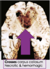
  • Grade IV/IV: Glioblastoma (this is malignant)
25
Q

Among the higher grade astrocytomas, presence of what mutation is associated with a better outcome?

A

Mutant form of IDH1

26
Q

What are Glioblastomas?

A

Most common primary brain tumor;

Rapidly progressive, Grade 4 malignant tumor of astrocytes the in the cerebral hemispheres.

27
Q

Where are Glioblastomas located?

A

Cerebral hemispheres: often cross the corpus callosum and form “butterfly gliomas”, which are necrotic and hemorrhagic

28
Q

Are glioblastoma primary tumors or secondary?

A
  1. Primary: new tumor in older people
  2. Secondary: progress in younger patients from a lower-grade astronoma.
29
Q

What mutations are associated with primary and secondary Glioblastomas?

A
  • Primary Glioblastomas (new tumor that occur in later people):
    • EGFR
    • PTEN
  • Secondary Glioblastomas (proceeds from a lower grade tumor in younger patients):
    • TP53
    • IDH1 / IDH2
    • (R132H = better prognosis)
30
Q

What mutation causing secondary Glioblastomas are associated with a BETTER prognosis?

A

R132H

31
Q

Glioblastomas

  1. ​Histological pattern (3)
A
  1. Serpentine pattern of necrosis
  2. Pseudo-palisading: ⬆︎ in # tumor cells around necrosis
  3. Vascular/endothelial proflieration that fill vascular lumen, producing a glomeruloid body (ball-like structure) due to ⬆︎ VEGF made by malignant astrocytes
32
Q

Ring-enhancing lesion on MRI => think ______

A

Glioblastoma: ring enhancing lesions iwth hypodense central necrosis

33
Q

Which tumor often crosses the corpus callosum and produces a “butterfly” appearance of hemorrhage and necrosis?

A

Glioblastoma

34
Q

Which brain tumor appears as a cystic mass with a mural nodule?

A

Pilocytic astronoma

35
Q

When are Oligodendrogliomas most commonly seen (decades)?

How are they graded?

A
  • Malignant tumor in oligodendrocytes that produces a calcified tumor in white matter, usually in the frontal lobe.
  • Adults 40-50 YO
  • Grade II/IV
36
Q

Where in the brain are Oligodendrogliomas most often seen?

A

White matter of cerebral hemispheres (frontal CTX)

37
Q

What are the most common mutations seen in Oligodendrogliomas; which have a better prognosis?

A
  1. IDH1 and IDH2 (better prognosis) = MC
  2. Loss of 1p19q (favorable prognosis)
  3. CDKN2A
38
Q

What are the distinguishing gross/histo morphological characteristics of Oligodendrogliomas?

A
  1. Gross Morphology:
    1. White matter TUMOR!!!
    2. Well circumscribed, gelatin, grey, CYSTIC and focal hemorrhage
    3. CALCIFICATION in the CTX (curvilinear or gyriform distribution)
  2. Histology:
    1. Perinuclear halos => creating “fried eggs” apperance (Artifact)
    2. Chicken wire vascular pattern: delicate anastomosing capillaries
39
Q

Which features distinguish an Anaplastic Oligodendroglioma (III/IV) from an Oligodendroglioma?

A
  1. Vascular hypertrophy and necrosis, but keeps geometric vascularity
  2. Nuclear anaplasia
  3. Poor prognosis
40
Q

What are ependymoma tumors?

A

Grade II/IV malignant tumors of ependymal cells, MC in children (first 2 decades of life = 0-20 YO)

41
Q

What are the MC sites/ situations for Ependymoma?

A
  1. Children = 4th ventricle => hydrocephalus
  2. Adults = SC central canal
  3. Neurofibromatosis 2, where NF2 gene is located
42
Q

When Ependymomas occurs in adults they are most often associated with what other condition; due to what mutation?

A

Neurofibromatosis 2 – due to mutation of NF2 gene on Chr22

43
Q

Well circumscribed tumors in the brain that often have cysts, focal hemorrhage, and calcification generally describes?

A

Oligodendrogliomas

44
Q

Histology of Ependymoma

A
  1. Uniform cells with round-oval nuclei and alot of clumped chromatin
  2. Epyndemal (true rosettes = tubular structure w central canal) or perivascular rosettes (ependymal cells project processes towards central BV)
    1. ​Ependymal are more diagnositc
45
Q

Which rosette type found in ependymomas are more diagnostic?

A

Ependymal rosettes (true rosettes)

46
Q

How do Grade III/IV Ependymomas present differently?

A
  1. Greater cell density and mitosis
  2. Microvascular proliferation
  3. Necrosis
  4. Clear cell variane
47
Q

Which tumors are the following Rosettes MC in?

A
  1. HW (neuropil in middle): medulloblastoma, neuroblastoma, PNET
  2. Flexner (true = lumen in center): ependymoma, retinoblastoma, Wilms
  3. Pseudo (vessel in center): astroblastoma and thymoma
48
Q

What is a frequent clinical manifestation of posterior fossa ependymomas?

A

Hydrocephalus (non-communicating) due to progressive obstruction of the 4th ventricle

49
Q

Which location of ependymomas have the worst overall prognosis in children?

A

- Posteior fossa = worst overall prognosis; 5 year survival is 50%

  • Completely resected supratentorial and spinal types are bette
50
Q

Choroid plexus papillomas (increase CSF) are most often found where in children and adults?

A
  1. Children – lateral ventricles
  2. Adults – 4th ventricle
51
Q

Clinical symptoms of a Choroid Plexus Papillomas

A

Obstuctrive hydrocephalus: make CSF

52
Q

Who is most often affected by Non-neoplastic Colloid Cysts of the 3rd Ventricle?

  1. Located?
  2. What is the clinical manifestation?
A
    • Young adults
    • Attached to roof of 3rd ventricle, can obstruct foramina of Monro => non-communicating hydrocephalus
    • HA (can be positional; worse when standinging) => rapidly fatal
53
Q

What is the most common malignant embryonal CNS tumor in children?

A

Medulloblastoma (Grade IV/IV) = poorly differentiated neuroectodermal tumor that ONLY occurs midline in the cerebellum

54
Q

What are medulloblastomas?

A

Malignant, poorly-differentiated PNET (peripheral neuroectodermal tumor) brain tumor, usually in children, that grows in the MIDLINE cerebellum => causing truncal ataxia.

In adults => grows more laterally

55
Q

Medulloblastomas are often located where?

A

Chilren; Midline of the vermis of the cerebellum => truncal ataxia

Adults = more lateral hemispheres

56
Q

Medulloblastomas can be divided into what 4 genetic groups?

Which have the best and worse prognosis?

A
  1. WNT mutation = best prognosis
  2. SHH mutation
  3. Group 3 = worst prognosis
  4. Group 4
57
Q
  1. Medulloblastomas that occur in older children tend to have what mutations and features?
  2. Prognosis?
A

Mutations in WNT signaling pathway

  1. Only have one Chr 6
  2. Nuclear expression of β-catenin

Best prognosis (90% 5 year survival)

58
Q
  1. Medulloblastomas due to mutations in SHH signaling: MC in who and associated with what other features?
  2. Prognosis?
A
  1. Infants - young adults
  2. Mutations in SHH sigaling + MYCN amplification
  3. 2nd best prognosis
59
Q
  • Group 3 medulloblastomas: MC in who and associated with what other features?
  • Prognosis?
A
  1. Infants and young children
  2. MYC amplification + isochromosome 17 (i17q)
  3. Worst prognosis
60
Q

Group 4 medulloblastomas: MC in who and associated with what other features?

Prognosis?

A
  • Isochromosome 17 (i17q) + without MYC (but does happen sometimes)
  • Intermediate prognosis; in general 17q = worse prognosis
61
Q

Gross Morphology of Medulloblastomas

A
  1. Well-circumscribed; solid gray, friable
62
Q

Histology of Medulloblastomas

A
  1. Sheets of anaplastic cells: hyperchromatic nuclei and abundant mitoses
  2. Homer-Wright Rosettes: small round blue cells around neuropil
63
Q

Clinical features/complications of Medulloblastomas

A
  1. Poor prognosis: Grow fast => NTRS 4th ventricle and occlude CSF => hydrocephalus.
  2. Spread from CSF => form masses far away, like cauda equina SC (drop metases)
  3. Infiltrate cerebral CTX and penetrate the pia => subarachnoid space
64
Q

Marker for Medulloblastoma

A

GFAP +

65
Q

What is a common complication due to Medulloblastomas propensity to form linear chains and infiltrate?

A

Infiltrate cerebral CTX and penetrate the pia => subarachnoid space

Dissemination through CSF is common complication, => nodular masses some distance from the priamry tumor (i.e., as far as the cauda equina) – Drop Metastases

66
Q

How well do Medulloblastomas respond to treatment?

A

- VERY radiosensitive

  • Total excision and irradiation => 5 year survival of 75%
67
Q

What is a CNS supratentorial PNET?

A

Primitive neuroectodermal tumor

68
Q

What is the difference between how Pilocytic Astronomas and Medulloblastomas look?

A
  • Medulloblastomas: well-circumscribed, solid, grey and friable; VERY cellular
  • Pilocytic astrocytomas: cystic, biphasic architecture and low cellularity
69
Q

What is the difference between Ependymoma and Medulloblastomas look?

A

Ependymoma =

  1. perivascular pseudorosettes
  2. less cellular
  3. more fibrillar
70
Q

Differentiate metastic carcinoma with Medulloblastomas?

A
  • Metastatic cancers are MC in adults
71
Q

Differentiate High-Grade Infiltrating Astrocytomas with Medulloblastomas

A

More pleiomorphic

72
Q

What are Atypical Teratoid/Rhabdoid Tumors (AR/RT)

  • MC in whom
  • Where are they seen in the brain?
  • Benign or malignant?
A
  • Very malignant (grade IV) tumor in young children (< 5 YO)
  • Posterior fossa and supratentorial compartments
73
Q

Mutations in AT/RT?

A

Chr 22 deletion, specially the HSNF5/INI1

74
Q

Cytoplasm of rhaboid cells found in Rhaboid tumors is immunoreactive for what?

A
  1. Epithelial membrane antigen (EMA)
  2. Vimentin
  3. *Other markers may include: smooth muscle actin and keratins; NO desmin and myoglobin
75
Q

Histology of AT/RT (Atypical Teratoid/Rhaboid Tumor)

A
  • Rhabdoid cells: eosinophilic cytoplasm with sharp borders & eccentric nucleus, resemble rhabdomyosarcoma
76
Q

Prognosis of AT/RT

A

Very aggressive, less than 1 yr survical

77
Q

What is the most common CNS neoplasm in immunosuppressed patients, such as those with AIDS or following transplantation?

A

Primary CNS lymphoma

78
Q

Most primary CNS lymphomas are of _____origin (immune cell) and latently infected by _____ virus

A

B-cell CD20+

EBV

79
Q
  • What is the morphology of the lesions associated with Primary CNS Lymphomas?
  • how do they spread?
  • Which stain shows their characteristic pattern?
A
  1. Many lesions (multifocal) in grey and white matter; rarely outside CNS
  2. Infiltrate parenchyma and accumulate around vessels
  3. Periventricular spread
  4. Stain: Reticulin + silver stain:hooping” = infiltrating cells seprated from one another by silver strain
80
Q

Germ Cell Tumors in the brain

  1. Occur where?
  2. MC in who?
A
  • Occur along the midline, most commonly in the pineal gland (M) and suprasellar regions.
  • Japanese M 0-20
81
Q

Which markers of CNS germ cell tumors may be helpful in assisting with diagnosis and tracking response to therapy?

A
  1. - α-fetoprotein
  2. - β- hCG
82
Q

Metastasis of which type of germ cell tumor to the CNS is common?

What is the significance of this in regards to classifying the tumor?

A

Gonadal germ cell tumors, thus, we have to rule out a non-CNS primary tumor before we exclude diagnosis of a primary germ cell tumor

83
Q

The tumor that is histologically similar to a seminoma in the testis is reffered to what in the CNS?

A

Germinoma

84
Q

What is the response of primary brain germ cell tumors to radiation therapy/chemotherapy?

A

Good response

85
Q

Pineal parenchymal tumors are derived from pineocytes;

What type do we see in the brain?

A

Well-differented lesions with neuronal differentiation (pinocytomas)

=>

high-grade tumors (pineoblastomas)

that spread throughout the CSF.

86
Q
  • High-grade pineal tumors (________) are MC in ________
  • Low-grade pineal tumors (________) are MC in ________
A
  • High-grade pineal tumors (pineblastoma) are MC in kids
  • Low-grade pineal tumors (pinecytoma = neuronal differentiation) are MC in adults
87
Q

Pineblastomas are high-grade pineal tumors w/ necrosis and mitosis that are MC in kids.

What is the mutation?

A

RB gene; spreads throughout CNS

88
Q

What is the most common pineal tumor?

A

Germinoma

89
Q

What are meningiomas?

A
  • Grade I/IV
  • MC benign tumors in adults (F >30YO) of arachnoid meningothelial cells (EMA +) and are attached to the dura, penetrating bone and compressing brain (do NOT infiltrate brain) => can cause seizures
90
Q

How do Meningiomas grow?

A
  • Enlarge slowly
  • DONT infiltrate brain.
  • But, they compress it and the penetrate the bone.
91
Q

What is a risk factor for the development of Meningiomas?

A

Prior radiation to the head and neck, typically decades earlier

92
Q

What is the severity of Meningiomas, who are they commonly seen in and where are they found?

A
    • Benign tumors of adults (>30 F)
    • Usually attached to the dura; found along any of the external surfaces of the brain as well as ventricular system
93
Q

What is the most common cytogenic abnormality seen in Meningiomas?

A
  • Loss of Chr 22, especially the long arm (22q), which has the NF2 gene, that encodes merlin.

= > seend more often in NF2

94
Q

Higher-grade Meningiomas are associated with what mutations?

A
  1. NF2 mutations,
  2. loss of chromosome 22
  3. Chromosomal instability **
95
Q

What is the most common mutation seen in Meningiomas without NF2 mutations?

What is the histologic grade of this type?

A
  1. TNF-receptor associated 7 (TRAF7)
  2. Lower histologic grade
96
Q

Gross morphology of Meningiomas

A
  1. Solitary, round/bossilated (looks like BLACKBERRIES) tumor with attached to dura that compress underlying brain, but easily separate from it.
    1. if many = think NF2
  2. Firm, no necrosis or extensive hemorrhage
  3. Calcified psammoma bodies
  4. En plaque: tumor has a sheet-like spread along the surface of the dura
97
Q

Histology of Meningiomas

A

Various histological patterns, with no prognostic significance.

  1. Syncytial
  2. Fibroblastic
  3. Transitional
  4. Psamomatous
  5. Secretory
  6. Microcystic
  7. Wholed clusters of monotonous cells
  8. Calcified psammoma bodies
  9. EMA +
98
Q

Which subtype of Meningiomas has PAS+ intracytoplasmic droplets?

Is associated with mutations in what genes?

A
    • Secretory subtype
    • TRAF7 and KLF4 genes
99
Q

What is the typical patient presentation for a Meningioma?

A

Present with vague non-localizing signs and symptoms or with focal findings due to compression of underlying brain

100
Q

Which receptors may be expressed by Meningiomas and which patient population can this potentially affect?

A
  • Progesterone (70% have) and Estrogen (31%) receptors, thus, more common in F
  • Pregnancy increases ⬆︎symptoms, NOT incidence
101
Q

If a patient has multiple meningiomas, what should we think of as the cause?

A

Neurofibromatosis 2

102
Q

Metastatic carcinomas are the MC metastatic brain tumo. They account for 1/2 of the intracranial tumors in hospitilized patients.

Where is the MC primary sites for potential metastasis to the brain?

A
    1. Lung (35%)
    1. Breast
    1. Skin (melanoma)
    1. Kidney
    1. GI
103
Q

Which rare tumor has a high likelihood of metastasing to the brain?

A

Choriocarcinomas

104
Q

Which common tumor, but RARELY metastasizes to the brain?

A

Prostate

105
Q

Where in the CNS is a frequent site of involvement for metastatic disease?

A
  1. - Meninges
  2. - Intraparenchymal = Gray/White junction
106
Q

Meningeal carcinomatosis (tumor nodules along the surface of the brain, SC and intradural nerve roots) most commoly occurs with which carcinomas?

A
  1. Lung
  2. Breast
107
Q

What are the common histologic findings of metastases to the CNS?

A

Nodules of tumor, often with central areas of necrosis, surrounded by reactive gliosis

108
Q

What are paraneoplastic syndromes?

A

Result from malignancy elsewhere in the body and are mostly due to anti-tumor immune responses that cross react with central/peripheral NS antigens.

109
Q
  • What is subacute cerebellar degeneration (paraneoplastic syndrome)?
  • Due to?
  • Common in which cancers?
A
  • PCA-1 AB (anti-Yo) destrory purkinje cells, cause gliosis, and a mild chronic inflammatory cell infiltrate
  • Commonly seen in
    1. Ovarian
    2. Uterine,
    3. Breast cancer
110
Q

Small cell lung carcinoma => cause limbic encephalitis (paraneoplastic) due to what AB?

A

ANNA-1 antibody (anti-Hu) recognized neuronal nuclei in the CNS and PNS

111
Q

Ovarian teratomas => can cause limbic encephalitis (paraneoplastic syndrome = subacute dementia associated with perivascular inflammation, microglial nodules, neuron loss and gliosis MC in anterior and medial temporal lobe) due to what AB?

A

NMDA-receptor AB attack neurons in hippocampus

112
Q

Which antibody is most commonly associated with peripheral neuropathy and may cause limbic encephalitis (paraneoplastic)?

A

VGKC-complex AB: voltage-gated potassium channel

113
Q

What is an important consideration in regards to limbic encephalitis and malignancy?

A

The syndrome appears before any malignancy is suspected

114
Q

What is a paraneoplastic syndrome commonly caused by Neuroblastomas, in kids?

A

Opsoclonus (eye movement disorder)

115
Q

Lambert-Eaton myasthenic syndrome is caused by antibodies against?

A

(VGCa2+ channels) in the presynaptic neurons

116
Q

Treatment of paraneoplastic syndroemes

A

Immunotherapy to remove circulating Ab and remove tumor

117
Q

Germ cell tumors are commonly located midline, at the pineal gland or suprasellar region.

What is the MC tumor of the pineal gland?

A

Germinoma, a GERM CELL tumor, not a pineocyte-derived tumor.

118
Q

F patient undergoes EXTENSIVE radiation. What brain tumor is she most likely to get?

A

Meningioma; often caused by radiation.

119
Q

Patient has a choriocarcinoma. What is the 1st step in imaging?

A

Typically goes to brain: make sure it didnt go there.

120
Q

Which metastatic carcinomas to the brain often hemorrhage?

A
  • 1. Melanoma
  • 2. Choriocarcinomas
  • 3. Renal cell carcinoma
121
Q

What familial syndromes cause pheochromacytomas?

A
  1. MEN2A/B
  2. NF1
  3. VHL (Von-Hippel Lindau syndrome)
122
Q

Cowden Syndrome

  • Inheritance
  • Chromosome
  • Gene/Protein
  • NL function/manifestation of loss
A
  • Inheritance = AD
  • Chromosome= 10q23
  • Gene/Protein= PTEN/PTEN => IK3/AKT signaling
  • NL function/manifestation of loss = lipid phosphatase /
    1. gangliocytoma tumors
    2. benign follicular appendages (trichelemmomas);
    3. internal adenocarcinoma (breast or endometrial)
123
Q

NF1

  1. Inheritance
  2. Chromosome
  3. Gene/Protein
  4. NL function/manifestation of loss
A
  1. AD
  2. 17q11
  3. NF1/neurofibromin
  4. (-) regulates RAS signaling / neurofibromas
124
Q

NF2

  1. Inheritance
  2. Chromosome
  3. Gene/Protein
  4. NL function/manifestation of loss
A
  1. AD
  2. 22q12
  3. NF2/merlin
  4. Cytoskeleton signaling/ neurofibromas and acoustic neuromas
125
Q

Tuberous Sclerosis

  1. Inheritance
  2. Chromosome
  3. Gene/Protein
  4. NL function/manifestation of loss
A
  1. AD
  2. 9q34 and 16p13
  3. TSC1/hamartin and TSC2/tuberin
  4. TSC1 and TSC2 form a complex => negatively regulate mTOR/ angiofibromas and mental retardation
126
Q

Li-Fraumeni syndrome is caused by a mutation of ____ => what tumor?

A

TP53 => medulloblastomas

127
Q

Turcot syndrome is caused by a mutation of ____ => what tumor?

A
  1. APC or mismatch repair genes
  2. Medulloblastomas or glioblastomas
128
Q

Gorlin syndrome is caused by a mutation of ____ => causes what tumor?

A
  1. PTCH mutation => upregulation of SHH pathway
  2. Medulloblastoma
129
Q

Tuberous Sclerosis Complex is characterized by the development of what?

Most frequent clinical manifestations are?

A
  • Development of benign hamartomas & benign neoplasms in brain and other tissues:
  1. Cortical tubers (=> cause seizures),
  2. Subependymal nodules
  3. Renal angiomyolipomas
  4. Cardiac rhadomyomas
  5. Shagreen patches (local thickening of skin)
  6. Ash-leaf patches (hypopigmented areas)
130
Q

Which tumors are associated with the Tuberous Sclerosis Complex?

A
  1. Cardiac rhabdomyoma
  2. Renal angiomyolipomas
  3. Cortical tubers
  4. Giant cell astrocytomas
131
Q

What are the cutaneous lesions associated with the Tuberous Sclerosis Complex?

A
  1. Shagreen patches (localized cutaneous thickenings)
  2. Ash-leaf patches (hypopigmented areas)
132
Q

Tuberous sclerosis causes multiple small tumor that are described as what?

A

Candle guttering: drop like masses that bulge into ventricles

133
Q

Von Hippel-Lindau disease

  • Inheritance
  • Chromosome
  • Gene
  • What is the function of the protein associated with this gene?
A
  • AD
  • Chr 3p25.3
  • VHL tumor supressor
  • VHL => ↓ regulates HIF1 => regulates expression of VEGF => regulates erythropoeitin
134
Q

VHL Disease causes what?

A
  1. Polycythemia due to ⬆︎ VEGF => erythropoeitin
  2. Hemangioblastomas in cerebellum and retina
  3. Pheochromacytomas - in adrenal gland
  4. Cysts in [pancreas, liver, kidneys]
  5. Renal cell carcinoma
135
Q

What is the mnemonic using VHL for Von Hippel-Lindau Disease?

A
    • V = VHL gene
    • H = Hemangioblastoma (cerebellum and retina)
    • L = Lots of catecholamines = pheochromocytoma
  • *VHL = 3 letters = RCC (renal cell carcinoma)
  • *VHL = 3 letters = Chr 3
136
Q

NF1 is more common and is characterized by which malignancies and other findings?

A
  1. Neurofibromas in peripheral nerve
  2. Optic nerve gliomas
  3. Lisch nodules: pigmented nodules of the iris
  4. Cafe au lait spots: cutaneous hyperpigmented macules
137
Q

NF2 is most commonly characterized by which malignancies?

A
  1. Bilateral schwannomas of CN VIII
  2. Multiple meningiomas and ependymomas of SC
138
Q

What are peripheral nerve sheath tumors?

A
  1. Schwannoma
  2. Neurofibromas
  3. MPNST
139
Q

Schwannomas

  1. Marker
  2. MC occur?
  3. Mutation:
  4. Histology
A
  1. S100+
  2. Cerebellar pontine angle => acoustic neuromas of CN8 (=> tinnitus and hearing loss)
  3. NF2 => loss of merlin
  4. Histology
    1. Antoni A bodies (spindle cells with nuclei aligned in palisading rows, leaving nuclear free zone and forming verocay bodies)
    2. Antoni B bodies = ECM with no cells
140
Q

Which type of neurofibromas look like a bag of worms and often have malignant transformation into MPNST?

A

Plexiform neurofibromas

141
Q

What are MPNST?

Who do we comonly see them in?

A

85% are high-grade tumors, with 1/2 arising in pts w NF1.