20. Sensory Systems 🧠Flashcards
What receptors are responsible for the sensory modalities of touch and proprioception?
Mechanoreceptors
Describe the structure of mechanoreceptors involved in touch and proprioception.
The receptor is NOT a separate entity but is actually the peripheral terminal of the peripheral axon of the primary sensory neuron.
Describe the structure of a Pacinian corpuscle and explain how this structure relates to its function.
RA1
There is an axonal ending in the middle and it is wrapped around several concentric circles of epithelial cells – this allows the receptor to be very sensitive to vibration.

What is the difference between slow adapting and fast adapting receptors?
- Slow adapting receptors continue firing impulses for as long as the stimulus is present
- Fast adapting receptors tend to fire at the start of the stimulus and sometimes when the stimulus switches off but they tend to fade in the middle
What type of receptors are mechanoreceptors?
Mixture of slow and fast adapting receptors
Describe how sensory neurons vary in their properties.
They vary in SIZE and CONDUCTION VELOCITY
What are the two classifications of axons?
- Anatomical = based on axon diameter (labelled using LETTERS)
- A
- B
- C
- Physiological = based on conduction velocity (labelled using ROMAN NUMERALS) As axon diameter and conduction velocity are related, there is a lot of overlap in the classifications

Describe the general structure of sensory neurons that convey touch and proprioceptive information.
- They are LARGE and have a FAST conduction velocity
- Touch
- A beta (II)
- Mechanoreceptors
- A beta (II)
- Proprioception
- A alpha (Ia and 1b)
- Muscle spindle and Golgi tendon organs
- A alpha (Ia and 1b)

What is a receptive field?
An area of skin that is innervated by one sensory axon and its branches
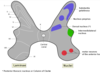
Describe how the receptive fields in the lips and mouth vary from the receptive fields of the upper arm.
Lips and Mouth – high-density innervation with very small receptive fields Upper arm – larger receptive fields and thinner innervation
Describe how neurons can code for the intensity of a stimulus.
It is coded by the FREQUENCY of the action potentials going down the sensory fibres
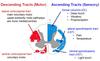
Which part of the spinal cord carries sensory axons for touch and proprioception?
Dorsal columns as part of the dorsal column-medial lemniscus pathway

What are the bundles of axons within the spinal cord that have come from above and below the waist called? Describe their spatial arrangement within the spinal cord (somatosensation)
- Above the waist
- Cuneate Fasciculus
- Below the waist
- Gracile Fasciculus
- Axons from below the waist are packed more medially in the dorsal column and above the waist are more lateral
- Lower = Medial
- Higher = Lateral

Where do the cuneata and gracilis fasciuli synapse?
They synapse in the Cuneate and Gracile Nuclei in the medulla

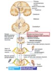
Describe what happens after the gracile and fasciculus neurons synapse int the medulla and the tract that they run in (somatosensation)
- The second order neurons then cross the midline (decussation) where they are then known as internal arcuate fibres
- They continue up the brainstem in the medial lemniscus where they then synapse at the ventral posterolateral nucleus at the medulla now becoming third order neurones
- They then continue to the primary somatosensory cortex in the thalamus

Which thalamic nucleus is responsible for relaying somatosensory information from the neck down?
Ventral Postero-lateral
Ventral Postero-medial nucleus = face
Describe the passage of the third order sensory neuron (in the dorsal column-medial lemniscus pathway)
The third order neurone travels from the ventral postero-lateral nucleus in the thalamus to the primary somatosensory cortex
What is the main sensory nerve of the face?
Trigeminal Nerve (CN V)
Where does the trigeminal nerve enter the brainstem and where does it synapse with a second order neuron?
- Enters the braisntem at the pons
- It synapses at the trigeminal nucleus
Describe the passage of the trigeminal nerve as a second order neurone after synapsing at the trigeminal ganglion (somatosensation)
The second order neuron crosses the midline (decussation) and joins the medial part of the median lemniscus
Which thalamic nucleus is responsible for relaying sensory information from the face?
Ventral Postero-medial

What is lateral inhibition?
- Lateral inhibition takes place in the cuneate and gracile nuclei
- Each axon has lateral branches that are inhibitory on neighbouring axons
- So each axon will stimulate a second order neuron and inhibit neighboring first order neurons
What is the purpose of lateral inhibition?
Improves the resolution of localising the stimulus
Name the three parts of the somatosensory cortex.
- Primary somatosensory cortex (S1)
- Secondary somatosensory cortex (S2)
- Posterior parietal cortex

What is the posterior parietal cortex mainly involved in?
Spatial relationships
Damage to the touch and proprioception pathway will halt sensory information going up to the primary somatosensory cortex. What effects will this have?
- Anaesthesia
- Complete cessation of sensation
- Parasthesia
- Sensation is there but it isn’t normal
What is the most common cause of peripheral neuropathy?
Diabetes mellitus
What are the pain receptors called?
Nociceptors
Can very high intensity stimulation of mechanoreceptors cause the feeling of pain?
No – only nociceptors can cause pain sensation
Describe some features of nociceptors.
- Polymodal
- different types of nociceptor respond to different stimuli
- Free nerve endings
- usually just free axonal endings of neurones
- High threshold
- higher activation threshold than touch receptors
- Slow adapting
- this is good because it means you are constantly reminded of the presence of a potentially harmful stimulus
What are the two main types of sensory neurone that carries sensory information? State some characteristics of each.
- A
- Large
- Fast conducting
- Fast adapting
- Produces pain fast
- C-fibre
- Smaller
- Produces a dull, aching pain
- It reminds you of the injury so that you guard this part of the body
- SLOW conducting -UNMYELINATED
Compare the receptive fields of nociception to those of touch.
Receptive fields for nociception are much LARGER because the nociceptive pathway is phylogenetically older than touch and you don’t need to be able to localise pain as well as touch.
Describe the method of coding intensity in nociception.
Same as touch – increase in frequency of impulses
Describe the passage of the central pathway, which carries information about pain and temperature.
- First order neurone enters the spinal cord and synapses in the dorsal horn with a second order neurone
- Second order neurone decussates immediately and travels up the white matter of the spinothalamic tract
- It then goes up to the thalamus where it synapses with a third order neurone, which then goes to the primary somatosensory cortex.
Describe the somatotopic arrangement of the fibres in the spinothalamic tract.
Lower fibres = Lateral
Higher fibres = Medial (Opposite of dorsal columns)
Sharp point arrow head that would hurt to sit on upwards
Which thalamic nucleus relays sensory information from below the neck?
Ventral Postero-lateral
Where does decussation occur in the pain and temperature pathway?
- At the same level as the information coming into the spinal cord
- Dorsal root ganglion
Which nerve carries nociceptive information from the face?
Trigeminal Nerve (CN V)
Where does the trigeminal nerve enter the brainstem?
Pons
Describe the passage of the trigeminal nerve from entry into the brainstem (nociception)
- It enters the trigeminal ganglion in the pons and then it moves DOWNWARDS along the trigeminal nucleus
- ​Trigeminal nerve nucleus = largest of the cranial nerve nuclei (collection on neurones) that extend through the whole of the midbrain, pons and medulla oblongata
- It then synapses in the lower part of the trigeminal nucleus in the medulla
- The second order neurone then decussates and joins the medial end of the spinothalamic tract.

Which thalamic nucleus relays pain information from the face?
Ventral Posteromedial
Which part of the trigeminal nucleus does the first order nociceptive neurones from the face synapse in?
Spinal Trigeminal Nucleus
What is the role of the primary somatosensory cortex in processing the nociceptive stimulus?
It registers the LOCATION and INTENSITY of the stimulus
As the spinothalamic tract projects towards the primary somatosensory cortex, it gives off collateral branches. Which structures do these branches go to?
BT LH
- Brainstem
- Reticular formation
- Thalamus
- intralaminar nuclei
- Limbic structures
- Hypothalamus
What is the point of these collateral connections?
The connections to the reticular formation (brainstem) and intralaminar nuclei (thalamus) allow the spinothalamic tract to increase your level of arousal to make sure that you are aware of potentially harmful situations
Which CNS structures are involved in signalling the unpleasantness of a stimulus?
- Limbic structures
- Hypothalamus
- Basal ganglia
- Cingulate gyrus
- Amygdala
- Hippocampus
- Hypothalamus
What are the two pathways that can reduce the amount of pain that you feel?
Central and Peripheral Inhibition Pathways
What is the main location of the central inhibition pathway?
Periaqueductal Grey Matter
Describe the arrangement and function of the central inhibition pathway.
- Increased brain activity will increase the impulses going down the central inhibition pathway, which goes to the dorsal horn at every level.
- These descending axons synapse with an interneurone and activate the interneurone
- The interneurone synapses with the first and second order nociceptive neurones and release ENKEPHALIN, which is inhibitory
- So enkephalin release will reduce the amount of information going up the spinothalamic tract hence you feel less pain.
What type of molecule is Enkephalin?
- Opioid
- Morphine mimics the action of this central inhibition system.
Other than a first order nociceptive neurone, what else has input into the second order nociceptive neurone?
- Non-nociceptive neurones
- Axons of non-nociceptive touch neurones will go into the dorsal horns
- Will also have collaterals that are capable of activating an inhibitory interneurone,
- ​can reduce the activity of the projecting neurone and hence reduce the activity going up the spinothalamic tract.
Describe the arrangement and function of the peripheral inhibition pathway.
Stimulation of touch receptors in the same area as the pain sensation will lead to increased activity of the non-nociceptive touch neurones meaning that there is increased activation of the inhibitory interneurone and hence reducing the activity going up the spinothalamic tract.
State two causes of loss of pain sensation.
-
Syringomyelia
- Cyst or cavity within the spinal cord
-
Charcot Joints
- due to peripheral neuropathy, you don’t realise that you are using your joints inappropriately or excessively – this leads to joint deformities
How can you get exacerbation of pain?
- Wind up in the dorsal horn If someone has chronic pain then certain peripheral nerves coming into the spinal cord will be carrying high levels of input for a long time
- The cells in the dorsal horn can lower their sensitivity or their synapses will change, which means that the information going into the spinothalamic tract is increased so this can actually increase the level of chronic pain.
Compare the receptive fields of nociception to those of touch.
Receptive fields for nociception are much LARGER because the nociceptive pathway is phylogenetically older than touch and you don’t need to be able to localise pain as well as touch.
Describe the method of coding intensity in nociception.
Same as touch – increase in frequency of impulses
Describe the passage of the central pathway, which carries information about pain and temperature.
- First order neurone enters the spinal cord and synapses in the dorsal horn with a second order neurone
- Second order neurone decussates immediately and travels up the white matter of the spinothalamic tract.
- It then goes up to the thalamus where it synapses with a third order neurone, which then goes to the primary somatosensory cortex
Describe the somatotopic arrangement of the fibres in the spinothalamic tract.
Lower fibres = Lateral Higher fibres = Medial (Opposite of dorsal columns)
Definition of rate coding
Signal strength is conveyed by impulse rates of the individual neurons
Template matching
Where a network of neurons can recognize a specific pattern of inputs from a population of presynaptic neurons
Extroception
Sensitivity to stimulu originating outside the body
What type of neurons are at the dorsal root ganglion?
- Pseudo-unipolar
- Axon of a dorsal root ganglion has two branches
- One projecting into the periphery
- One projecting to the CNS
- Axon of a dorsal root ganglion has two branches
TRP ion channels
- Non-selective cation receptor channels similar in structure to voltage-gated channels.
- They have four protein subunits each of which contain 6 transmembrane domains.
- Pore between the 5th and 6th segment
TRPA1
- Only expressed in low-threshold cold receptor terminals
- Threshold below 17°C
TRPV4
Threshold above 27°C
TRPV3
Threshold above 35°C
TRPV1 & TRPV2
Threshold above 45°C
Thermal nociceptors
Activated by extremes in temperature greater than 45°C (
- TRPA1 = < 17°C
- TRPM8 = < 25°C
- TRPV4 = > 27 °C
- TRVP3 = > 35°C,
- TRPV1 & 2 = > 45°C
Mechanical nociceptors
Respond to excess pressure / mechanodeformation
Polymodal nociceptors
Can be activated by high-intensity, mechanical, chemical or thermal stimuli
Silent nociceptors
Found in the viscera, not normally activated by noxious stimuli instead by inflammation and various chemical agents which dramatically reduce their firing threshold resulting in secondary hyperalgesia and central sensitization
Nociceptive pain
Activation of nociceptors in the skin or soft tissue in response to tissue injury
Neuropathic pain
Pain caused by damage or disease affecting the somatosensory nervous system
Dysesthesia
Abnormal sensations
Allodynia
Pain from normally non-painful stimuli
Lamina 1
Many neurons in this most superficial lamina respond to noxious stimuli conveyed by A-delta and C fibers

Lamina 2
- Substantia gelatinosa
- Receives input from C fibers that are activated selectively by the cold
- Site of nociceptive modulation
- Densely spaced layer that contains many different classes of local interneurons, some excitatory and some inhibitory

Lamina 3 & 4
- Contains a mixture of local interneurons and supraspinal projection neurons
- Many of these neurons receive input from A-beta afferent fibers that respond to innoccous cutaneous stimulation such as the deflection of hairs and light pressure

Lamina 5
- Contain neurons that respond to a wide variety of noxious stimuli and project to the brain and thalamus
- Neurons in lamina 5 also receive input from nociceptors in visceral tissue
- The convergence of somatic and visceral nociceptive inputs onto individual lamina 5 neurons provides one explanation for referred pain
Lamina 6
- Receive inputs from large diameter fibers that innervate muscles and joints
- 1 - A alpha
- Muscle spindle
- Golgi tendon organ
- 1 - A alpha
- These neurons are activated by innocuous joint movement and do not contribute to the transmission of nociceptive information

Lamina 7
- Respond to stimulation on either side
- Activation contributes to the diffuse quality of many pain conditions

What are the main landmarks of the visual pathway?
- Eye
- Optic nerve
- Optic chiasm
- Optic tract
- Lateral geniculate nucleus
- Optic radiation
- Primary visual cortex (striate cortex)
Where do retinal ganglion axons coming down the optic nerve synapse?
Lateral Geniculate Nucleus
Where is the lateral geniculate nucleus found?
Thalamus
What are the fibres leaving the lateral geniculate nucleus called?
Optic Radiation

Which order neurones are these and where do they terminate?
4th Order Neurones They terminate in the primary visual cortex
What are the first, second and third order neurones in the visual pathway?
First Order – photo-receptors (rods and cones) Second Order – bipolar cells Third Order – retinal ganglion cells
What happens as the retinal ganglion cells enter the optic nerve, which improves the transmission of the signal?
They become myelinated
What percentage of retinal ganglion cell fibres crosses the midline at the optic chiasma?
53%
Describe the convergence and receptive field sizes of rods and cones.
Rods have high convergence and large receptive fields Cones have low convergence and small receptive fields
Describe how the convergence of the rod system differs across different parts of the retina.
The rod system near that macula has lower convergence than in the peripheral retina
What is the benefit of having high convergence and a large receptive field?
High light sensitivity
What is the benefit of having low convergence and a small receptive field?
Fine visual acuity
Retinal ganglion cells can be described as on-centre or off-centre. What do these two terms mean?
On-centre – they are stimulated by light falling on the centre of the receptive field and inhibited by light falling on the edge of the receptive field Off-centre – they are stimulated by light falling on the edge of the receptive field and inhibited by light falling on the centre This is important in contrast sensitivity and enhanced edge detection
Where do the fibres that decussate at the optic chiasma originate?
The nasal part of the retina These fibres are responsible for the temporal half of the visual field
What effect do lesions anterior to the optic chiasm have on vision?
Affects only ONE eye
What effect do lesions posterior to the optic chiasm have on vision?
- Affects BOTH eyes
- Right-sided lesion = left homonymous hemianopia
- Left-sided lesions = right homonymous hemianopia

What is the effect of a lesion at the optic chiasm?
Bitemporal hemianopia
Which part of the brain does the upper division of the optic radiation travel through and which parts of the visual field is it responsible for?
- Parietal lobe
- Responsible for the inferior visual quadrants
Which part of the brain does the lower division of the optic radiation travel through and what part of the visual field is it responsible for?
- Temporal lobe
- Responsible for the superior visual quadrants
The lower division loops inferiorly and anteriorly before going posteriorly towards the primary visual cortex. What is this loop called?
Meyer’s Loop
What would be the consequence of a lesion in Meyer’s loop?
Superior homonymous quadrantopia

What would be the consequence of a lesion of the upper division of the optic radiation?
Inferior homonymous quadrantopia
What is homonymous hemianopia typically caused by?
Strokes and other cerebrovascular accidents
Where is the primary visual cortex located?
Along the Calcarine Fissure in the occipital lobe
Describe which parts of the primary visual cortex are responsible for the different fields of vision.
- The macula has a disproportionate representation in the primary visual cortex
- The left primary visual cortex is responsible for the right visual field from both eyes
- The right primary visual cortex is responsible for the left visual field from both eyes
- Visual cortex above the calcarine fissure is responsible for the inferior visual field
- Visual cortex below the calcarine fissure is responsible for the superior visual field
How is it possible for the macula to be spared by a stroke in the primary visual cortex leading to homonymous hemianopia?
The area representing the macula in the primary visual cortex has adual blood supply (from both right and left posterior cerebral arteries) meaning that it is less vulnerable to ischaemia
What are the two pathways of the extrastriate cortex and what are they responsible for?
- Dorsal Pathway – deals with motion detection
- Ventral Pathway – handles detailed object recognition and face recognition
Describe what happens to the eyes in the light.
Iric circular muscle contracts Constriction of pupillary aperture Reduced rate of photopigment bleaching Increased depth of field
Describe the pathway that is responsible for the consensual lightreflex.
Retinal ganglion cells from the retina send they axons back via the optic nerve The fibres that are responsible for the pupillary reflex will get passed the optic chiasm and then leave the posterior 1/3 of the optic tract before it reaches the LGN The axons then go to the pretectal nucleus in the dorsal brainstem The afferent pathways from each eye then synapse on the Edinger-Westphal nuclei on both sides of the brainstem. A parasympathetic nerve from the Edinger-Westphal nuclei to the ciliary ganglion forms the efferent pathway Short ciliary nerves travel from the ciliary ganglion to the pupillary sphincter Summary: Retinal Ganglion Cell –> Pretectal Nucleus –> Edinger-Westphal Nucleus –> Ciliary Ganglion –> Short Ciliary Nerves –> Sphincter Pupillae
What would the consequences be of a right afferent defect?
Light shone in right eye: no direct or consensual response Light shone in left eye: direct and consensual response present
What would the consequences be of a right efferent defect?
Light shone in right eye: no direct response, consensual response present Light shone in left eye: direct response, no consensual response
What does RAPD mean?
Relative Afferent Pupillary Defect A partial pupillary response is still present despite damage to an eye and its pupillary reflex pathway – there is some degree of constriction
What test would you do to identify RAPD? What would you expect to see in a patient showing a RAPD?
Swinging Torch Test When the light is shone on the good eye, there will be a direct and consensual response When the light is then swung and shone at the bad eye, there will be a paradoxical dilation of the iris in the bad eye This is because the constriction response elicited by the bad eye is weaker than the consensual response elicited by the good eye
Define: a. Duction b. Version c. Vergeance d. Convergeance
a. Duction Movement of one eye b. Version Simultaneous movement of both eyes c. Vergeance Simultaneous movement of both eyes in opposite directions d. Convergeance Simultaneous adduction of both eyes when viewing a near object
What is the term for the elevation of one eye?
Supraduction
What is the term for the depression of both eyes?
Infraversion
What is the technical term for right gaze?
Dextroversion
What is the technical term for left gaze?
Levoversion
What are the two types of eye movement and how are they different?
Saccade – short fast burst Smooth Pursuit – sustained slow movement
What reflex is used to assess visual acuity in preverbal children?
Optokinetic Nystagmus Reflex It is a form of physiological nystagmus triggered by the presentation of a constantly moving grating pattern
State which nerve innervates each of the extrinsic eye muscles.
Lateral Rectus = Abducens (CN VI) Superior Oblique = Trochlear (CN IV) Medial Rectus, Superior Rectus, Inferior Oblique, Inferior Rectus and Levator Palpebrae Superioris = Oculomotor (CN III)
Where do all the rectus muscles originate?
Common tendinous ring at the apex of the orbit
Where do the rectus muscles insert?
Into the sclera anterior to the globe equator
In what position would the eye have to be to get maximum elevation/depression due to: a. Superior and Inferior Recti b. Superior and Inferior Obliques
a. Superior and Inferior Recti Abducted b. Superior and Inferior Obliques Adducted
Explain why this is with respect to the anterior-posterior axis of the eye.
The anterior-posterior axis of the eye is aligned with the axis of the vertical recti when the eye is abducted If the eye is adducted, the axes are not aligned and contraction of the vertical recti would cause torsion
Where do oblique muscles attach to the eye?
Into the sclera posterior to the globe equator They pull the eye forwards and nasally This is because of the pulley system established by the trochlea and the oblique muscles
The oculomotor nerve has two branches. State what each of these branches innervates.
Superior Oculomotor Nerve ï‚· Superior Rectus ï‚· Levator Palpebrae Superioris Inferior Oculomotor Nerve ï‚· Inferior Rectus ï‚· Medial Rectus ï‚· Inferior Oblique ï‚· Parasympathetic nerve that causes pupil constriction
How would you test the extraocular muscles?
Isolate the muscle to be tested by maximising its action and minimising the action of the other muscles E.g. to test the superior rectus, make the patient abduct and elevate their eye
Describe and explain what you would see in a patient with 3rd nerve palsy.
Their affected eye would point down and out This is because of the unopposed contraction of lateral rectus and superior oblique Ptosis – because of the loss of innervation of levator palpebrae superioris Pupil dilation – loss of parasympathetic innervation to the eye via CN III
Describe and explain what you would see in a patient with 6thnerve palsy.
When asked the abduct the affected eye, they eye will stop around midline This is because the lateral rectus isn’t functioning and can’t abduct the eye This can lead to blurred vision
What is Hering’s Law of Equal Innervation?
Muscles from both eyes involved in conjugate movement receive equalinnervation
What structure in the brainstem acts as a synchronising link between the eyes, allowing paired eye movements?
Medial Longitudinal Fasciculus
What can damage to the MLF cause?
Internuclear Opthalmoplegia E.g. right abduction wont be accompanied by left adduction Could be accompanied by nystagmus on right gaze
What is Sherrington’s Law of Reciprocal Innervation?
Agonist muscles contract while antagonist muscles relax
State a condition in which Sherrington’s Law is violated.
Duane’s Syndrome – congenital absence of abducens (CN VI) Both lateral and medial recti are innervated by CN III (oculomotor)
What scale is used to measure how loud a sound is?
Decibels (logarithmic scale)
What is the audible range for humans in terms of frequency?
20-20,000 Hz
What is the name given to the wing shaped flap skin and cartilage that makes up the outer ear?
Pinna
Describe the shape of the outer ear and its importance.
It is conical – starts off wide at the external auditory meatus and narrows to the tympanic membrane This focuses the noise and increases the pressure on the tympanic membrane
Is the tympanic cavity fluid-filled or air-filled?
Air-filled
State 2 ways in which the ossicles increase the pressure of vibration of the tympanic membrane.
Focussing the vibrations from the large surface area of the tympanic membrane to the small surface area of the oval window – this decrease in surface area means that the pressure is increased The incus has a flexible joint with the stapes, such that the ossicles use leverage to increase the force on the oval window This amplifies the sound by 30 dB
What is the point of the middle ear? Why isn’t the tympanic membrane continuous with the cochlea?
The cochlea contains fluid, in which you are trying to induce a pressurewave If the tympanic membrane was continuous with the cochlea, you would go straight from air to fluid and 99% of the energy will bounce back due to impedance Sound waves require more energy to travel through fluid than air so the increase in pressure of vibration allowed by the ossicles is crucial for this conduction
What 2 muscles are involved in making sure that the ossicles aren’t damaged by excessive vibration due to loud noise?
Tensor Tympani Stapedius
What is the name given to this reflex?
Auditory reflex
What is the latency period of this reflex?
50-100 ms
What is hyperacusis?
Painful sensitivity to low intensity sounds – can occur in conditions that lead to flaccid paralysis of the auditory reflex muscles (e.g. Bell’s Palsy)
Which test is used to determine the site of damage to the auditory system, that is causing hearing loss?
Weber Test
What are the 2 specialised membranes of the cochlea?
Oval Window Round Window
What are the three compartments of the inner ear?
Scala Vestibuli Scala Media Scala Tympani
Which types of fluid do each compartment contain?
Scala Vestibuli + Scala Tympani = perilymph Scala Media = endolymph
What structure connects the two perilymph compartments?
Helicotrema
Describe how the cochlea functions.
The vibration of the tympanic membrane is conducted and amplified to a vibration of the oval window by the footplate of the stapes. This vibration induces a pressure wave in the perilymph in the scala vestibuli. This vibrates the scala media leading to vibration of the basilar membrane. The round window vibrates as well to equalise the pressure in the cochlea.
Describe the difference in sensitivity of different parts of the basilar membrane.
Higher frequency sounds = base Lower frequency sounds = apex
What is the Organ of Corti?
The sense organ of the cochlea, which converts sound signals into nerve impulses that are transmitted to the brain via the cochlear nerve
Where is the Organ of Corti found?
It lies on top of the basilar membrane and beneath the tectorial membrane
What are the two types of cell in the organ of corti?
Inner and outer hair cells
Describe the features and function of inner hair cells.
Found on their own Not in contact with the tectorial membrane Send impulses to the brain They have stereocilia that move in response to the movement of endolymph in the scala media Roughly 3500 in the body
Describe the features and function of outer hair cells.
Found in groups of three They are in contact with the tectorial membrane They receive input from the brain Electromotile so can expand and contract to amplify the amount of vibration (this is the basis of the cochlear amplifier) Damage can result in sensorineural hearing loss Roughly 20,000 in the body
Which compartment of the cochlea does the stereocilia of the hair cells project into?
Endolymph (base is in the perilymph)
What internally generated sounds are the outer hair cells responsible for?
Otoacoustic emissions
What are stereocilia connected by?
Tip links
What bony conical structure is found at the middle of the cochlea?
Modiolus
Describe what happens when the basilar membrane is displaced upwards.
Depolarisation Stereocilia move away from the modiolus K+ channels open K+ enters from the endolymph
Describe what happens when the basilar membrane is displaced downwards.
Hyperpolarisation Stereocilia move towards the modiolus K+ channels close
Describe the difference in K+ and Na+ concentration in the different compartments of the cochlea.
Scala Media = High K+ and Low Na+ Scala Tympani = High Na+ and Low K+ NOTE: stria vascularis maintains this concentration
Describe the auditory pathway from the cochlea to the primary auditory cortex.
Spiral ganglion -> cochlear nuclei -> superior olive -> inferior colliculus -> medial geniculate nucleus -> primary auditory cortex
Up to what point is the auditory pathway from one ear ipsilateral?
Cochlear nuclei Beyond this point there is bilateral representation
The inferior colliculus receives input from both cochlea. What is the inferior colliculus responsible for?
Reflex associations – turning your head towards loud noise
Describe a phenomenon that is involved in sharpening the signal coming from the cochlea.
Lateral inhibition
To which parts of the CNS do collaterals from the auditory pathway go?
Reticular formation Cerebellum
In which lobe is the primary auditory cortex?
Temporal
What is the secondary auditory cortex responsible for?
Responding to sounds coming off/on Responding to the duration of sound
What is the name given to the axons that project from the medial geniculate nucleus to the primary auditory cortex?
Acoustic radiations (they travel via the internal capsule)
How do you localise short sound burst?
Interaural time delay
How do you localise continuous sound?
Interaural intensity difference
What is conductive hearing loss?
When diseases of the middle ear damage the ossicles or stiffen theirjoints so that the amplification system is eliminated – results in conductive hearing loss
What is sensorineural hearing loss and what can it be caused by?
When the cochlea or cochlear nerve get damaged, the signal transmitted to the primary auditory cortex is reduced or lost It can be caused by acoustic schwannoma (tumour of the cochlear nerve) or cerebellar tumours expanding and putting pressure on thecochlear nerve
What is the term used to describe loss of hearing due to the death of hair cells in normal ageing?
Presbyacusis
What the three types of cell that makes up the olfactory epithelium?
Bipolar Olfactory Neurones Sustentacular Cells – support cells mainly providing metabolic support Basal Cells – there is some regeneration in olfactory neurones
Where is the olfactory bulb found?
Sitting on top of the cribriform plate
Which cells synapse within the olfactory bulb?
The bipolar cells pass their axons through the cribriform plate to synapse with the second order neurones (olfactory bulb mitral cells) in the glomerulus within the olfactory bulb
What structure do the second order neurones form and what does this structure split into?
Olfactory tract It splits to form the medial and lateral olfactory stria
Where does higher processing of smell take place?
Piriform Cortex Orbitofrontal Cortex
What is a clinical deficit in the olfactory system called?
Anosmia
What is a common cause of anosmia?
Mid-face trauma Impact with enough force could cause a fracture of the cribriform plate, shearing the neurones going from the olfactory epithelium to the olfactory bulb
The piriform cortex is found within the temporal lobe. Explain thesignificance of this with regards to epileptic patients.
Epilepsy is often focused in the temporal lobe This means that some people with epilepsy will experience PRODROMAL AURA (they are made aware of an imminent seizure because they’ll smell something that’s not there)
Neurodegenerative disease is a relatively common cause of anosmia. State two neurodegenerative diseases that could cause anosmia.
Alzheimer’s disease Parkinson’s disease
What is the limbic system?
A rim of cortex adjacent to the corpus callosum and diencephalon
What are the roles of the limbic system?
Homeostasis (mainly hypothalamic functions such as regulation of food intake and pituitary hormone release) Agonistic behaviour (fight or flight) Sexual and reproductive behaviour Memory
State two important parts of the limbic system that are found within the temporal lobes.
Hippocampus and Amygdala
What circuit are these structures a part of?
Papez Circuit
What is the cortical representation of the limbic system?
Cingulate Cortex
What is the function of the Papez circuit?
It is a neural circuit for the control of emotional expression
Describe/draw the papez circuit.
Hippocampus –> Fornix –> Mammillary Bodies –> Mammillo-Thalamic Tract (MTT) –> Anterior Nucleus of the Thalamus –> Cingulate Cortex –> Cingulum Bundle –> Hippocampus
What conditions could damage the mammillary bodies leading to amnestic issues?
Chronic Alcoholism Wernicke-Korsakoff Syndrome
What is our emotional expression ‘coloured’ by?
Neocortex
What form of imaging is used to study the limbic system?
Digital Tensor Imaging – shows co-instant activity in different parts of the brain thus showing which parts of the brain are working together
Describe the afferent pathway of the hippocampus.
Afferent Pathway = Perforant Pathway The entorhinal cortex is linked to the hippocampus via the afferent pathway (perforant pathway) The entorhinal cortex receives input from all other parts of the neocortex
What is the efferent pathway of the hippocampus called?
Fimbria/Fornix
What are the functions of the hippocampus?
Memory and Learning
What happens to the hippocampus in Alzheimer’s disease?
It shrinks severely
Describe the spatial relations of the hippocampus and the fornix to other important brain structures.
The hippocampus is found on the floor of the lateral ventricles The fornix comes out of the hippocampus and passes under the corpus callosum It then dives inferior and anteriorly towards the mammillary bodies
Describe the appearance of advanced Alzheimer’s disease on a CT head scan in the coronal plane.
There will be extensive cortical atrophy The ventricles would appear enlarged There will also be widening of sulci
State two microscopic hallmarks of neurodegeneration.
Tau Immunostaining  Intracellular pathology – the cytoskeleton has been compromised  The tau proteins show up in the staining and allow the damaged neurones to be seen Senile Plaques  Extracellular pathology  Lumps of protein sitting in between cells in the neuropil
Describe the anatomical progression of Alzheimer’s disease, including the symptoms experienced.
Early ï‚· Hippocampus and entorhinal cortex affects ï‚· Short-term memory problems Moderate ï‚· Parietal lobe (where you have your procedural memory) ï‚· Example of effects: dressing apraxia Late ï‚· Frontal lobe ï‚· Loss of executive skills
Where is the amydala found?
In the white matter of anterior temporal lobe
What are the afferent connections of the amygdala?
Olfactory Cortex Septum (septal nuclei) Temporal Neocortex Hippocampus Brainstem
What is the main output pathway of the amygdala?
Stria terminalis
What is the function of the amygdala?
Fear and Anxiety (fight or flight)
In Alzheimer’s and Parkinson’s disease, the amygdala starts showing pathology early on. What are the possible outcomes of this?
Patients could either become terrified of everything or they could become totally disinhibited and become quite aggressive
State and describe a syndrome affecting the amygdala.
Kluver-Bucy Syndrome Bilateral lesions of the anterior temporal lobe (including amygdaloid nucleus) Symptoms ï‚· Hyperorality ï‚· Hypersexuality ï‚· Loss of Fear ï‚· Visual Agnosia
State three structures associated with aggression.
Hypothalamus Brainstem (periaqueductal grey matter) Amygdala
What are the main afferent connections of the septum?
Amygdala Olfactory Tract Hippocampus Brainstem
What are the functions of the septum?
Reinforcement and Reward
Name another structure that is important in the reward system.
Nucleus Accumbens
Describe another dopaminergic pathway other than the nigro-striatal pathway that is affected in Parkinson’s disease.
Ventral Tegmental Area (VTA) of the midbrain –> Median Forebrain Bundles –> Cortex + Nucleus Accumbens + Amygdala
Name a structure that is important in drug dependence.
Nucleus Accumbens
What effect do all drugs of abuse have on the nucleus accumbens?
They all increase dopamine release in the nucleus accumbens
Which structures in the body are responsible for angular (rotational) motion of the head?
Semi-circular canals
Which structures in the body sense the acceleration of the headand the strength of gravity?
Otolith organs
What are the two otolith organs?
Saccule Utricle
What are the main functions of the vestibular system?
Subserve perception of motion in space and tilt (with respect to gravity) Provide reflex balance reactions to sudden instability of gait/posture (vestibulo-spinal reflexes) Stabilise the eyes on fixed targets during head movement, preserving acuity (vestibulo-ocular reflexes) Assist in control of heart rate and blood pressure during rapid up-down tilts Assist synchronisation of respiration with body reorientations Provokes motion sickness
Define vertigo.
False perception of movement in space
Define vestibular ataxia
Instability of gait or posture
What happens to the ability of the brain to stabilise the eyes in unilateral vestibular lesions?
Vestibular nystagmus The eyes start moving in the direction of the lesion
What happens to the ability of the brain to stabilise the eyes in bilateral vestibular lesions?
Oscillopsia Everything appears to be shaking – the ability to stabilise the eyes is lost
What are some other consequences of vestibular loss?
Slight impairment of orthostatic control Severe nausea and vomiting Loss of coordination on directional reorientation, motion intolerance, oversensitivity to visual motion in the environment
What type of cell is involved in the detection of movement in the vestibular system?
Hair cells
Describe the cilia of these hair cells.
There is one kinocilium and the rest are stereocilia
What does the hair cell fibre synapse with and where does it project?
It synapses with a primary neurone dendrite (cell body in Scarpa’s ganglion) They project to the vestibular nuclei in the brainstem
What stimulates hair cells?
In Otoliths: deflection by forces of inertial resistance to acceleration In Semi-circular Canals: endolymphatic fluid rotation
Describe how the hair cell receptor potential can be changed.
Depolarisation = movement towards the kinocilium Hyperpolarisation = movement away from the kinocilium
Describe how ganglion cell discharge can be changed.
Towards the kinocilium = increased firing frequency Away from the kinocilium = decreased firing frequency
Describe the orientation and sensitivities of the saccule.
Saccule is oriented vertically with the hair cells projecting normal to the plane They are sensitive in all combinations of vertical and antero-posterior directions
Describe the orientation and sensitivities of the utricle.
Utricle is oriented almost horizontally with the hair cells projecting vertically Directional sensitivities in all combinations of lateral and antero-posterior directions
How do the otolith organs give a signal of linear acceleration in all 3-dimensional directions?
Vector sum of utricular and saccular stimulation patterns gives signal of linear acceleration in all 3-dimensional directions
Describe the structure and function of the semi-circular canals.
Hair cells project from the ampulla in the wall of the canal and are uni-directionally oriented so that acceleration to a particular side stimulates the canals on that side (e.g. rotation of the head to the right stimulates the right canal, rotation in the other direction inhibits the right canal activity) When head rotation decelerates to a stop, the canal on the other side (left side) is stimulated
Describe the firing of the canals when the head is still.
Each canal has a tonic firing rate so that they equal out when the head is still
What are the effects of loss of canal function on one side?
There is unopposed signal coming from the intact side meaning that there is partial impairment of sensitivity to rotation in the ‘on’ direction of the defunct canal
Why would a unilateral canal lesion cause vertigo?
The unopposed tonus of the intact canal gives a signal as if the head is rotating to the intact side. Patient may feel like they’re spinning even though they’re not.
Why would acute unilateral vestibular disorder cause vestibular nystagmus?
Unopposed tonus of the intact canal causes the eyes to be driven to the lesioned side – this is a vestibulo-ocular reflex (because it thinks that your head is rotating towards the intact side)
Where do superior and medial vestibular neurones project?
They project to the motor nuclei supplying extraocular muscles.
Describe the path of medial vestibular neurones.
The axons of medial vestibular neurones cross the midline and project to the contralateral abducens (VI) nucleus to abduct the eye on the opposite side (in the opposite direction to head rotation) Axons from the abducens nucleus ascend in the MLF to the contralateral oculomotor nucleus (III) to adduct the other eye (in theopposite direction to head rotation)
Describe the path of superior vestibular neurones.
Project ipsilaterally to the oculomotor and trochlear nuclei to generate VERTICAL vestibulo-occular reflexes
What is oscillopsia?
Everything appears to be oscillating This is due to marked loss of vestibular function impairing eye stabilisation during rapid head movements. The vestibulo-ocular reflex is lost.
How would you test if a patient has oscillopsia?
Tell the subject to look at a fixed target and then rapidly move their head. If they have bilateral loss of vestibular function then their eyes will be taken off target by the head swing.
What are the effects of bilateral vestibular disorder on gait?
Mild gait ataxia
What are the effects of unilateral vestibular disorder on gait?
Tendency for the body and head to lean or fall to the lesioned side
Describe the path and function of the lateral vestibulo-spinal tract.
Descends ipsilaterally in the ventral funiculus of the spinal cord Axons terminate in lateral part of ventral horn Influence motor neurones to limb muscles
Describe the path and function of the medial vestibulo-spinal tract.
Descend bilaterally in MLF to cervical and upper thoracic spinal cord Axons terminate in medial part of ventral horn Influence motor neurones to back and neck muscles
State a common cause of vestibular vertigo that lasts: a. Seconds b. Minutes c. Hours d. Days e. Fluctuating/continuous f. Silent
a. Seconds Benign Paroxysmal Positional Vertigo (BPPV) b. Minutes Vertebrobasilar insufficiency c. Hours Meniere’s Syndrome d. Days Vestibular neuritis e. Fluctuating/continuous Uncompensated vestibular lesion f. Silent Acoustic neuroma
What is BPPV and how is it treated?
Benign paroxysmal positional vertigo It is caused by otoconial debris in the canals and is provoked by head movement Debris floating in the canal stimulates the ampulla and generates falsesignals of head rotation Cured by turning the head vigorously in the opposite direction to that which provokes the vertigo, through 360 degrees, flushing out the debris Treatment name: particle repositioning manoeuvre
Define Local Anaesthetic.
Drugs that reversibly block neuronal conduction when applied locally
What is the rapid depolarisation stage of the action potential caused by?
Voltage-gated sodium channels
What are the three components that make up all local anaesthetics?
Aromatic region Basic amine side-chain Amide or ester link
What are the two types of local anaesthetics? Give an example of each.
Ester = COCAINE Amide = LIDOCAINE
Name a local anaesthetic that doesn’t fit the structure of all other local anaesthetics.
Benzocaine – it has an alkyl group rather than the basic amine side chain NOTE: this means that it is relatively weak but highly lipid soluble (good for surface anaesthesia)
What are the two pathways of local anaesthesia? State which one is more important.
HYDROPHILIC – most important Hydrophobic
Describe the hydrophilic pathway.
Unionised LA from the blood crosses the axon membrane and gets into the axon Within the axon it forms the cation form of the LA This cation form then binds to the inside of the voltage-gated sodium channels (when they open) and block sodium entry This blocks action potential conduction
What feature of local anaesthetics helps make it more selectivefor nociceptive neurones?
Use-dependency
Describe the hydrophobic pathway.
Some very lipophilic local anaesthetics will move into the cell membrane (in unionised form) and then drop straight into the sodium channel It will then become the cation form in the sodium channel And it will block sodium influx
What effect do local anaesthetics have on resting membrane potential?
No effect on resting membrane potential
Explain the effect of local anaesthetics on channel gating.
There is some suggestion that local anaesthetics bind more strongly to the sodium channels in their inactive state Once bound to the sodium channel, it then holds it in the inactive stage for longer thus increasing the refractory period and reducing the frequency of action potentials
Explain the effect of local anaesthetics on surface tension.
They lodge into the plasma membrane and reduce surface tension of the membrane This leads to non-selective expansion of the lipid membrane and leads to non-specific inhibition of ion channels
Describe the selectivity of local anaesthetics.
Preference for small diameter axons (e.g. nociception neurones) Tend to block non-myelinated axons
Describe the pKa of all local anaesthetics.
8-9 All local anaesthetics are WEAK BASES
Explain why it is difficult to anaesthetic infected tissue.
Infected tissue is ACIDIC So there will be less anaesthetic that is unionised
What are the 6 methods of administration of local anaesthetics?
Surface anaesthesia Infiltration anaesthesia Intravenous regional anaesthesia Nerve block anaesthesia Spinal anaesthesia Epidural anaesthesia
What are the consequences of using high doses in local anaesthesia?
It can cause systemic toxicity
What is infiltration anaesthesia?
Injection of anaesthetic directly into the tissue near the sensory nerve terminals It is used for minor surgery
What is often coadministered with infiltration anaesthesia andwhat are the benefits of this?
Adrenaline – this causes vasoconstriction and increases the duration of action of the anaesthetic meaning that a lower dose can be used It also slows bleeding at the site of injection and reduces the amount of local anaesthetic going into the systemic circulation NOTE: felypressin (V1 agonist) can also be used
What is intravenous regional anaesthesia and how can this cause systemic toxicity?
Pressure cuff is used to cut off the blood supply downstream of it Anaesthetic is administered intravenously Removing the pressure cuff too early can lead to a bolus of anaesthetic entering the systemic circulation
What is nerve block anaesthesia? Describe the dosage and onset.
Inject anaesthetic close to the nerve trunks Low doses and slow onset
What is coadministered with nerve block anaesthesia?
A vasoconstrictor e.g. adrenaline
What is another name given to spinal anaesthesia?
Intrathecal
Where is the anaesthetic inserted in spinal anaesthesia?
Into the subarchnoid space (into the CSF)
Which parts of the body can be anaesthetised effectively with spinal and epidural anaesthesia?
Abdomen, pelvis, lower limbs
How does spinal anaesthesia affect blood pressure and why does it have this effect?
It can cause a drop in blood pressure because it anaesthetises the nerve roots and the preganglionic sympathetic nerves are particularly sensitive to blockade by local anaesthetics This leads to reduced sympathetic output and hence a drop in blood pressure
What trick can anaesthetists do to get better control over the location of the spinal anaesthesia?
Add glucose to the anaesthetic mixture This increases the specific gravity of the local anaesthetic meaning that the patient can be tilted to move the bolus of anaesthetic to the right place
Describe the difference in metabolism of lidocaine and cocaine.
Lidocaine – hepatic – N-dealkylation Cocaine – hepatic and plasma by non-specific cholinesterases
Describe the difference in half-life between lidocaine and cocaine.
Lidocaine – 2 hours Cocaine – 1 hour
What are the CNS side-effects of lidocaine? Explain why it has these effects.
CNS stimulation Restlessness Confusion Tremor This is because the GABA system (inhibitory effect on CNS) is very sensitive to local anaesthetics
What are the CVS side-effects of lidocaine?
Myocardial depression Vasodilation Decrease in blood pressure All because of sodium channel blockade
What are the CNS side-effects of cocaine?
Euphoria and excitation Because of blockade of monoamine transporters
What are the CVS side effects of cocaine? Explain why it has these effects.
Increased cardiac output Vasoconstriction Increased blood pressure Due to increased sympathetic drive caused by blockade of monoamine transporters