17 - Interstitial/Acute lung injury cases Flashcards
What are 3 causes of lacrimal gland swelling?
- infectious (bacterial, viral, TB, syphilis, mumps)
- inflammatory (sarcoid, pseudotumor)
- malignancy (solid tumor, leukemia)
What is a major lung disorder that does NOT cause finger clubbing?
Emphysema/COPD
**if you see clubbing in COPD think lung cancer
What are some causes of finger clubbing?
- lung disease (e.g. cystic fibrosis, lung cancer)
- heart disease (e.g. cardiac shunt, bacterial endocarditis)
- cirrhosis of the liver
- inflammatory bowel disease (e.g. Crohn’s)
- congenital clubbing

Contrast obstructive and restrictive PFT results
- obstructive= cannot get air OUT but have plenty of it in your chest
- FEV1/FVC ratio < 70%
- restricted= can get air out but you have LESS in your chest than normal
- FEV1 and FVC reduced but ratio > 70%
What is the mnemonic for causes of restrictive lung disease?
PAINT:
- Pleural
- Abdomen (diaphragm pushed up)
- Interstitial lung disease
- Neuromuscular
- Thoracic abnormality (kyphosis)
What could cause an elevated residual volume?
Obstructive lung disease or neuromuscular weakness
What does this chest xray show?

“Potato nodes” of sarcoid stage I
What is the mnemonic for interstitial infiltrates?
SAFE PITCH (use when scratchy CXR)
- Sarcoid
- Asbestosis
- Fungal
- Eosinophil granuloma (langerhans cell histiocytosis)
- Pharmacia (drugs)
- Interstitial lung disease (pulm fibrosis most common)
- Tumor/TB
- Collagen vascular disease (SLE, rheumatoid arthritis)
- Hypersensitivity pneumonitis
What does this image show?

Non-caseating granuloma consistent with sarcoidosis
What does this chest xray show?

Sarcoid stage II (hilar adenopathy + scratchy infiltrate)
What does this chest xray show?

Sarcoid stage III (more scratchy infiltrate, no nodes)
What does this chest xray show?

Sarcoid stage IV (BAD fibrosis, may need transplant)
What does this chest xray show?

“Fluffy” alveolar infiltrate
What is the mnemonic for alveolar infiltrates?
PC PIE “pecan pie” (use when fluffy CXR)
- Pus
- Cells (blood, eosinophils, tumor)
- Protein
- Inflammatory
- Edema (cardiogenic/increased hydrostatic pressure or non-cardiogenic/increased permeability)
What are the 4 types of respiratory failure?
- type I= hypoxemic (PaO2 < 60)
- type II= hypercapnic (PaCO2 > 45)
- type III= perioperative respiratory failure
- type IV= shock
What are the berline definititions of mild, moderate, and severe ARDS?

What does this chest xray show?

“Sail sign” of left lower lobe collapse
What are the 3 main etiologies of atelectasis?
- obstructive (mucus plug, mass)
- non-obstructive (effusion/fluid, pnemothorax/air, ARDS)
- post-operative (diaphragm takes several days to fully recover from anesthesia and surgery)
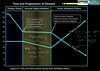
Describe the progression of acute ventilatory failure
- disease onset
- alveolar hyperventilation
- acute ventilatory failure
What therapy can help a patient with atelectasis?
CPAP mask (positive pressure to open lungs)
