15 - AKI, CKD and Glomerulonephritis Flashcards
What is the definition of an AKI?
Decreased renal function over a short period of time defined by a rise in serum creatinine from patients normal baseline, OR drop in urine output
- Rise in serum creatinine >26 umol/L within 48h
- Rise in creatnine >1.5 baseline within 7 days
- Urine output <0.5ml/kg/h for >6 consecutive hours

How is the severity of an AKI graded?
Stage 1: 1.5-1.9x rise in creatinine to baseline OR >26.5umol/L increase
Stage 2: 2-2.9 x rise in creatinine to baseline
Stage 3: >3x rise in creatinine to baseline OR renal replacement therapy initiated OR >353.6 umol/L increase

What are some limitations of the use of serum creatinine to define an AKI?
- Muscle mass dependent
- Dilution
What are some risk factors for AKI?
- Diabetes
- CKD
- IHD/CCF
- Aged>75
- Sepsis
- Medications e.g ACEi, NSAIDs, ARBs, Abx
MEASURE SERUM CREATININE DAILY IN HOSPITAL FOR THESE PATIENTS!!

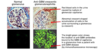
What are some causes of an AKI? (commonest first)
(N.B look at picture)
Pre-renal: sepsis, cardiogenic shock, hypovolemia, heart failure, myeloma, hepatorenal syndrome, rhabdomyolysis, contrast induced, urate nephropathy
Renal: drugs, contrast, abx,
Post-renal: obstruction e.g stones, BPH

What are some complications of an AKI?
- CKD
- Hyperkalaemia
- Fluid overload
- Metabolic acidosis
What are some investigations you should do if there is an AKI to establish the cause?
- URINE DIPSTICK before catheter to look for proteinuria and haematuria
- US KUB within 48 hours if risk of obstruction to rule out
- LFTs for hepatorenal syndrome
- Check platelets, if low need to look at blood film for haemolysis (HUS/TTP)
- If blood on urine dipstick suspect intrinsic renal disease so check immunoglobulins, paraprotein, complement (C3/C4), autoantibodies
- FBC, U+Es, Bone profile, CRP, CK, Serum bicarbonate

What are some autoantibodies you should look for if you suspect nephritic disease is causing an AKI?
- Anti-GBM
- ANA
- p-ANCA
- c-ANCA
Also do myeloma screen, look at C3/C4 if suspect lupus nephritis and immunoglobulins
If you suspect an AKI is due to post-steptococcal GN, what investigation should you do?
Anti streptolysin O titres
What is involved in a haemolysis screen?
- Blood film
- LDH
- Bilirubin
- Reticulocytes
- Haptoglobin
CALL RENAL SpR URGENTLY
What are some things you should monitor in a patient with AKI?
- Daily creatinine until falls
- Fluid balance with catheter and hourly urine out put
- K+ until creatinine falls
- General observations every 4 hours
- Lactate if signs of sepssi
How is an AKI managed in general?
- Treat underlying cause
- Consider referral to renal/critical care for dialysis
- Send off investigations to find out cause
- Stop any nephrotoxic drugs and change dose of any drugs e.g antibiotics
- Check volume status and correct if too high or low
- Monitor urine output and daily bloods
- Avoid hyperglycaemia

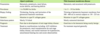
How are the different types of AKI managed in general?
Treat underlying cause. For all types manage fluid balance, hyperkalemia and consider those who may need renal replacement
Pre-renal: correct volume depletion, correct any sepsis, cardiac support
Renal: refer for biopsy and treatment of intrinsic renal disease
Post-renal: catheter, nephrostomy or urological intervention
How should you treat patients with an AKI that have fluid overload?
- IMMEDIATE REFERRAL TO RENAL/CRITICAL CARE FOR RENAL REPLACEMENT THERAPY
- Monitor weight daily
- Oxygen supplementation if required
- Fluid restriction
- Loop diuretics if symptomatic overload
What are dangers of giving sodium bicarbonate to correct a metabolic acidosis caused by an AKI?
- Generates CO2 so need adequate ventilation
- May precipitate fluid overload due to the sodium in it
When should you refer a patient with an AKI to the renal team?
e.g if they developed an AKI on cardiology ward when do you escalate?
- AKI not responding to treatment
- AKI with complications e.g fluid overload, acidosis, rising K
- AKI stage 3
- AKI with difficult fluid balance e.g heart failure
- AKI due to intrinsic renal disease
- AKI with hypertension

What are some indications for renal replacement therapy in an AKI?
- Fluid overload refractory to diuretics
- Metabolic acidosis refractory to treatment
- Hyperkalaemia refractory to treatment
- Uraemic pericarditis
- Uraemic encephalopathy
- Intoxications e.g methanol, salicyclates, lithium

What are some possible complications of using RRT to treat an AKI?
- Risks of catheter insertion e.g pneumthorax, infection
- Procedural hypotension
- Bleeding due to need for anticoagulation
- Altered nutrition
- Drug clearance
What are some causes of a raised serum urea?
- AKI
- Upper GI bleed (not lower)
- Dehydration

How can you distinguish malena due to a upper and lower GI bleed?
Upper GI cause will have raised serum urea
What is the definition of CKD?
Presence of kidney damage (abnormal structure or function) for >3 months.
Measured using eGFR and albuminuria
Need to have markers of kidney damage or decreased function on 2 occasions in 3 months

How is chronic kidney disease classified?
Using eGFR and ACR on KDIGO score
Stage 1: eGFR>90 with proteinuria
Stage 2: eGFR <90 but more than 60 with proteinuria
Stage 3A: eGFR<60
Stage 3B: eGFR<45
Stage 4: <30
Stage 5: <15 kidney failure

What does the KDIGO score calculate?
Risk of adverse outcomes with CKD based on ACR/Albuminuria and eGFR

The G part of the KDIGO score is for eGFR. What does the A score stand for?
ACR or albuminuria
A1
A2
A3