12. Histology of Accessory Organs (Pancreas, Liver, and Glands) Dennis Flashcards
3 main digestive glands are?
- Major salivary glands
- Exocrine pancreas
- Liver
What type of secretory cell is shown here; secretes what?

- Mucous acini
- Glycoprotein-rich product

What type of secretory cell is shown here?

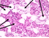
- Serous acini

What type of secretory cell is shown here?

Mucoserous
What type of gland is this, distinguishing features, and label the arrows!
- Parotid Gland
- Pyramidial cells w/ basally located nuclei

What type of gland is this?

Parotid gland
*Pyramidal shaped cells!*
What type of gland is this?

Sublingual gland
What type of gland is this, distinguishing features, label arrows!

- Sublingual gland
- Mixed gland, but predominantly mucous

What type of gland is this?

Submandibular gland
What type of gland is this; distinguishing features (all the arrows)?

- Submandibular
- Serous cells are predominant
- Mucous cells are capped by serous demilumes (‘bonnet’)

What is shown here?

- Hepatic lobule

What is shown here, the ‘stick’ like structures?

Bile canaliculi
What is shown here and what do each of the letters represent?

- Hepatic lobule
C = central venule
S = sinusoids
H = hepatocytes
What is depicted by this picture?

Portal Triad (hepatic artery, bile duct, and portal vein)
What is a hepatic lobule, what shape is it?
- Structural unit of kidney
- Hexagonal in shape

What is a portal lobule based on; what is the central axis?
- Bile drainage pathway from adjacent lobules toward the same bile duct
- Portal triad is the central axis, so it consists of 3 hexagons connected to one another

What are the liver acinus based on, and what determines the boundaries; establishes what?
- [O2] gradient along sinusoids of adjacent lobules
- Boundaries determined by a terminal branch of the hepatic a
- Establishes zones I, II, III
Which zone is closest and furthest from the portal triad?
Zone I = closest
Zone III = furthest away
What is the Perisinusoidal space of Disse; contains; what takes place here?
- Separates the basolateral domain of the hepatocyte from blood circulating in hepatic sinusoid
- Contains Type I, III, and IV collagen fibers
- Protein absorption and secretion
How does blood, bile and lymph flow within the liver?
Blood/lymph flow in the opposite direction from bile
Excess fluid in the space of Disse is collected where and then what happens?
Collected in the space of mall and then drained by lymphatic vessels
Describe what happens in Zone 1 of the liver?
- Closest to blood supply (so will be affected by immune response 1st)
- First to receive oxygen, nutrients, and toxins
- First to regenerate
- Last to die

Describe what happens in Zone 3 of the liver?
- First to show necrosis
- Last to respons to toxins and bile stasis
What type of cell is shown here and its function?

- Kupffer cells
- Specialized macrophages along the sinusoids, detect and phagocytose effete erythrocytes


