1.1 Anatomy of gut Flashcards
List 3 functions of the abdomen
1) Protects and supports viscera within
2) Assists in breathing
3) creates changes in intra-abdominal pressure
Where is McBurney’s point and what is its significance in appendicitis?
Location: 1/3 along a line from the right ASIS to the umbilicus
Significance: In appendicitis pain initiates near the umbilicus in the epigastric region. This is because Infection begins irritating the visceral peritoneum -> poorly localised pain that is felt where the organ was embryologically derived.
Over- time, as the infection spreads to the surrounding abdominal wall, it begins to irritate the parietal peritoneum -> more localised pain that is felt closer towards the appendix’s adult position -> migrates towards McBurneys point
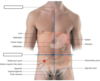
What are the 4 quadrants of the Abdomen? Which significant abdominal organs lie in each quadrant?
R Upper: Liver, gallbladder
R Lower: Appendix
L Upper: Stomach, spleen
L Lower: descending colon, sigmoid colon

Label the 2 planes shown below

What are the boundaries of the abdomen?
Superior: diaphragm
Inferior: Pelvic inlet
Lateral: serous membranes
What are the 9 regions of the abdomen?
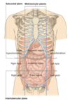
Lable the planes shown below


List the 3 types of mesentery in the abdomen
- Mesentery proper
- Trasverse mesocolon mesentery
- Sigmoid mesocolon mesentery
What is mesentery and what is its significance?
Double layer of the peritoneum: tissue that houses blood vessel, nerves, and lymphatics to gut as vessels cannot directly penetrate the peritoneum
What is the peritoneal cavity and what is its clinical significance?
A potential space between parietal and visceral peritoneum ➞ contains serous fluid that lubricates the GI tract allowing for movement during digestion
Inflammation and infection can lead to increased production of fluid which can put pressure on the organs ➞ ascities
Compare the parietal vs visceral peritoneum
Parietal ➞ lines abdominal walls
Visceral ➞ covers suspended organs
(These layers slide freely against one another)
What is the nervous innervation to the Parietal peritoneum?
What impacts does this have on “feeling pain” in the abdomen?
Innervated by somatic afferent fibres, sensitive to well localised pain ➞ can feel pain here and perceive it being there
What is the nervous innervation to the Visceral peritoneum?
What imapact does this have on “feeling pain” in the abdomen?
Innervated by visceral afferent fibres, activation of these can lead to referred pain ➞ poorly localised sensations of discomfort
(reffered pain localises to region of embryological origin)
What is meant by an intraperitoneal organ?
Give 4 examples of organs which are intraperitoneal
Intraperitoneal: completely invested by visceral peritoneum and suspended by mesenteries
- stomach, spleen, jejunum, ileum, caecum, appendix, transverse colon, sigmoid colon
What is meant by a retroperitoneal organ?
Give 4 examples of organs which are retroperitoneal
Retroperitoneal: NOT suspended in the abdominal cavity by mesentery, lay between visceral peritoneum (anteriorly) and abdominal wall (posteriorly)
- kidney, adrenal glands, ureters, IVC, abdominal aorta
Give 4 organs which are considered ‘Inbetween’ structures (retro/intraperitoneal)
Duodenum, ascending colon, descending colon, pancreas
State which areas are covered primarily with serosa and others with adventitia and why?
Intraperitoneal regions are covered with serosa (visceral peritoneum) which secretes fluid to lubricate the outside of the GI tract
Retroperitoneal regions requiring anchoring are covered with adventitia
What is Peritonitis and what does it commonly result from?
Inflammation and infection of the peritoneum
Commonly the result of:
- burst appendix
- penetrating wound
- perforated duodenal ulcer
What landmarks the beginning of the abdominal aorta?
Aortic hiatus of the diaphragm, T12
What are the 2 terminal branches of the Abdominal aorta
R and L common iliac arteries
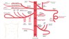
What are the 3 major trunks off the abdominal aorta supplying the gut?
Which vertebral level AND embryological division does each correspond to?
- Celiac Trunk: T12, foregut,
- Superior mesenteric artery: L1, midgut
- Inferior mesenteric artery: L3 hindgut
Label the image


State where reffered pain is felt for organs embryologically derived from the:
- Foregut
- Midgut
- Hindgut
Foregut: Epigastric region
Midgut: umbilical region
Hindgut: Pubic region
What are the 3 major branches of the Celiac Trunk?
Which branches go L vs R, and what do they collectively supply (7)?
- L gastric artery
- Splenic artery (goes L)
- Common hepatic artery (goes R)
Collectively: supplies the stomach, spleen, liver, gall bladder, abdominal esophagus, pancreas and duodenum
What is the main cause of a celiac aneurysm
Weakness to the wall of the abdominal aorta causes it to balloons out ➞ high pressure can cause it to burst
Why is the common hepatic artery so essential?
What are its 2 terminal branches?
It is the sole arterial supply to the liver
Gives rise to the proper hepatic artery and gastroduodenal arteries
Which artery is likely to be affected in a posterior duodenal ulcer and why?
Gastroduodenal artery (duodenal ulcers tend to erode posteriorly)
What are the borders (3) and contents (3) of the triangle of calot?
Borders:
- Superior - inferior boarder of the liver
- Medial - common hepatic duct
- Lateral - cystic duct
Contents:
- Cystic artery
- R hepatic artery
- Accessory ducts
What is the importance of the triangle of Callot?
Important in a cholecystectomy: removal of gall bladder
The cystic duct and artery are ligated. Care must be taken to ensure the R hepatic artery and accessory ducts are not damaged as these are located within the triangle
Name the 5 major branches off the SMA
- Jejunal branches
- Ileal branches
- Ileocolic branchs:
- R and middle colic artery
- Inferior pancreaticoduodenal artery

Which organs do the SMA collectivley supply?
Distal duodenum, jejunum, ileum, colon (until the splenic flexure)
The SMA is part of the _____ artery of _____.
What is this?
Marginal artery of drummond; a continuous arterial circle along the inner border of the colon
Name the 3 major arterial branches that come off the IMA
- L colic artery
- Sigmoidal arteries
- Superior rectal artery; (rectum and upper half of anal canal)
Which organs does the IMA collectivly supply?
Distil transverse colon, descending colon, sigmoid colon, rectum, upper part of rectal canal
List the 3 major veins that drain the gut and state where these all eventually drain
- SMV
- IMV
- Splenic vein
All drain into the liver via the portal vein which is the final pathway for venous blood from the spleen, pancreas, gallbladder, and abdominal part of the GIT. Blood is then returned to the heart via the IVC
The portal vein is formed by the union of what 2 veins?
Splenic v. + SMV
Gut lymphatics follow _____ supply. All lymph from contents of the digestive tract drain as _____ into the _____ which then drains into the thoracic duct
arterial, chyle, cisterna chyli
Give an example of 4 lymph nodes which drain the gut
- Celiac nodes
- Paracolic nodes
- Superior mesenteric nodes
- Gastric nodes
Where does sympathetic innervation to the gut arise?
List 3 actions of sympathetic innervation to the GI tract
From sympathetic trunk
Actions:
- decrease motility
- decrease gland secretions
- contraction of sphincter muscles
Where does parasympathetic innervation to the gut arise?
List 3 actions of parasympathetic innervation to the GI tract?
From Vagus nerve (CN X) and sacral plexus
Actions:
- increase motility
- increase gland secretions
- relax sphincter muscles
What/where are the ‘paracolic gutters’?
What is their significance?
Paracolic gutters are located lateral to the ascending and descending colons. They are depressions formed between the lateral margins of the intestine and posterolateral abdominal walls and are a relatively blood-free zone
Important for spread of infection! fluid/puss/infection can pass from one region of peritoneal cavity to another via the gutters
