11/29 Presenting Signs/Sx of Pulm Disease - Corbett Flashcards
hypoxemia vs hypoxia vs cyanosis
hypoxemia: low arterial O2 tension (PaO2 low)
- free O2 dissolved in plasma; NOT a measure of O2 content
- measurement
- assess via pulse oximeter : detects amt of O2 bound to Hb in blood (infrared=oxy, red=deoxy)
- reading can be disrupted by changes in bloodflow (vasoconstriction, lack of pulsatile bloodflow)
- assess via arterial blood gas : invasive procedure at radial/femoral/brachial a
- assess via pulse oximeter : detects amt of O2 bound to Hb in blood (infrared=oxy, red=deoxy)
hypoxia: low O2 delivery
cyanosis: increase in deoxygenated Hb level above 5g/dL
(normal Hb: 13.5-15 g/dL)
A-a gradient
shortcut eqn
normal gradient
A-a gradient = PAO2 - PaO2
[150-PaCO2/.8] - PaO2
normal range:
A-a gradient in diff types of hypoxemia
normal gradient x2
elevated gradient x3
normal A-a gradient: decrease in O2 intake but no issues with diffusion
- either:
- low O2 inspired
- PCO2 elevation
elevated A-a gradient: issue with diffusion OR shunt
*

causes of hypoxia
inadequate level of tissue oxygenation for cellular metabolism
- low arterial O2 sat
- decr oxygen content
- inadequate O2 delivery (DO2 = CaO x CO)
- impaired ability of cells to utilize O2 (ex. cyanide poisoning)
cyanosis
what is it?
when do you see it?
how reliable?
bluish/purplish tinge to skin and mucous membranes (lips, buccal mucosa, tongue, etc)
- can be central or peripheral
key: drop of at least 5 g/dL deoxyHb in capillaries
therefore…possible to be hypoxemic and NOT cyanotic!
- that said, central cyanosis increases probability of hypoxemia
patients with normal level of Hb manifest cyanosis at higher SaO2 values than patients with anemia!
- easier to get the required drop of 5 g/dL

peripheral cyanosis
decreased local circulation AND incr oxygen extraction in peripheral tissues
conditions assoc with:
- peripheral vasoconstriction
- stasis of blood in extremities (CHF, circ shock, cold temp exposure, abnormalities of periph circ)
presentation of:
dyspnea
“shortness of breath”
- tachypnea (RR > 20) is not necessarily dyspnea (ex. could be acidosis!)
occurs when ventilatory demand exceeds capacity for ventilation → imbalance between motor drive to breath and afferent feedback from mechanoreceptors of resp system
“length-tension in appropriateness”
“neuroventilatory dissociation”
pathophys correlates
- structural or mechanical interference with vent
- obstruction to flow (emphysema, asthma, chronic bronchitis, upper airway obst)
- restriction to lung or chest wall expansion
- extrensic diseases (not involving lung parenchyma)
- kyphoscoliosis, obesity, ascites, pregnancy, pleural disease
- intrinsic diseases (involve lung parenchyma)
- ARDS, CHF
- extrensic diseases (not involving lung parenchyma)
- incr in dead space ventilation
- emphysema, PE
- incr in resp drive

respiratory info processing centers
- cortex: can override any peripheral signal
- central chemoreceptors : CSF pH (secondary to incr pCO2)
-
carotid bodies (CN IX) : severe hypoxemia (O2 < 60mmHg)
* pH/pCO2 dependent! → sensitized by incr in pCO2 - mechanoreceptors in chest
- muscle spindles in resp muscles : mechanical load
- vagal sensory fibers (CN X) : stretch (can be reflection of interstitial disease)
receptors send info to respiratory centers AND to sensory areas to be able to compare supply and demand of breathing
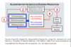
how is info processed/where is it sent?
- dorsal respiratory group : INSPIRATORY neurons
- receive info from chemoreceptors and stretch receptors
- send info to phrenic nerve
-
ventral respiratory group : INSPIRATORY/EXPIRATORY neurons
* hit upper airways, intercostals, etc - apneustic center/pontine center
presentation of:
cough
most common sx of lung disease → often dismissed as a result
normal defense mech of lungs
- clears larynx/trachea/lg bronchi of mucus, particles, organisms
- protects airways from foreign bodies
presistent cough (> 3wk) needs investigation
three phases of cough
- inspiratory phase
- closure of glottis and diaphragmatic relaxation
- rapid contraction of expiratory muscles causing rise in intra-abd and intrapleural pressures followed by opening of glottis
cough triggers
sensory receptors in larger airways (bronchioles and bronchi)
- nonmyelinated C type fibers
- respond to acid, infl signals, etc
cough reflex mediated by infl signals
timing and etiology of 3 types of cough
acute cough ( < 3wk)
chronic ( > 8wk)
- 90% of the time, one of the following three:
- upper airway cough syndrome (postnasal drip)
- asthma
- gastroesophageal reflux disease (GERD)

cough as a side effect of ______
and why
ACE inhibitors
ACE metabolizes bradykinin in lungs → ACE inhibitors lead to buildup of kinins and substance P → cough fibers sensitized
presentation of:
hemoptysis
coughing or spitting blood derived from lungs or bronchial tubes (secondary to pulmo or bronchial hemmorhage)
classified according to volume:
- blood-tinged sputum
- life-threatening amt (> 500cc in 24h, 100cc/h)
- tends to be bronchial in origin (90% bleeding originates from bronch circ and collaterals)
- recall: bronchial aa are part of systemic circ → much much higher pressure than pulmo circ
tracheobronchial origins:
- bronchitis (acute or chronic)
- bronchogenic carcinoma, endobronchial metastatic tumor, Kaposi’s sarcoma, bronchial carcinoid
- bronchiectasis (infl of airways, ex. cystic fibrosis)
most common cause of hemoptysis in US: 60-7-% of cases from infection
- bronchitis
- pneumonia
- tuberculosis (prob leading cause worldwide)
second most in US: primary lung cancers
keys:
- repeated small hemoptyss or blood-streaking of sputum
- fever, night sweats, weight loss
- “rust” colored sputum
- massive bronchial hemorrhage
repeated small hemoptyss or blood-streaking of sputum : cancer
fever, night sweats, weight loss : tuberculosis
“rust” colored sputum : pneumococcus
massive bronchial hemorrhage : bronchiectasis and mycetomas (ex. aspergillomas)
chart of rate/depth of breathing

hyperpnea
hypopnea
Kussmaul’s
Cheyne-Stokes
hyperpnea: incr TV, often incr RR, acidosis
hypopnea: shallow resp (often impending resp failure)
Kussmaul’s: incr rate and depth of resp
Cheyne-Stokes: constant rate, variable depth; apneic period
- assoc with neuro disorders, CHF, high altitude, normal aging
tripod position
why it works
hands on knees → helps accessory muscles of resp
works because allows pectoralis major m to fix shoulder and therefore helps elevate rib cage

paradoxical breathing
what? why?
what: chest expands, abdomen moves inward
when: respiratory fatigue or diaphragmatic weakness
- the neg pressure generated in chest on inhale pulls a weak diaphragm up with it → see abd move inward
sign of pending resp failure!
in picture: B normal inhale, C paradoxical inhale

purse lip expiration
works to maintain positive airway pressure during expiration
- incr aterial O2 and reduced CO2 retention
seen in obstructive lung disease (ex. emphysema)
barrel chest
normal ratio, BC ratio
normal ratio of AP diameter : transverse diameter = 1 : 2
in barrel chest = 1 : 1

clubbing
most commonly acquired (80%)
often assoc with pulmo or CV disease
- lung cancer
- COPD
- interstitial pulmo fibrosis
- lung abscess
- pulmo TB

tactile fremitus
asymmetric tactile fremitus is the concerning pathologic finding
can be caused by: OBSTRUCTION ANYWHERE
- bronchial obstruction
- pleural effusion (interesting finding: tactile fremitus will be decr over effusion, incr over the consolidated lung area)
- pneumothorax
- thick chest wall (obesity)
breath sounds
normal vs adventitious
normal
- bronchial
- vesicular
adventitious
- stridor: high-pitched musical sound, assoc with upper airway obstruction
-
wheezes: long duration musical sound (usually exp, but can be biphasic), assoc wth obstructive disease
- always assoc with airway limitation
-
rhonchi: low-pitched wheeze (sounds like snoring)
- assoc with secretions, can be cleared with coughing
- crackles (aka rales): discont nonmusical sounds caused by opening of collapsed distal airways/alveoli; fine or coarse

