1. Anatomy Flashcards

1
Q

how many bones are in the foot?

A

26

(*not including sesamoids)

2
Q

how many joints are in the foot?

A

35

3
Q

name the accessory ossicle:

between 1st cuneiform and 1st & 2nd metatarsal bases

A

os intermetatarsium

4
Q

name the accessory ossicle:

proximal 5th metatarsal base

A

os vesalianum

5
Q

name the accessory ossicle:

accessory navicular

A

os tibiale externum

6
Q

name the accessory ossicle:

dorsal aspect of navicular

A

os supranaviculare

7
Q

name the accessory ossicle:

sesamoid bone in PB tendon

A

os peroneum

8
Q

name the accessory ossicle:

dorsal, anterior process of calcaneus

A

os calcaneus secondarius

9
Q

name the accessory ossicle:

posterior aspect of sustentaculum tali

A

os sustentaculi

10
Q

name the accessory ossicle:

posterior aspect of talus (Steida process)

A

os trigonum

11
Q

name the accessory ossicle:

distal to medial malleolus

A

os subtibiale

12
Q

name the accessory ossicle:

distal to lateral malleolus

A

os subfibulare

13
Q

name avascular necrosis of:

tibial sesamoid

A

Renandier

14
Q

name avascular necrosis of:

fibular sesamoid

A

Trevor

15
Q

name avascular necrosis of:

phalanges

A

Theiman

16
Q

name avascular necrosis of:

metatarsal heads

A

Freiberg

17
Q

name avascular necrosis of:

5th metatarsal base

A

Iselin

18
Q

name avascular necrosis of:

cuneiforms

A

Buschke

19
Q

name avascular necrosis of:

navicular

A

Kohler

20
Q

name avascular necrosis of:

cuboid

A

Lance

21
Q

name avascular necrosis of:

talus

A

Diaz

22
Q

name avascular necrosis of:

calcaneus

A

Severe (?)

23
Q

name avascular necrosis of:

proximal, medial tibial epiphysis

A

Blount

24
Q

name avascular necrosis of:

tibial tuberosity

A

Osgood-Schlatter

25
Q

name avascular necrosis of:

femoral epiphysis

A

Legg-Calve-Perthes

26
Q

what attaches periosteum to bone?

A

Sharpey fibers

27
Q

what are the 3 different types of coalitions?

A

syndesmosis, synchondrosis, synostosis

  • synDESMOSIS - fibrous
  • synCHONDROSIS - cartilaginous
  • synOSTOSIS - osseous
28
Q

what is the difference between a coalition and a bar?

A
  • coalition - intra-articular fusion of 2 bones
  • bar - EXTRA-articular fusion
29
Q

what is the most common coalition in the foot?

A

distal and middle phalanx of 5th digit

30
Q

what is the most common coalition in the rearfoot?

A

talocalcaneal

31
Q

what is a steida process?

A

enlarged Os Trigonum

32
Q

what is the only bone in the foot

without any muscle origin or tendon insertion?

A

talus

33
Q

what are the plantar muscle layers of the foot

from superficial to deep?

A
  1. (superficial) abductor hallucis, flexor digitorum brevis, abductor digiti minimi
  2. quadratus plantae, 4 lumbricals
  3. flexor hallucis brevis, adductor hallucis, flexor digiti minimi
  4. 3 plantar interossei, 4 dorsal interossei (deep)
34
Q

what layer of the foot does the Flexor Digitorum Longus run?

A

2nd layer;

it is the origin of the lumbricals AND the insertion of quadratus plantar

35
Q

what deformity will result from cutting Quadratus Plantae (QP)?

A

digits 4 and 5 will become adductovarus

(because action of QP is to assist FDL with flexion of DIP joints)

36
Q

how is Extensor Digitorum Longus (EDL) attached to the proximal phalanxes?

A

sling wraps around the capsule –>

which attaches to plantar plate, deep transverse metatarsal ligament (DTML), and flexor sheath –>

thus attaching to plantar proximal phalanx

*there is NO DIRECT INSERTION to proximal phalanx*

37
Q

what is the origin and insertion of the

capsularis tendon?

A

o: extensor hallucis longus (EHL) muscle or tendon

i: first metatarsophalangeal joint capsule (1st MTPJ capsule)

38
Q

Master Knot of Henry:

define

A

fibrous connection between the FHL and FDL tendons

39
Q

which structures attach to the FIBULAR SESAMOID?

A

6 total attachments

  1. plantar metatarsal-phalangeal ligament
  2. lateral metatarsal-sesamoidal ligament
  3. intersesamoidal ligament
  4. phalangeal-sesamoidal ligament
  5. FHB (flexor hallucis brevis) tendon
  6. ADH (adductor hallucis tendon)
40
Q

are the sesamoids capsular or extra-capsular?

A

CAPSULAR

sesamoids are found within joint capsule

41
Q

Lisfranc ligament:

define

A

attaches lateral aspect of medial cuneiform

to medial base of 2nd metatarsal

42
Q

what structures in the Lisfranc joint are NOT connected by ligaments?

A

1st and 2nd metatarsals

43
Q

spring ligament:

true name

A

plantar calcaneonavicular ligament

(plantar CN ligament)

44
Q

which ligaments compose the

bifurcate ligament

A

dorsal calcaneonavicular, and

calcaneocuboid ligaments

(bifurcate lig = dorsal CN and CC ligaments)

45
Q

bifurcate ligament:

other names, and course

A

aka internal calcaneocuboid, interosseous ligament, or bifurcated ligament

  • strong band, attached behind to the deep hollow on the upper surface of the calcaneus and
  • dividing in front in a Y-shaped manner into a calcaneocuboid and a calcaneonavicular part
46
Q

which is stronger?

deltoid ligament or lateral ankle ligaments

A

deltoid ligament

(deltoid is stronger than lateral ankle ligaments, and therefore provides more medial ankle stability –> this is why lateral ankle sprains are more common than medial)

47
Q

components of deltoid ligament

A
  • Superficial: tibionavicular, tibiocalcaneal, posterior tibiotalar
  • Deep: anterior tibiotalar

(4 total: 3 superficial, 1 deep)

48
Q

what tendons pass OVER the deltoid ligament?

A

tibialis posterior & flexor digitorum longus (FDL)

(2 tendons: PT, FDL)

49
Q

lateral ankle ligaments

list

A
  • anterior talofibular
  • calcaneofibular
  • posterior talofibular
50
Q

what angle do the ATFL and CFL create?

A

105 degrees

51
Q

what is the strongest lateral ankle ligament?

A

posterior talofibular (PTFL)

(lateral ankle ligaments rupture in order of ATFL, CFL, PTFL - most common to least commonly ruptured)

52
Q

which ankle ligaments are EXTRA-CAPSULAR versus CAPSULAR?

A

calcaneofibular ligament (CFL) is EXTRA-CAPSULAR;

all others are capsular

53
Q

which tendons pass over the:

lateral ankle ligaments?

A

PB and PL

(peroneus brevis and peroneus longus pass over lateral ankle ligaments)

54
Q

which ligaments support the:

ankle syndesmosis

A
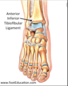
  • anterior-inferior tibiofibular ligament
  • posterior-inferior tibiofibular ligament
  • interosseous tibiofibular ligament

(3: AITFL, PITFL, Interosseous ligaments)

55
Q

Basset ligament

define

A

anterior-inferior tibiofibular ligament

(AITFL is Bassett ligament)

56
Q

another name for flexor retinaculum

A

laciniate ligament

(origin: Mid 18th century from Latin lacinia meaning β€˜fringe, hem, flap of a garment’)

57
Q

another name for superior extensor retinaculum

A

transverse crural ligament

58
Q

another name for inferior extensor retinaculum

A

cruciate crural ligament

59
Q

plantaris:

insertion

A

i: medial aspect of tendo-achilles into calcaneus
* (o: Lateral supracondylar ridge of femur above lateral head of gastrocnemius,*
* a: Plantar flexes foot and flexes knee)*

60
Q

peroneus quartus:

incidence

A

7%

(only 7% of patients have a QP)

61
Q

Hoke tonsil (or Hoke’s):

define

A

a fibrous, fatty plug within the sinus tarsi

62
Q

pes anserinus

define

A

insertion of Sartorius, Gracilis, and Semitendinosus (anteromedial aspect of proximal tibia) where bursa may cause knee pain

(CC: pes anserinus bursitis)

63
Q

Baker’s cyst

define

A

swelling of the bursa between the tendons of the medial head of the gastrocnemius and the semimembranosus muscles

64
Q

fabella

define

A

sesamoid bone occasionally found in the tendon of lateral head of gastrocnemius

65
Q

which nerves form the sural nerve

A
  • medial sural cutaneous nerve - branch of the tibial nerve
  • sural communicating branch - branch of the lateral sural cutaneous nerve, which originates from the common peroneal nerve
66
Q

does a neuroma lie dorsal or plantar to the deep transverse intermetatarsal ligament?

A

plantar

67
Q

where do these muscles run in relation to the deep transverse intermetatarsal ligament?

(interossei, lumbricals)

A
  • Interossei – dorsal
  • Lumbricals – plantar
68
Q

in which layers of the foot do the plantar nerves run?

A
  • Medial plantar nerve – in the 1st layer (between FDB and abductor hallucis)
  • Lateral plantar nerve – between the 1st and 2nd layers
69
Q

what is the innervation to the plantar muscles of the foot?

blood supply?

A

(Never LAFF at A FAD)

  • N – medial plantar Nerve
  • L – 1st Lumbrical
  • A – ABH
  • F – FHB
  • F – FDB (innervated by both medial and lateral plantar nerves)
  • A – medial plantar Artery
  • F – FDB
  • A – ABH
  • D – 1st Dorsal interossei
70
Q

what are the branches of the femoral nerve?

A
  • *-Nerve to femoral artery
  • Small muscular branch to pectineus
  • Anterior division (cutaneous)**
  • Anterior femoral cutaneous
  • Nerve to sartorious
  • Intermediate femoral cutaneous nerve
  • Medial femoral cutaneous nerve

-Posterior division (muscular)

  • Saphenous nerve
  • Infrapatellar branch
  • Medial crural cutaneous nerve
  • Nerve to rectus femorus
  • Nerve to vastus medialus
  • Nerve to vastus intermedialus
  • Nerve to vastus lateralus
71
Q

what are the branches of the femoral artery?

A
  • Superficial epigastric artery
  • Superficial circumflex iliac artery
  • Superficial external pudendal artery
  • Deep femoral (profunda femoris) artery
  • Medial femoral circumflex artery
  • Lateral femoral circumflex artery
  • Descending genicular artery
  • Femoral artery continues as the popliteal artery
72
Q

Trace the path of a drop of blood from left ventricle to the hallux

A
  • Ascending aorta β†’
  • aortic arch β†’
  • descending aorta β†’
  • thoracic aorta β†’
  • abdominal aorta β†’
  • common iliac artery β†’
  • external iliac artery β†’
  • femoral artery β†’
  • deep femoral artery β†’
  • popliteal artery β†’
  • anterior tibial artery β†’
  • dorsalis pedis β†’
  • 1st dorsal metatarsal artery β†’
  • 1st dorsal common digital artery β†’
  • 1st dorsal proper digital artery
73
Q

what are the sources of blood supply to the talus?

A

Essentially the 3 major blood supplies to the foot:

  • Superior surface of head and neck – artery of sinus tarsi and branch from anterior tibial artery or dorsalis pedis
  • Medial side of body – artery of tarsal canal and posterior tibial artery
  • Lateral turbercle – anastamosis of branch of peroneal artery with medial calcaneal branch
74
Q

what are the sources of blood supply to tendons?

A
  1. myotendinous junction
  2. paratenon
  3. at the insertion to bone