Wrist and Hand Flashcards
Active ROM
Wrist
- Flexion (85-90°)
- Extension (70-90°)
- Ulnar deviation (30-45°)
- Radial deviation (15°)
- Pronation (90°)
- Supination (90°)
Finger
- Flexion (MCP 85-90°, PIP 100-115°, DIP 80-90° )
- Extension (MCP 30-45°, PIP 0°, DIP 20°)
- Abduction (20-30°) and Adduction (0°)
Thumb
- Flexion (CMC 45-50°, MCP 50-55°, IP 85-90°)
- Extension (MCP 0°, IP 0-5°)
- Abduction (60-70°) and adduction (30°)
- Opposition of little finger and thumb
Passive ROM
Wrist
- Pronation and supination (tissue stretch)
- Flexion (tissue stretch)
- Extension (tissue stretch or bone-to-bone)
- Ulnar and radial deviation (bone-to-bone)
Finger
- Flexion and extension (tissue stretch)
- Abduction and adduction (tissue stretch)
Thumb
- Flexion and extension (tissue stretch)
- Abduction (tissue stretch) and adduction (tissue approximation)
- Opposition (tissue stretch)
Fanning and Folding of the Hand
Purpose
- To assess conjunction rotation of the hand
Procedure
- The examiner holds the scaphoid and trapezium with the index and middle finger of one hand and the pisiform and hamate the index and middle finger of the other hand
- The practitioner holds the patient’s capitate and trapezoid with their right and left thumbs on the dorsum of the hand
- The examiner folds and fans the hand, feeling the movement while monitoring and feeling for crepitus and joint motion
Indication of positive
- Symptom reproduction or abnormal moving of shifting of joints is an indication of a positive result
Clinical note
- The test is used as a general screening examination, not to identify a specific pathological condition

Finkelstein Test
Purpose
- To assess for irritation of the extensor pollicis brevis and abductor pollicis longus tendon as they pass deep to the extensor retinaculum in the first dorsal compartment in the wrist
Procedure
- The patient is asked to make a fist with the thumb inside the fingers
- The examiner stabilizers the patient’s forearm with one hand and then passively ulnar deviates the patient’s wrist while keeping the patient’s thumb enclosed in the fist
Indication of Positive
- Pain or reproduction of symptoms over the abductor pollicis longus and extensor pollicis brevis tendons at the wrist is a positive test result and indicates a paratendonitis or tendinosis of these two tendons
Clinical Note
- Because the test can cause some discomfort even in normal individuals, the examiner should compare the pain caused on the involved side with that on the unaffected side
- The test result is considered positive only if greater pain or symptom reproduction is noted on the affected side

Tinel’s Sign: Wrist
Purpose
- To assess for median nerve involvement at the carpal tunnel of the wrist
Procedure
- The examiner supinates the patient’s hand and wrist and stabilizes the forearm with the secondary hand
- The primary hand is used to tap along the median nerve pathway
- The examiner taps over the carpal tunnel at the wrist with the index and middle finger, working up the arm and following the path of the median nerve
Indication of Positive
- A positive test result is indicated by tingling or paraesthesia into the thumb, index finger (forefinger), and middle and lateral half of the ring finger (median nerve distribution)
- For a positive test result, the tingling or paraesthesia must be felt distal to the point of tapping
Clinical Note
- The test gives an indication of the rate of regeneration of sensory fibres of the median nerve
- The most distal point at which the normal sensation is felt represents the limit of nerve regeneration
- The manoeuvre can be applied at any of the common entrapment sites in the wrist and hand

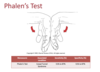
Phalen’s (Wrist flexion) Test
Purpose
- To assess for median nerve involvement through the carpal tunnel of the wrist
Procedure
- Patient is sitting or standing
- The patient flexes the wrist maximally and then presses the dorsum of the hands together
- The patient then holds this position for 1 minute
Indication of Positive
- A positive test result is indicated by tingling in the thumb, index, middle and lateral half of the ring finger
- A positive test is indicative of carpal tunnel syndrome caused by pressure (compression) in the carpal tunnel
Clinical Note
- Tests for neurological dysfunction are highly suggestive of a particular nerve lesion if they produce a positive result, but they do not rule out the problem if the results is negative. In fact, tests results may be negative 50% of the time or more often even when the condition actually exists. Electrodiagnositc tests are more conclusive.
Carpal Compression Test
Purpose
- Part I: This test is a pressure provocation test over the carpal tunnel to assess for median n. involvement
- Part II: This test is a pressure provocation test over the Tunnel of Guyon to assess for ulnar n. involvement
Procedure
- Part I: The practitioner applies direct thumb pressure over the carpal tunnel for up to 30 seconds
- Part II: The practitioner applies direct thumb pressure over the Tunnel of Guyon for up to 30 seconds
Indication of Positive
- Reproduction of the patients symptoms if combined with relief when the pressure is released
- Paraesthesia in the associated n. distribution

Reverse Phalen’s Test
Purpose
- To assess for median nerve involvement through the carpal tunnel of the wrist
Procedure
- The patient is standing or sitting
- The patient is asked to put the palms of the two hands together and bring the hands down towards the waist while keeping the palms in full contact (prayer test), thus causing extension of the wrist
- This position is held for approximately 1 minute
Indication of Positive
- A positive test result is indicated by tingling in the thumb, index, middle and lateral half of the ring finger
- A positive test is indicative of carpal tunnel syndrome caused by stretching the median nerve

Allen Test
Purpose
- To assess the patency of the ulnar and radial arteries and to determine which artery is providing the main blood supply to the hand
Procedure
- The patient is asked to open and close the hand quickly several times and then to squeeze the hand tightly so that the venous blood is forced out of the palm
- The thumb of the practitioner’s primary hand is placed over the radial a. and the thumb of the secondary contact are placed over the ulna a. and they press the arteries against the underlying bones with the aim of occluding the arteries
- With the arteries still occluded the patient is asked to open the hand, the palm should be pale and white
- One artery is tested by releasing the pressure over the artery while keeping the pressure on the other artery
- The procedure is then repeated with the other artery being released first
Indication of Positive
- Normally, after releasing one artery the hand should flush immediately
- If it flushes slowly or not at all the artery may be partially or totally occluded
- The same situation should occur when the other artery is released
Clinical Note
- Both hands should be tested for comparison
- The test limb can be held in an abducted position in the initial phase of the test to maximise the circulatory block

Digital Blood Flow (Perfusion) Test
Purpose
- To determine the patency of the digital arteries in the fingers
Procedure
- The examiner compresses the nail bed and then releases it and notes the time taken for colour to return to the nail
- Compare to normal side
Indication of Positive
- Normally, colour should return to the nail bed within 3 seconds
- If return takes longer, it may indicate possible arterial insufficiency to the fingers

Fovea Sign
Purpose
- A provocative manoeuvre for the assessment of disassociations of the distal radio-ulnar ligaments from the fovea region of the ulnar head or injury to the ulnocarpal ligaments
- Ulnotriquetral lig.
- Ulnocapitate lig.
- Ulnolunate lig.
- TFCC tear
Procedure
- Patient sitting with the elbow on the table and arm flexed to 90°-100° and forearm in neutral
- The practitioner pushes their thumb into the soft spot between the ulnar styloid, and the flexor carpi ulnaris tendon, volar surface of the ulna head and the pisiform
Positive
- Exquisite tenderness compared to the contralateral side
- Replication of the patient’s complaint/symptoms
- Facial grimacing
Indication of Positive
- Dysfunction of the distal radio-ulnar ligament or the ulnocarpal ligaments or the TFCC

TFCC Load Test
Purpose
- To assess the integrity of the TFFC in the wrist
Procedure
- The examiner stabilisers the patient’s wrist by contacting the patients hand with the primary contact in a ‘handshake’ position
- The examiner stabilises the patient’s elbow with the secondary contact
- The practitioner passively ulnar deviates and applies an axial load through the patient’s wrist
- The wrist may also be rolled from flexion to extension while maintaining ulnar deviation which is referred to as ‘grinding’ or ‘scouring’
Positive
- Pain, clicking, crepitus in the area of the TFCC
Indication of Positive
- Disruption of the TFCC

Finger Extension (‘Shuck’) Test
Purpose
- General screening tool for wrist/hand pathological
- Radiocarpal instability
- Midcarpal instability
- Scaphoid instability
- Inflammation
- Keinbock’s Disease
Procedure
- The examiner holds the patient’s pronated wrist in approximately 45° of wrist flexion
- The examiner contacts the patient’s metacarpals and asks the patient to actively extend the wrist and hand against resistance
Positive
- Pain
- Possible radiocarpal, midcarpal or scaphoid instability
- Possible inflammation
- Possible Kienbock’s disease

Watson (Scaphoid Shift) Test
Purpose
- To asses the integrity and stability of the scapholunate joint:
- Scapholunate lig. sprain/tear
- Scapholunate joint instability
- Scapholunate joint subluxation
Procedure
- The patient places their flexed elbow on the table with their palm facing the practitioner
- The examiner contacts over the patient’s metacarpals with the secondary contact, and creates full ulnar deviation and slight extension at the wrist
- The examiner then contacts the patient’s scaphoid with the primary hand using a pincher grip with the thumb over the volar scaphoid
- The practitioner then applies slight A-P pressure to the volar scaphoid while radially deviating and flexing the patient’s hand over the thumb contact
Positive
- Dorsal scaphoid shift on flexion and a ‘clunk’ on release of the thumb contact
- Indicating scapholunate instability

Lunotriquetral Ballottement Test
Purpose
- To assess the integrity of the and stability of the lunotriquetral joint:
- Lunotriquetral lig. sprain/tear
- Lunotriquetral joint instability
- Lunotriquetral joint subluxation
- Distal Radio Ulnar Joint (DRUJ) instability
Procedure
- The examiner grasps the triquetrum with the thumb and index finger of the primary hand and grasps the lunate between the thumb and index finger of the secondary hand
- The examiner then moves the lunate P-A and A-P while stabilising the triquetrum
Positive
- Laxity, crepitus and pain
- Indicating lunotriquetral instability
- Indicating DRUJ instability if performed at the DRUJ

Dorsal Capitate Displacement Apprehension Test
Purpose
- This test is used to assess the stability of the capitate bone
Procedure
- The examiner holds the patient’s distal forearm in neutral with the secondary hand
- The practitioner contacts the patient’s capitate using a pincher grip with the primary hand (thumb on the volar surface of the capitate and the fingers stabilising the dorsal capitate)
- The primary thumb contact pushes the capitate posteriorly
Positive
- Reproduction of symptoms, pain or apprehension
- The examiner notes if a click occurs
- Indicates capitate instability

Thumb: Valgus/Varus Stress Test
Purpose:
- To assess the integrity of the ulnar and radial collateral ligaments of the MCP joint of the thumb:
- 1st MCP ulnar collateral lig. injury: accessory and/or proper portions
- 1st MCP radial collateral lig. Injury: accessory and/or proper portions
Procedure:
- Part I:
- The practitioner stabilises the patient’s hand at the 1st metacarpal using a pincher grip with the secondary contact
- The practitioner holds the patient’s 1st MCP joint in 0° extension and applies valgus stress to the MCP joint with the primary contact
- The test is then repeated with the thumb flexed to 30°
- Part II:
- The practitioner stabilises the patient’s hand at the 1st metacarpal using a pincher grip with the secondary contact
- The practitioner holds the patient’s 1st MCP joint in 0° extension and applies varus stress to the MCP joint with the primary contact
- The test is then repeated with the thumb flexed to 30°
Positive:
- Laxity with or without pain
- Empty end feel
Indication of a Positive:
- Laxity greater than 30-35° = complete tear
- Laxity less than 30-35° = partial tear (usually with pain)
- Varus/valgus testing in extension tests both the joint capsule and the collateral ligaments
- Flexion of the MCP joint localises the test to the collateral ligament (both ulnar and radial)

Finger: Valgus/Varus Stress Test
Purpose:
- To assess the patency of the collateral ligaments of the MCP and IP joints of the fingers:
- Collateral lig.sprain
- Avulsion fracture
Procedure:
- Part I:
- The practitioner stabilises the patient’s finger proximal to the joint being assessed with the secondary contact
- The patient’s MCP joints should be in flexion when assessing the IP joints
- With the primary contact the practitioner applies a valgus force while the joint is held in a fully extended position
- Part II:
- The practitioner stabilises the patient’s finger proximal to the joint being assessed with the secondary contact
- The patient’s MCP joints should be in flexion when assessing the IP joints
- With the primary contact the practitioner applies a varus force while the joint is held in a fully extended position
Positive:
- Laxity with or without pain
- Empty end feel
Indications of Positive:
- Collateral lig. Injury
- Avulsion fracture
Clinical note:
- The tests should be performed in varying degrees of flexion to test different aspects of the collateral ligaments
- Ideally the finger should be tested at 0 and 30 degrees of flexion

Profundus Test
Purpose:
- To assess for disassociation of the flexor digitorum profundus from the distal phalanx
Procedure:
- The practitioner holds the patient’s affected finger (MCP and PIP joints) in extension
- The other fingers should be flexed at the MCP and PIP joints
- The patient is then asked to flex only the DIP joint of the affected finger
Positive:
- Inability to flex the affected DIP joint
Indication of Positive:
- Rupture of flexor digitorum profundus

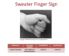
Sweater Finger Sign
Purpose:
- To assess for disassociation of the flexor digitorum profundus from the distal phalanx
Procedure:
- The patient is instructed to make a fist
- The practitioner observes for the patient’s inability to flex the affected finger into the fist shape
Positive:
- An inability of the patient to flex the distal interphalangeal joint
Indication of Positive:
- Rupture of the flexor digitorum profundus tendon from the distal phalanx
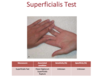
Superficialis Test
Purpose:
- To assess for disassociation of the flexor digitorum superficialis from the middle phalanx
Procedure:
- The practitioner holds the patient’s unaffected fingers in extension
- The patient is asked to flex the affected finger
Positive:
- Inability to flex the affected PIP joint
Indication of Positive
- Rupture of flexor digitorum superficialis
Test for Extensor Hood Rupture
Purpose:
- To test the integrity of the extensor hood
Procedure:
- Patient is sitting with the pronated forearm resting on the table
- The PIP joint of the finger to be examined is flexed to 90° over the end of the examining table or a thick textbook
- The practitioner firmly holds the patient’s finger in this position by contacting distal to the PIP joint on the patient’s middle phalanx
- From this position the patient is asked to gently extend the finger at the PIP joint againstresistance
Positive:
- The practitioner is unable to feel or feels minimal extension force acting to move the middle phalanx
- The distal phalanx does not remain flail and may extend as the patient attempts to extend the finger from the PIP joint
Indication of Positive:
- Complete rupture of the central slip of the extensor mechanism
Clinical Note:
- Long standing central slip rupture will present as a Boutonierre Deformity
- Also known as Elson Test

Bunnel-Littler Test
Purpose:
- To assess for potential causes of limited PIP joint flexion
- Can be used to examine individuals with swan neck deformity
Procedure:
- The MCP joint of the affected finger is held in extension
- The examiner attempts to passively move the PIP joint of the affected finger into flexion
- If the PIP joint is unable to flex the examiner then slightly flexes the MCP joint and again tries to flex the patient’s PIP joint
- The patient remains passive throughout the procedure
Positive:
- Inability to flex the PIP joint with an extended MCP joint indicates contracture of the PIP joint or tight intrinsic muscles of the hand
- Inability to flex the PIP joint with the MCP slightly flexed indicates PIP joint contracture

Test for Trigger Finger
Purpose:
- To assess for nodules or thickening of regions of the flexor tendons in the hand
Procedure:
- The examiner grasps the patient’s affected finger with a pincher grip with the index finger or thumb over the location of the proximal pulley
- The patient then flexes their finger over the examiner’s contact
Positive:
- The examiner may feel a small nodule pass beneath their fingers with the patient complaining of pain
Indication of Positive:
- Stenosing Tenosynovitis (Trigger finger)
- Early stage Dupuytren’s Contracture

