Week 9 Flashcards
components of urinary system
- kidneys (2)
- ureters (2)
- bladder
- urethra
retroperitoneal space
space that lies posterior to the peitoneum (lining of GI tract and other abdominal organs) in abdomen
positioning of kidneys in body
left kidney = T11 - L2
right kidney = T12 - L3
Upper half of kidney is protected by the rib cage
Right kidney positioned lower due to liver
located lateral to vertebral bodies in paravertebral gutters
why is it more common to biopsy the inferior pole of the kidney rather than the superior pole?
Biopsy of superior pole could cause pneumothorax
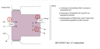
supporting structures around the kidneys

hilus of kidney contains (anterior to posterior order):
- renal vein
- renal artery
- ureter
3 major sections on kidney (internal)

interdigitations of cortex and medulla of kidney include:
- renal columns
- medullary rays

renal sinus in kidney is made up of:
minor calyx
major calyx
renal pelvis
order is from medulla to ureter

how to tell right kidney from left kidney
from anterior to posterior the hilus of the kidney goes renal vein, renal artery, then ureter; hilus points toward medial aspect; can figure out direction of kidney by location of ureter
“lobe” of the kidney
renal pyramid and its associated cortex

“lobule” of kidney
one central collecting duct and associated nephrons that drain filtrate into the central duct

smallest functional unit of the kidney
nephron
how many nephrons per kidney
1-4 million
components of renal corpuscle
- glomerulus (tuft of capillaries)
- glomerular capsule (Bowman’s capsule)
- visceral layer – completely surrounds tuft of capillaries; podocytes
- parietal layer
afferent vs efferent arteriole and glomerulus
afferent brings blood that needs to be filted to the glomerulus; efferent arteriole bringing filtered blood away from glomerulus
the two poles of a renal corpuscle
afferent pole and urinary pole
podocytes of nephron
specialized epithelial cells
have primary and secondary processes
pedicals envelop the endothelium
blood in kidney is being filtered through what 3 components?
- fenestrated endothelium – capillary with lots of openings, allows molecules to leave the bloodstream
- basement membrane
- Combined basal lamina of both the endothelium and the podocytes
- Filtration slits between the pedicels
Proximal Convoluted Tubule (PCT) of nephron anatomy
- At urinary pole of renal corpuscle
- Modified simple cuboidal cells
- Cells have “brush border”
- Abundant microvilli and canaliculi
- Absorb macromolecules
anatomy: Loop of Henle in nephron
- Extends into the medulla
Thick descending limb
- Similar cells to PCT (simple cuboidal cells with brush border)
Thin limb
- Simple squamous epithelium
Thick ascending limb
- Simple cuboidal cells without brush border

anatomy: macula densa of nephron
- Region of densely concentrated nuclei at end of thick ascending loop
- Adjacent to afferent arteriole
- Cells are sensitive to filtrate flow and ion content
- Part of juxtaglomerular apparatus

anatomy: Distal Convolueted Tubule (DCT)
- Begins after macula densa
- Simple cuboidal cells without brush borders
anatomy: collecting duct
- Drains filtrate from many nephrons
- Center of the lobule
- Simple cuboidal cells
- Traverses medulla to renal papillae
histology of collecting system components of kidney: Minor calyx; major calyx; renal pelvis
- ALL transitional epithelium
- “Urothelium”
- Cells can break and reform junctions
- Cells store plasmalemma intracellularly that can be added to cell surface as needed
histology of ureter
- Transitional epithelium
- Contraction propagated by surrounding smooth muscle in minor calyces
- peristaltic wave continues through ureter
anti-reflux mechanism in urinary bladder
as bladder fills, the hole where ureter is entering closes up in response to the weight; prevents urine from moving back up the system
trigone of urinary bladder
triangle between ureteric orifice and urethral orifice
does not stretch/contract
detrusor muscle of urinary bladder
smooth muscle found in the wall of the bladder. The detrusor muscleremains relaxed to allow the bladder to store urine, and contracts during urination to release urine.
histology of urethra
Distally, the urothelium transitions to nonkeratinized stratified squamous epithelium
Female urethras are much shorter, UTIs are more common
Urethral sphincters
Internal urethral sphincter
- Base of bladder
- Smooth muscle
- Autonomic
External urethral sphincter
- Perineum of pelvis
- Skeletal muscle
- Somatomotor
how much of blood supply from aorta do kidneys receive?
Receive ~ 25% of the blood supply from the aorta
kidneys receive blood supply from what vessels?
left and right renal artery (paied branch of abdominal aorta) then to lobar (segmental) arteries – 70% of population
30% of population – polar arteries; remnants of ascent of kidney into abdomen from pelvic from development
blood supply to adrenal glands (suprarenal glands)
Superior suprarenal artery
- Branch of inferior phrenic a.
Middle suprarenal artery
- Branch of aorta
Inferior suprarenal artery
- Branch of renal a.
how are adrenal glands associated with kidneys
Anatomically, but not functionally, related to kidneys
pathway of arterial blood supply to the kidney
arteries: renal, lobar, interlobar, arcuate, interlobular
No collateral circulation/anastomoses in the kidney
Occlusion of a lobar artery will cause pyramid(s) to become necrotic

afferent arteriole to glomerulus comes from which artery?
interlobular artery
venous return from kidneys
- IF efferent arteriole is closer to capsule, it will enter peritubular capillaries in the cortex
- IF efferent arteriole is closer to medulla, it will enter the vasa recta in the medulla
- interlobular vein, arcuate evin, interlobar vein, lobar vein, renal vein
difference betwen left and right renal veins
IVC runs down right side of midline; to get to the IVC the left renal vein has to cross the midlinel
pathway of left renal vein
- left side has some veins that drain into the left renal vein so that they don’t also have to cross the midline: left suprarenal vein, left gonadal vein
- left renal vein runs underneath the SMA (superior mesenteric artery); Travels between superior mesenteric artery (SMA) and abdominal aorta
Nutcracker syndrome
Left renal vein travels between superior mesenteric artery (SMA) and abdominal aorta
If vein is compressed, patient will present with hematuria, left flank pain, varicosities, could present with pelvic congestion

blood supply to the bladder - pathway/branching
abdominal aorta
- Internal iliac artery
- Umbilical artery
- Superior vesicle artery
- Superior bladder (males and females)
- Superior vesicle artery
- Inferior vesicle artery (males)
- Inferior bladder, prostate
- Uterine artery (females)
- Uterus, inferior bladder, vagina
- Umbilical artery

sympathetic innervation to the abdominal organs
- Thoracic and lumbar splanchnic nerves will synapse in collateral ganglia (prevertebral, preaortic )
- Celiac ganglion, SMA and IMA ganglion
- Postganglionic sympathetic fibers travel to target organ (kidney, ureter)

sympathetic innervation to pelvic organs
Lumbar splanchnic nerves synapse in superior hypogastric plexus (collateral ganglia)
Sacral splanchnic nerves synapse in inferior hypogastric plexus
Plexuses communicate with each other via left and right hypogastric nerves
From autonomic plexus the postganglionic sympathetic fibers travel to target organ (bladder, urethra)

parasympathetic innervation to abdominal organs
- Vagus nerve provides parasympathetics to majority of abdominal organs
- Preganglionic parasympathetic fibers
- Terminal ganglia in organ
parasympathetic inneration to pelvic organs
- Cell bodies in lateral horn at S2-S4 levels
- Pelvic splanchnics
- Preganglionic parasympathetics run through inferior and superior hypogastric plexus to synapse on terminal ganglion of organ.
- Bladder, urethra, urethral sphincters
Autonomic plexus
A nerve plexus of sympathetic or parasympathetic axons, often containing autonomic neurons or ganglia. Such a plexus typically extends along major arteries and is named for its underlying artery
- Preganglionic sympathetic fibers (sacral splanchnics)
- Postganglionic sympathetic fibers
- Preganglionic parasympathetic fibers (pelvic splanchnics)
- Viscerosensory
sacral plexus
- Lumbosacral trunk (L4- L5) and sacral spinal nerves
- 3 S’s (somatomotor, somatosensory, sympathetic)
- NOT autonomic
- S2-S4 of sacral plexus
- Pudendal nerve
- External urethral sphinctor

definition of Chronic Kidney Disease (CKD)
When kidneys do not work as they should
condition characterized by a gradual loss of kidney function over time
definition of End State Renal Disease (ESRD)
no kidney function
need dialysis or a transplant to survive
fraction of people in US with kidney disease
1 out of 9 people
risk factors associated with CKD
age >50
males
smoking
hypertension
diabetes
abdominal obesity
pre-term birth
Seven Major Functions of the Kidney
- Regulation of water and electrolyte balance
- Regulation of acid base balance
- Excretion of metabolic waste and bioactive substances
- Maintenance of blood pressure
- Regulation of red blood cell production
- Regulation of Vitamin D production
- Gluconeogenesis
glomerular filtration
Fluid flows from area of high pressure to area of lower pressure • Inorganic ions filtered • Proteins left behind • 180 L per day
cells that make up the juxtaglomerular apparatus
- Granular cells (secrete renin); also called juxtaglomerular cells
- Macula densa cells (detect Na+ flow, can help regulate blood flow); distal part of thick ascending loop
- Mesangial cells (phagocytic/contractile)
glomerulus and blood flow
each glomerulus has the ability to change the amount of blood coming to the glomerulus – can modify how much filtration is going on
amount of fluid filtered by kidney that is reabsorped into system
99% of filtered
(rest is secreted/excreted)
two types of nephrons
cortical nephrons and juxtamedullary nephrons (descend deep into medulla, role in concentration)
percentage of glucose reabsorbed in healthy person in kidneys
100% should be reabsorbed (none excreted)
proximal tubule reabsorption of filtrate
reabsorbs about 2/3 of filtrate; mainly sodium and water
major site of reabsorption of sodium and water
major characteristic of thick ascending limb of nephron
impermeable to water; sodium, potassium, and chloride ions can be reabsorped
highlight channel of distal convoluted tubules
sodium-chloride transporter
lot of the energy in tubular system used here
here about hydrochlorothiazide in context here
collecting duct system clinical significances
where aldosterone and ADH work
this is where blood pressure is adjusted; via control of amount of sodium reabsorped vs excreted
Compare the differential blood supply of the renal cortex and medulla
all blood first goes to the cortex; the osmolarity of the cortical interstitium matches rest of body
Small percentage flows into the medulla (<10%); restricted flow borders on hypoxia and allows interstitial fluid composition to be varied
renal blood flow vs. renal plasma flow
take blood flow and calculate plasma flow using hematocrit; gives us the RPF which is used to calculate the filtration fraction
filtration fraction
filtration fraction = GFR / RPF
GFR: glomerular filtration rate; plasma going to bowman space
RPF: renal plasma flow
filtration fraction usually 20% (about 125 mL/min.)
significance of low pressure in peritubular capillaries?
this is the location of reabsorption; low pressure allows for reabsorption whereas high pressure would make it hard
site of largest vascular resistance in renal blood flow
the afferent and efferent arterioles are the site of largest vascular resistance due to their smooth muscles
role of afferent and efferent arterioles on blood pressure control in glomerular capillary blood pressure
glomerular capillary blood pressure can be cahgned by changes in arteriole diameter of afferent and efferent arterioles
glomerular filtration definition
movement of fluid (plasma) across filtration barrier
layers that fluid passes through/between in filtration barriert to get to Bowman space from capillary
capillary
between capillary endothelial cells
pass glomerular basement membrane
Bowman Space (now fluid gets referred to as “filtrate”
charge of filtration barrier
filtration barrier is negatively charge
effect of high blood sugar (i.e. diabetes) on glomerular filtration
glycoproteins can attach to the negative charge on filtration barrier leading to clumping and decreased glomerular filtration
features of filtration barrier that allow us to keep cells and proteins within glomerular capilarries (keep them from getting filtered)
Size Barrier
- <7,000 daltons can be filtered easily
- up to 70,000 daltons has some filtration
Electrical Charge
- filtration barrier has (-) charge; lots of plasma proteins also have negative charge which causes repulsion of charge and, therefore, decreased filtration of negatively charged proteins
glomerular filtration of substances that bind to plasma proteins
plasma proteins don’t really get filtered, so substances that bind to plasma proteins are also not filtered (some calcium, hydrophobic hormones)
equation for Glomerular Filtration Rate
GFR = Kf x NFP
Kf = hydraulic permeability x surface area
NFP = net filtration pressure in glomerulus (sum of starling forces)
equation for Net Filtration Pressure (NFP
NFP = PGC - PBC - πGC
PGC: glomeruluar capillary blood pressure
PBC: Bowman Space hydrostatic pressure
πGC: osmotic force from protein in plasma
values for starling force components of net filtration pressure
PGC = 60 (constant)
PBC = 15 (constant)
πGC = variable; increases along the length of the glomerular capillary (due to plasma protein conc increasing); usually average 29 mmHG
πBC = 0
how does filtration coefficient in glomerular capillaries compare to typical systemic capillaries?
100 times greater in glomerular capillaries – due to fenestrations allowing more to filter through capillary walls
what changes the glomerular filtration rate? (general variables)
GFR = Kf x NFP
changing either these variables change GFR
NFP includes PGc, PBC, and πGC
factors that can change filtration coefficient in glomerular filtration
- contraction of mesangial cells reduces surface area (not a muscle but has contractile properties); decrease in Kf
- decrease in functioning nephrons due to age and disease decrease Kf
factors that can change glomerular capillary pressure (PGC)
Increases
- increase in renal artery pressure
- increase in efferent arteriole resistance
- dilation of afferent arteriole (decrease resistance)
Decreases
- increase in afferent arteriole pressure
- dilation in efferent arteriole pressure (decrease resistance)
factors that change pressure of bowman’s space (PBC) in glomerular filtration
altered in pathophysiology when there is urinary tubule occlusion; examples are prostate hypertrophy, benign tumor, kidney stones
factors that change oncotic pressure of glomerular capillaires (πGC) in glomerular filtration
decrease in protein conc (such as in liver disease) increase NFP
decrease in renal plasma flow –> concentrated plasma protein = decreases NFP
what is filtered load?
the quantity of a substance that gets filtered to the glomerular capillaries per unit time; what get’s presented to the nephron to handle (some can be reabsorped)
equation for filtered load
GFR x [S]plasma where [S] is our substance of interest that is being measured
defintion of renal autoregulation
the process by which kidneys resond to changes in systemic pressure in order to maintain GFR and keep a constant flow by adjusting resistance
two mechanisms for renal autoregulation of RBF and GFR
1. Arteriole Myogenic Mechanism
- Increase in mean arterial pressure => increase in smooth muscle stretch in afferent arteriole => triggers channels to release calcium to smooth muscle => contraction
2. Tubuloglomerular Feedback
- macula densa cells in juxtaglomerular apparatus have transporters for salts
- in increased pressure more salts get filtered to nephron; macula densa cells working harder to reabsorp these (Na+, Cl-, K+) => Na+/K+ pump working harder leads to incrase in ADP and adenosine
- ADP and adenosine bind to receptors on smooth muscle surrounding afferent arteriole => increase in intracellular calcium => vasoconstriction
reabsorption in the proximal convoluted tubule
HUGE reabsorption of Na+, Cl- , K+, HCO3 - , and water back into peritubular caps; about 67% of what was filtered
filtration is iso-osmotic; reabsorbing a proportional amount of water with the solutes (300mOsm)
amount of filtrate that goes past PCT per dau (proximal convoluted tubule)
60 L/Day
about 180 L/day enters glomerulus but 2/3 is reabsorbed through the PCT so only 60 L/day goes past the PCT
apical membrane of epithelial cells around nephron tube
the apical membrane is the mebrane of the epithelial cell that faces toward the lumen of the nephron tube
basolateral membrane of epithelial cells around nephron tube
the basolateral membranes are the membranes of the epithelial cell that are not the apical membrane
mechanism of paracellular reabsorption at nephron tube epithelial cells
absorption occurs between the epithelial cells; passive transport down electrochemical gradient through tight junctions that filter for size and charge (selectivity of tight junctions varies – depends on the nephron segment); proximal tubule tight junctions are leaky
Epithelial Salt and Water Reabsorption in the Proximal Tubule
Step 5: Bulk flow into peritubular capillaries favored by low Pc, high Pisf and high peritubular capillary πc.
Exit of water concentrates many solutes, like urea, K+ , Cl- , Mg2+, and Ca2+; they then diffuse down their concentration gradients through tight junctions

limits to tubule reabsorption
Gradient-Limited:
If a solute can leak back through the paracellular path, that limits the size of the gradient that can be generated (e.g., Na+)
Tubular Maximum-Limited
If a solute cannot leak through the paracellular path, the limit on transport is set by the number and activity of transporters (glucose, Glu for example)
Osmole
Number of moles of free solute particles in the solution
Osmolarity
Number of osmoles of solute per liter of solution
Osmolality
Number of osmoles of solute per kilogram of solvent
equation for osmolality
Osmolality ≈ 2[Na] + [glucose]/18 + [BUN]/2.8
division of water in the body

substance that is the main contributor to osmolality in the blood
sodium contributes the most
how to calculate total body water?
take the patient’s and multiply by a fraction
- 60 x weight in men
- 50 x weight in women
osmolality between body compartments
have consistent osmolality
Properties of the Intracellular Fluid (ICF)
- water content in person determines ICF volume
- 2/3 of total body water
- major solutes are K+, phosphates and proteins
how does ICF affect osmolality in body?
osmolality = total osmoles in body / total water in body
total water in body is proportional to ICF; increases in ICF means a decrease in osmolality
major properties of ECF
- All fluid outside the cell
- Major solutes include Na, Cland HCO3
- Changes in sodium content (NOT concentration) affect ECF
components of the ECF
intrvascular volume (plasma) and extravascular volume (interstitial)
signs and symptoms of low ECF
Symptoms:
- Thirst
- Lightheadedness
- Palpitations
Signs
- Orthostasis
- Urine output
- Dry mouth/moist membranes
- Dry axilla
- Low JVP/high JVP
- Skin turgor/edema
which part of the nephron is the most susceptible to change in absorption of sodium (in terms of percentages)
collecting duct
how does chloride absorption relate to sodium absorption?
Chloride absorption is dependent on sodium reabsorption and the percentages of filtered chloride absorbed are the same as that of sodium
minimal urinary water loss we need to get rid of solutes in body
0.4 liters/day
in which party of the nephron is the most water reabsorbed?
proximal tubule
If you drink a gallon of water, where would the reabsorption of water be affected (in terms of percentages)?
Collecting duct
types of transporter for sodium in proximal tubule
Sodium passes from the lumen to epithelial cell via a Sodium-Hydrogen antiporter (H+ balances the charge of moving Na+)
Also, Organic nutrient reabsorption with Na+ via a variety of specific transporters
where the second most sodium absorbed in the nephrone loop?
thick ascending limb (25%)
sodium absorption on distal convoluted tubule
5% of total sodium that’s reabsorbed
Sodium-Chloride cotransporter
sodium reabsorption in collecting duct system
70% of the cell are called principal cells. Reabsorb sodium via epithelial sodium channels
Aldosterone influences Na absorption here
water reabsorption in collecting duct system
The cortical collecting duct cells are not permeable to water unless aquaporins are present. Aquaporins are controlled by ADH secretion
what are the effects of adding an isotonic solution to the body?
ECF osmolality increases
TBW increases by the amount of fluid given
ECF volume increases
ICF volume and osmolality does not change
what effect does adding salt to the body have on body fluid compartments?
ECF osmoles increases
ECF volume increases
ICF volume decreases
osmolality in both ICF and ECF increases (and equal each other)
what effect does adding pure water have on the body fluid compartments of the body?
osmolality decreases (same osmolality for ECF and ICF)
both ICF and ECF increase
(2/3 of added water to ICF, 1/3 to ECF)
equation for filtered load
Px x GFR
Px = PX is the plasma concentration of X (mg/mL)
GFR is the glomerular filtration rate (mL/min)
equation for Excreted Load (mg/min)
UX x V
Ux = UX is the urinary concentration of X (mg/mL)
V is the rate of urine formation (mL/min).
fractional excretion
Fractional Excretion = Excreted Load/Filtered Load
what is renael plasma clearance (RPCx)?
the amount of blood plasma from whicht the kidney completely removes a substance
equation for RPCx (renal plasma cleraance)
(Ux x V) / Px
PX is the plasma concentration of X (mg/mL)
UX is the urinary concentration of X (mg/mL)
V is the rate of urine formation (mL/min).
Uses for knowing the Renal Plasma Clearance (RPCx) in a patient
- determine how well a kidney is functioning by looking at the RPC of a substance that is well-studied and normal values are known for
- determine how a healthy kidney handles a novel substance (like new drug)
how can inulin be used to determine GFR?
the excreted load of inulin exiting the body via the urine derives entirely from that portion of the plasma filtered at Bowman’s capsule. So if you determine the RPC for inulin, you have simultaneously determined the GFR
Inulin isn’t secreted or reabsorbed in nephron
This method is problematic for clinical assessments, however, because it is rather invasive; inulin must be administered at a constant rate via intravenous infusion to achieve a steady plasma concentration, and a urinary catheter must be inserted to get an instantaneous measure of urine flow rate and inulin concentration
creatine and calculation of GFR
A single measurement of plasma creatinine concentration, accompanied by a 24-hour determination of urinary flow rate and creatinine concentration, can give a fairly good estimate of GFR.
most commonly used clinical method to measure GFR
measure plasma concentration of creatinine; if function is cut into half then the plasma concentration would be doubled to compensate
measuring renal blood flow using PAH (para-aminohippuric acid)
RPCPAH = (UPAH x V)/ PPAH= Renal Plasma Flow
P = plasma concentration
U = urine concentration
V = rate of urine formation
Then to calculate RBF: (RPF) / (1-HCT)
hematocrit
what does the RPC tell us about absorption and secretion of a substance by the nephron?
the RPC can only tell us the net movement, not how much of each process was involved
type of drugs favored for elimination by kidneys and liver
kideys – favor elimination of hydrophilic drugs, nonionic, protein-bound
liver – favors elimination of lipophilic drugs; cannot be protein-bound
kidney ability to clear synthetic drugs
Most synthetic drugs are ~ 500 daltons or less, so potentially can be cleared by the kidney because small enough, but need to be hydrophilic and not highly bound to protein
definition of biotransformation
the alteration of a substance, such as a drug, within the body to make it easier to process/eliminate
primary site of biotransformation in the body and why
liver
Liver enzymes convert substances to more polar, more hydrophilic metabolites that enhance renal elimination
Polar substances can cross cell membranes and the filtration barrier in the kidney easier than non-polar substances
Name two drug-binding proteins in plasma and their drug-binding characteristics
Albumin binds acidic compounds (major blood protein contributing to drug binding)
α-1 acidic glycoprotein binds basic compounds
Proximal tubular secretion of drugs
Drugs that are not filtered at the glomerulus can be transported into the lumen of the proximal tubule • Protein bound drug dissociates, then the drug can be transported
- Renal organic acid (anion) transport system
- Renal organic base (cation) transport system
Renal organic acid (anion) transport system
Active transport of negatively charged acidic drug across basolateral membrane, then efflux across apical membrane into tubular lumen
Renal organic base (cation) transport system
Passive movement of positively charged basic drug across basolateral membrane, then active transport across apical membrane into tubular lumen
mechanisms of Tubular reabsorptionof substances (vitamins, drugs, enzymes)
- Passive reabsorption
- Lipophilic substances (undissociated molecules of weak bases and acids) can move from lumen into tubular cell, then into interstitium
- Ionized drugs are trapped in the lumen and eliminated in the urine
- Urine pH can be altered to facilitate urinary elimination
- Active reabsorption
- Vitamins e.g. ascorbic acid
- Endocytosis (active)
- Insulin
normal urine pH physiological range
4.5 to 8.5
effect of urinary pH on a weak acid drug
Weak acid
RCOOH (lipid soluble) ↔ RCOO- (trapped) + H+
As urine pH rises above pKa of drug, percentage ionized form increases, trapping drug in urine
Acidifying urine will enhance reabsorption; alkalinizing urine will enhance elimination
effect of urinary pH on weak base drug
RNH3 + (trapped) ↔ RNH2 (lipid soluble) + H+
As urine pH falls below pKa of drug, percentage ionized form increases, trapping drug in urine
Alkalinizing urine will enhance reabsorption; acidifying urine will enhance elimination
bioavailability
mathematically representation of how much of a drug dose reaches the systemic circulation; extent of absorption
Bioavailability (F) is the fraction of administered dose that is delivered to the systemic circulation
pharmokinetics parameters
Bioavailability
Volume of distribution
Elimination rate constant
Half-life
Clearance
bioavailability difference in IV dosing vs. oral dosing
Intravenous (IV) dosing bypasses GI absorption and yields 100% bioavailability
Oral drug formulations need to pass multiple barriers before getting into the systemic circulation – less bioavailability

Volume of distribution (Vd) of a drug and how to calculate it
Vd determines the dose that should be given to a patient
Published Vd (L/kg) x patient weight (kg) = Vd (L) for the patient
After the first dose of a drug, the Vd(L) can be estimated for the drug in the individual patient:
Vd(L) = Dose (mg) / Cpost
Where Cpost is the highest concentration achieved after the first dose
Zero Order drug:
percentage of drugs of this order, plasma conc over time, characteristic, rate, general example of drug

First Order Drug
percentage of drugs of this order, plasma conc over time, characteristic, rate, general example of drug

Elimination rate constant in first order drug and how it can be altered by changes in clearance
The amount of drug in the body diminishes logarithmically over time (first order elimination); Ke is the elimination rate constant
Ke = Cl/Vd
t1/2 = 0.693/Ke
If 𝐶𝑙↑, then 𝐾e will ↑ and 𝑡𝑡 ½ will ↓
rate of elimination relating to clearance and plasma concentration
Rate of Elimination = Cl x Cp
where Cp is plasma conc and Cl is clearance
steady state of a drug
when the rate of drug infusion becomes equal to rate of elimination
drug in = drug out
how long to reach the steady state of a drug?
about 3 to 5 half lives if there is not loading dose
way to reach a steady state sooner in drugs with a long half life
can give a loading dose – a dose of higher concentration/frequency to front load and try to reach therapeutic state sooner
how to calculate a loading dose
loading dose = (Vd)(Cp) / (F)
where F is bioavailability
Cp is desired plasma concentration
Vd is volume of distribution in liters
Vd = populatio Vd x patient weight in kg
when to use intermittent infusions
Drugs that work by maximizing plasma concentration for a short period of time (for efficacy) but require the drug to clear from the plasma (to prevent toxicity) require intermittent infusions
when to use continuous infusion
Drugs that need constant exposure at receptors in the body will
require a continuous infusion
Steady state relationship to time and dosase
time to steady sate independent to dosade; the dosage applied will determine how high the steady state is, but not how long it takes to reach the steady state, no matter what level it happens to be at
sodium regulation in a response to body volume
- Volume status mediates sodium balance
- Body: Volume status is up
- Kidney: Response is to waste sodium
- Body: Volume status is low
- Kidney: Response is to reabsorb sodium
baroreceptors that can effect sodium reabsorption
- arterial baroreceptors
- cardiopulmonary baroreceptors
- intrarenal baroreceptors

where is renin secreted from?
granular cells in the juxtaglomerular apparatus
triggers for renin release
- sympathetic nervous system actiation of B1 adrenergic receptors
- decrease in afferent arteriole stretch
- macula densa sensing less sodium in filtrate
Renin-Angiotensin system: renin release to formation of Angiotensin II

effects Angiotensin II has on the body

what is the rate-limiting step of the renin-angiotensin system?
Release of Renin is the rate-limiting step
role of Natriuretic Peptides in kidney and blood pressure maintenance (where produced, what it’s produced in response to, effects on body, purpose)
produced in the heart; response to stretch
effects natriuretic peptide has on the body: vasodilation, decrease PCT reabsorption of Na+, offset effect of aldosterone
purpose: get rid of excess sodium
pressure natriuresis mechanism
high blood pressure leads to increases in renal perfusion pressurelead to decreases in sodium reabsorption and increases in sodium excretion.
effect of volume contraction on body (overview map)

systemic effects of volume expansion (overview map)

Assessment of Volume Status: history and physical exam
- Thirst, diarrhea, vomitting
- JVP (venous bed)
- Skin turgor and presence/absence of edema (interstitial bed)
- Low BP (arterial bed)
- Urine sodium and chloride for effective circulating volume
effect of aldosterone level on urine sodium and chloride
high serum aldosterone would lead to low sodium and chloride in patient’s urine
where are plasma proteins synthesized
liver
the effects of Na depletion on effective circulating volume, ECF volume, plasma volume, and cardiac output

the effects of heart failure on effective circulating volume, ECF volume, plasma volume, and cardiac output

the effects of advanced hepatic cirrhosis on effective circulating volume, ECF volume, plasma volume, and cardiac output

In the setting of a high sodium diet, what would happen to total extracellular fluid volume
Will Increase
In the setting of a high sodium diet, what would happen to Effective Circulating Volume
Will Increase
In the setting of a high sodium diet, what would happen to urine sodium excretion
Will Increase
In the setting of an acute myocardial infarction, what will happen to the total extracellular fluid volume?
Will remain unchanged
In the setting of an acute myocardial infarction, what will happen to the effective circulating volume?
Will decrease
In the setting of an acute myocardial infarction, what will happen to the urine sodium excretion?
Will decrease
In the setting of heart failure, what would happen to total extracellular fluid volume
Will Increase
In the setting of heart failure, what would happen to Effective Circulating Volume
Will decrease
In the setting of heart failure, what would happen to urine sodium excretion
Will decrease
Treatment of Volume Contraction
priority is to replace volume
give isotonic solution
(increases the ECF without increasing the ICF bc same tonicity)
will provide sodium
Treatment of Volume Overload
Treatment = Remove sodium
decrease intake and/or increase output (furosemide)
If a patient has low blood pressure and high edema, how should you treat them?
priority would be to address blood pressure at first and then look at how to treat edema
definition of diuretic vs natriuretic
Diuretic: A compound that increases the excretion of urine (volume)
Naturetic: A compound that increases the renal excretion of sodium
note – The action of most diuretics is to increase renal sodium excretion
main action of diuretics
Decrease reabsorption of filtered sodium by blocking sodium transporters
three main types of diuretics
- Loop diuretics
- Thiazide diuretic
- Potassium sparing diuretics
normal mechanism of reabsorption in thick ascending loop of henle

mechanism of loop diuretic
NET EFFECT: Na+, K+, Cl- , Ca2+, Mg2+ lost in urine
Inhibit Na-K-2Cl carrier in the thick ascending LOH (Compete with Cl- at its binding site (within the tubular lumen)
Decrease urinary Ca2+ reabsorption in the LOH
examples of loop diuretic meds
Furosemide (Lasix), bumetanide (bumex)
indications for loop diuretics
HTN, edema
Hypercalcemia
major side effects of loop diuretics
- large reduction of effective circulating volume
- hypokalemia and metabolic alkalosis
- hypocalcemia
- ototoxicity (Na/2CL/K channels in ear also blocked by med)
mechanism of reabsorption in the distal convoluted tubule
- ) Decrease in intracellular [Na+], increase in intracellular [K+]
- ) Decrease in intracellular [K+] and [Cl-] via basolateral channels
- ) Reabsorption of filtered Na+ and Cl- down their concentration gradients via NCC channel
NET EFFECT: Na+, Cl- reabsorbed
mechanism of thiazide diuretics
Inhibit NaCl transport in distal tubule
Compete with Cl- at its binding site (within the tubular lumen)
Can theoretically lead to excretion of 5-7% of filtered Na+
Increase urinary calcium reabsorption
Likely due to mild volume depletion
Increased passive reabsorption of calcium in proximal tubule

examples of thiazide diuretics medications
Chlorothiazide, hydrochlorothiazide, chlorthalidone, metolazone
indications for thiazide diuretics
hypertension, hypercalcuria, diabetes insipidus (a condition characterized by large amounts of dilute urine and increased thirst)
major side effects of thiazide diuretics
- volume depletion
- hypokalemia and metabolic alkalosis
- hypercalcemia
mechanism of reabsorption in the distal tubule/collecting duct

mechanism of potassium sparing diuretics
two types of potassium sparing diuretics
- mineralocorticoid receptor blocker
- aldosterone never gets to bind to its receptor in the cell
- Na+ not absorbed, K+ and H+ not secreted
- block ENaC channels
- blocks Na+ channel on basal lumenal membrane
- Na+ not absorbed, K+ and H+ not secreted
examples of medications that are potassium sparing diuretics
type 1: spironolactone
type 2: amilioride, triamterene
indications of potassium sparing diuretics
hypertension
hypokalemia
metabolic alkalosis
major side effects of potassium sparing diuretics
- hyperkalemia
- metabolic acidosis
- nephrotoxicity (triamterene)
- systemic anti-adrogen (spironolactone)
mechanism of carbonic anhydrase reabsorption in the proximal tubule

effect and mechanism of carbonic anhydrase inhibitors
NET EFFECT: Na+ and HCO3- are lost in the urine
Inhibits carbonic anhydrase enzyme (catalyst) (Critical enzyme involved in movement of CO2 in/out of cells)
example medication of carbonic anhydrase inhibitor
Acetazolamide (Diamox)
main indications for carbonic anhyrase inhibitors
- Edema in a patient with metabolic alkalosis (rare)
- Extra-renal
- glaucoma, altitude sickness, epilepsy
Major Side Effects of Carbonic Anhydrase Inhibitors
- METABOLIC ACIDOSIS
- Sedation
- Paresthesias
- Bone marrow depression
mechanism of action for osmotic diuretics
Increases osmolality of tubular lumen
Prevents water and (and some sodium) reabsorption
example of used osmotic diuretic
Mannitol (Freely filtered, non-reabsorbable polysaccharide)
major side effects of mannitol (osmotic diuretic)
Causes water loss in excess of sodium (water moves without sodium)
- Hypernatremia
Can cause increases in ECV due to increased serum osmolality
conditions to consider if a patient is demonstrating refractory edema
Poor GI absorption
- Bowel wall edema can impact oral medications intestinal absorption
Decreased drug entry into the tubular lumen
- Renal failure – competition with organic anions for luminal secretion
- Nephrotic syndrome – tubular protein binding and third spacing
Heart failure – poor renal perfusion increases proximal Na+ reabsorption
how to treat patient with refractory edema
Use combination of diuretics with different site of action
Different classes of diuretics work at different segments of the nephron
Remember: Blocking sodium reabsorption proximally causes increased sodium reabsorption distally
diuretics used together in combonation for diuretic synergy
Loop + thiazide
• Most potent diuretic effect
Loop + K+ sparing
• Help limit alkalosis and hypokalemia
Thiazide + K+ sparing
• Help limit alkalosis and hypokalemia
can you assess a patient to see if they need to restrict their sodium intake?
Can be assessed by 24 hour urinary collection for sodium
If UNa+ is > 100-150 mEq/day, need better dietary control
magnitude of effectiveness of different diuretic types
Loop diuretics – Most effective – ~30% of sodium reabsorbed in LOH
Thiazide diuretics – Moderately effective – ~7% of sodium reabsorbed in distal tubule
Potassium Sparing diuretics – Mildly effective – ~2% of sodium reabsorbed in collecting duct
general properties of organic solute transport
- each organic solute does not have its own designated channel; one channel can move many different solutes
- majority of organic solute transport happens in the proximal tubule – Organic solutes not reabsorbed there are (almost) always excreted
- transport cascade always starts with Na-K ATPase
- Neutral or negatively charged organic solutes reabsorbed with sodium via symporters
- Positively charged organic solutes reabsorbed along electrical gradient
glucose reabsorption in the kidney
all glucose is reabsorbed when plasma glucose is less than 375 mg/min; if higher than 375 then glucose will be excreted (found in urine)
Transporter capacity is overwhelmed so excess glucose ends up in the urine
glucose transport inhibitors in kidney
SGLT2 channel inhibitors used to block glucose reabsorption (increase excreted glucose) for people with excess glucose
rarely used; interesting concept
how larger filtered proteins, such as albumin, reabsorbed back into circulation?
- albumin binds to receptor
- gets endocytosed
- vesicle merges with lysozyme in cell
- enzyme degrades protein to its amino acids
- amino acids transported out of cell across basolateral membrane via transporters
- **normally no protein ends up in urine but this process can be easily saturated
how are filtered peptides reabsorbed back into circulation?
- Peptides are catabolized within the lumen via peptidases on apical membrane
- Amino acids are transported into cell via transporters on apical membrane
- Amino acids are transported out of cell across basolateral membrane via transporters

changes in concentration of peptide hormones as a result of kidney disease
Kidneys are a major site of catabolism of many peptide hormones
decreased rates of degradation in kidney disease may increase plasma concentrations
the three mechanisms of proteinuria
Glomerular
- increased filtration of proteins due to loss of charge/size selectivity; Tm is reached and excess protein excreted
Overflow
- excess amounts of plasma proteins increase amount in filtrate (more being made); Tm is reached and excess protein is excreted
Tubular
- tubular damage or dysfunction inhibits normal reabsorption
- protein released from damaged tubular cells
Organic cation secretion (where occurs, nature of transporters, mechanism)
- Primarily in the proximal tubule
- Transporters are generally non-selective
high Tm

Organic anion secretion (where occurs, nature of transporters, modifications, process)
Primarily in the proximal tubule
Transporters are generally non-selective; high Tm
Many anions are poorly water soluble
- Dependent on conjugation with glucoronate or ulfate
- Occurs in liver
- Conjugated anion removed by kidney

properties of urate
- Freely filterable
- Reabsorbed, secreted, and again reabsorbed
- Blood levels are normally maintained relatively constant; secretion can increase to keep levels normal
what is urate and what is it’s significance?
- Organic anion, base form of uric acid
- elevated serum levels can cause gout
- removal from the blood by kidneys is important for preventing disease
elevated serum urate/uric acid can be caused by:
decreased GFR
excessive reabsorption
decreased secretion
pH of typical western diet
generally more acidic
what is urea?
converted from ammonium in the liver
byproduct of amino acid metabolism
not toxic, but is osmotically active
must be excreted
properties of urea
- freely filtered
- polarized – cannot cross lipid bilayer
- can cross tight junction in some segments of nephrons
- reabsorbed, secreted, reabsorbed
- urea recycling is important in countercurrent mechanism
what is BUN?
blood urea nitrogen; a reflection of plasma urea concentration
proportion of urea in urine
makes up about half the solute in urine
how much of filtered urea ends up in the urine?
about 50%
what does urea excretion try to match?
tries to match hepatic production
definition/purpose of .Countercurrent multiplication in the kidneys
the process of using energy to generate an osmotic gradient that enables you to reabsorb water from the tubular fluid and produce concentrated urine
