Week 5: Lymphoid Flashcards
(28 cards)
Where are lymphoid cells formed, and specifically what kind of cells form there?
White blood or immune cells initially form in the bone marrow (B cells) and thymus (T cells). They circulate into the system, and are often contained in large part within the encapsulated lymph nodes and/or spleen, which monitor lymph and blood. The unencapsulated lymphoid organs include MALT (single, diffuse, aggregated MALT tissues)that are present onmucosal surfaces.

B cells form in the bone marrow and T cells form in the thymus
What are the two types of lymphoid organs, and what are some examples of each?
Encapsulated organs include the lymph nodes (monitor lymphatic flud) and spleen (monitor blood)
Unencapsulated organs include MALT (Mucosa Associated Lymphoid Tissue), single (one cell), diffuse (a few cells), transitory (develop and then regress), and aggregated lymphatic tissue. These are on mucosal surfaces like the tonsils of the mouth and Peyer’s patches of the small intestines.
What accounts for the strong majority of lymphatic tissue?
MALT (Mucosa Associated Lymphatic Tissue) accounts for 85% of all lymphatic tissue
Where are B cells mostly located in lymphatic tissues?
Located in lymph nodes, lymphoid nodules, and MALT

Where are T cells mostly located in lymphatics?
Paracortex, PALS (PeriArterial Lymphatic Sheath), blood and thoracic duct of the lymphatic system
What are the relative amounts of each kind of leukocyte in circulating blood?
T-cells: 75% (specific immunity)
B-cells: 10% (specific immunity)
NK cells: 15% (non-specific immunity)
Where is MALT mostly found?
The lamina propria of mucosal tissues, like the small intestine
What kind of cells are the MALT lymphatic cells?
B-cells that can secrete IgA to protect the mucosa from infection
How is IgA secred into the lumen of tissues from the basal membrane?
(1) IgA produced by plasma cells in the basal lamina passes into the cytoplasm of the epithelium, beginning transcytosis
(2) During transcytosis, epithelial cells add a secretory component to IgA-containing vesicles, protecting them from degradation by lysosomes
(3) The secretory component is clipped ate the lumenal surface, so IgA can be released to interact with pathogens
How is IgA involved in conferring immunity from mother to child postpartum?
Plasma cells secrete IgAs into the mammary glands, which then secrete milk and allow IgAs to be passed onto the child through ingestion
What do the primary nodules of MALT contain? Describe their appearance and developmental function, if any.
Naive B cells, appear uniform in density with NO germinal centers
What do the secondary nodules of MALT contain? Describe their appearance and developmental function, if any.
Secondary nodules contain a mantle/cap (dark-staining cells) that contains naive B cells, as well as a germinal center (GC) that contains B cells and other active immune cells (T cells, dendritic cells, and tingible bodies–macrophages that digest improperly developed cells). The germinal center stains lighter because the activated B-cells have abundant cytoplasm

What are the aggregated follicles of lymphoid tissue? Where are they found, and how would you describe their structure andn function?
Aggregated nodules are MALT tissue found in the tonsils of the mouth, and Peyer’s Patches of the ileum. They are partially encapsulated on the basal surface, and have an exposed, uncapsulated side composed of stratified squamous epithelium. Large secondary nodules aggregate when infections are present.
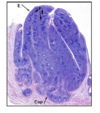
What are Peyer’s Patches, and how do we ID them? What is their function?
The Peyer’s Patches (PP) are found in the lamina propria of the ileum–we recognize the Peyer’s Patches compared to the vermiform appendix from the villi in the GI tract cross-sections. They contain microfold cells, which bind antigens and pass them directly through to lymphocytes for an immediate immune response.

What is the vermiform appendix? What is it’s function?
It is a worm-looking (vermiform) pouch that hangs from the cecum. It’s function is unknown. However, it contains immunocompetent cells that decline over time. This is called immunosenescence.

What are the four specific lymphatic organs?
Bone marrow, thymus, spleen, and lymph nodes
What is the function of the bone marrow as it relates to the lymphatic system?
The bone marrow is the site of production and maturation of B cells, which moderate and modulate the production of antibodies.
What is the structure and function of the thymus?
It is a bilobed, encapsulated organ that is located in the superior mediastinum. It contains a vast amount of T-lymphocytes. It contains an outer cortex, an inner medulla, and connective tissue capsules that can branch and form trabeculae.

Where are Hassall’s corpuscles found, and what is their function?
Hassall’s corpuscles contain matured thymocytes that have developed from epithelial reticular cells. They are unique to the THYMUS!

What is the function of the thymus? Why is it inefficient?
The thymus helps T-cells mature properly. They use positive selection (development of cells that MODERATELY bind MHCs), and then negative selection (DO NOT allow development of cells that STRONGLY bind MHCs, as they may attack self cells). This process is inefficient because about 95% of T-cells improperly self-recognize and are destroyed.

Why is the blood-thymus barrier important?
Since 95% of T-cells improperly self-recognize, we don’t want them binding improperly to blood cells.
What is thymic involution?
The process of declining function and size of the thymus with age (immunosenescence)
What is the structure and function of lymph nodes?
The 500+ lymph nodes of the body are encapsulated organs that serve as connecting points for lymphatic vessels. They function to filter pathogens out of the blood. They have multiple lymph nodules formed in the cortex by trabeculae. The cortex is subdivided into a superficial and deep layer. The medulla is the deepest part, and contains plasma B cells, the paracortex is the middle section that contains T cells and antigen presenting cells (APCs), and the cortex contains B cells and APCs.


