Week 4: Nervous System Intro II Flashcards
What is the difference between white and gray matter? What are the different cell components of each?
White matter is comprised of axons/nerve terminals covered by myelin. Myelin is a lipid formed by oligodendrocytes in the CNS and Schwann cells in the PNS. The cell components of white matter include oligodendrocytes and myelinated axons.
Gray matter is composed of cell bodies, unmyelinated axons, astrocytes/microglia and dendrites/axon terminals.
What are the morphologies and functions of glial and other cells within the CNS?
The glial cells are: Astrocytes, Oligodendrocytes, and Microglia. We remember these and others via:
Any Old Man Can Play Every Violin
Astrocytes (gray matter): guide neurons during development, produce/secrete growth factors, form the BBB, and control bloodflow/ionic concentration
Oligodendrocytes (white matter): can myelinate multiple axons, helping form myelin sheath around neurons. Cannot myelinate the same neuron twice, but can myelinate multiple kinds of neurons.
Microglia***NOT CNS-derived: WBCs of the CNS that derive from mesoderm/early leukocyte progenitors. Phagocytose problem cells/toxins in the CNS.
Choroid plexus: makes CSF
Pial cells: form a protective barrier for the brain and SC, including the pia mater, arachnoid space, and dura mater
Epindymal cells: form the inner layer that lines the ventricles and central canal of the CNS, endothelial in nature
Vascular cells: provide blood to the CNS, contain endothelial cells
What are the structural features of the different neuron types? Describe the four main kinds with respect to presynaptic/peripheral terminals, cell body, axons, and dendrites. Match each type with the cells in the photo.

(1) Multipolar: Dendrites stem from cell body, single axon extends outwards ending in presynaptic terminals (classic depiction of neuron)
(2) Bipolar: Cell body in the middle of the axon, with dendrites at one end that receive information, and presynaptic terminals (usually innervate the CNS) at the opposite end
(3) Pseudounipolar: Much like bipolar, but the cell body stems off of the main axon. Peripheral terminals and dendrites are at either end of a common axon, with a branch for the cell body. Found in dorsal root/spinal ganglia
(4) Unipolar: Cell body at far end, presynaptic terminals at opposite end, and dendrites in between
What are the components of neuron cell bodies?
The cell body supports metabolism, transcription and translation. Has a prominent nucleolus to produce mRNA. Also extensive Golgi apparatus to produce/package mRNA and VESICLES. High number of mitochondria due to need for production of ACh in cholinergic neurons.
What are the components of dendrites?
Dendrites have small spines to increase contact with nearby cells. They contain many receptors to receive signals. They are usually extensions of the cell body and are not myelinated. They have RER to produce proteins for use in constructing ion channels.
What are the cellular components of axons?
The axon hillock is where an axon stems from/begins. It also prevents ribosomes from moving into the axon.
The initial segment is high in ion channels and is the site of action potential initiation
The cytoplasm/axoplasm contains smooth ER, and no ribosomes, unlike dendrites.
Axons also contain mitochondria, microtubules for transporting proteins down the axon, as well as transport vesicles and neurofilaments
How does axonal transport work?
Axonal transport is bidirectional and thus has two main functions:
Anterograde/orthograde (OUTPUT) has a slow phase of cytoplasm wave extension, where unused material is later broken down and recycled, as well as a fast phase of vesicular transport
Retrograde (INPUT) occurs via fast transport of endocytic vesicles from the synaptic terminal to the cell body. This removes debris from the axon terminal, and transports it to the cell body for disposal.
How are neurons regulated during development to match the size of their target structures?
In early neurogenesis, there is an overproduction of motor neurons in the spinal cord. Optimal contact with targets allows neurons to release trophic factors in retrograde fashion from the synaptic terminal to the cell body via endocytosis. The neuron cell bodies that don’t receive trophic factors are “pruned” off eventually, leaving ONLY THE STRONGEST LIL’ BUDDIES.
What are neurotrophins and how do they work? What are some examples?
Neurotrophins (NTs) are examples of trophic factors. Trk receptors maintain the survivability of neurons, and p75 receptors induce cell death when signaled alone. Deletion of various trk receptor genes produced offspring without pain receptors and, alternatively, a lack of innervation to muscle spindles. Ouchies!
Describe the molecules that regulate axonal growth and guidance to their appropriate target cells
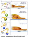
- *Microtubules** at the end of axons extend and add tubulin to their edges. As they reach the central zone, axons form two main elements from different structures that make the end of the axon look like a “webbed hand”:
- *Lamellipodia**: contain actin, microtubules, and neurofilaments. Actin filaments from the filapodia are broken down at the lamellipodia, which are like the webbing portions of the “hand”
Filapodia: composed of actin filaments, which divide into actin subunits and globular actins. The globular actins polymerize at the leading end of the filapodia to extend actin filaments and allow axons to grow.
What are some examples of axonal directional movement?
Axon filapodia touching other axons. The filapodia tugs on other axons, as both grow.
Axons contacting laminin-–the axon binds laminin and grows towards it preferentially
Guidepost cells help axons develop a pathway for long-distance development
Myosin in the leading filapodia exert tension to slide the actin filaments towards targets. Actin filaments tether to substrate and pull the growth cone foward. Myosin inside of the axon tugs on the inner wall via clutch, and once the myosin filaments reach their target spot, they release clutch in order to start another round of pulling.
What are some common guidance cues that axons use to grow and form connections?
Long-range chemoattractive (netrins)
Long-range chemorepulsive (semaphorins, some netrins)
Contact attraction (Ig CAMs, cadherins, ECM/laminins)
Contact repulsion (Eph ligands, semaphorins, ECM/tenascins)
What percentage of our overall oxygen is used by our brains, and why?
20% of O2 in the body is used by the mitochondria in the brain. This is to produce large volumes of ATP in order to power the extensive amount of biosynthetic machinery involved in axonal cells.
What is the name for the axonal region where action potentials are generated?
The initial segment
What are some examples of trophic factors, and how do they relate to the critical period of programmed cell death?
Neurotrophins (NT) are signaling molecules released by target cells into the PRESYNAPTIC ends of axons. These signals then travel in a retrograde fashion UP the axon, towards the cell body, and keep the nerve cell alive. If the nerve cell receives enough of these signals, they survive to be a part of the developed organism’s neural system. Some examples are:
Trk receptors: maintain survivability of neurons
p75 receptors: when signaled alone, induce cell death
What is the trophic factor hypothesis?
Target cells release limiting molecules essential for neuronal cells to remain viable. These enter the presynaptic bouton/terminal, move up the axon to the cell body (retrograde) via endocytosis. The neurons that don’t receive trophic factors die.
What are the two main components of the growth cone?
(1) Lamellipodium - “web” of the growth cone contains actin, microtubule, and neurofilaments. This is where actin filaments from the filapodia are broken down.
(2) Filopodia - composed of actin filaments, which further divide into actin subunits, and globular actins. The globular actins polymerize at the leading end of the filopodia to extend actin filaments, allowing axons to grow.

What are some ways actin grow towards substrates?
(1) Axon filapodia can touch other axons. It then “tugs” on that axon, and both of them grow together.
(2) Axons contact a substrate with laminin on it. The axon preferentially binds to the laminin-rich substrate and grows into it.
(3) Guidepost cells help axons develop a pathway for long-distance travel

How do filapodium grow into nearby substrates of interest?
(1) Actin is linked to a substrate as a “clutch” while microtubules and myosin sit “farther back” towards the lamellopodia
(2) Myosin pull on the actin fibers, moving fowards to grow and then releassing clutch
(3) Actin fibers extend via growth (polymerization), then engage clutch to attach to substrate once again. The cycle repeats. The cycle of life.

What are the elements of the nervous system?
(1) Somatic sensory - touch, external pain, proprioception, pressure, in the skin, body wall, and limbs (also hearing, vision, smell, and equilibrium) (EXTERNAL)
(2) Visceral sensory - pain, motion, nausea, stretch, temperature, chemical agitation, hunger (taste) (INTERNAL)
(3) Somatic motor - skeletal muscles/voluntary motor (EXTERNAL)
(4) ANS/visceral motor - involuntary motor, mostly smooth muscles, cardiac muscles, glands (branchial arch muscles) (INTERNAL)


