Week 2 - D - Myeloma, MGUS, AL AMyloidosis, Waldenstroms - B/Plasma cells/Ig - electropheresis/immunofixation, Signs&Tx Flashcards

1
Q

Bcells are derived in the bone marrow from haemopoietic stem cells Which immune system are they part of? What is the dual function of the Bcells?

A

They are part of the adaptive immune system B cells have the dual function of producing antibodies and acting as an antigen presenting cell

2
Q

Antibodies are produced by B cells and plasma What is the difference in antibody produced by both these cells?

A

When B cells produce antibodies, they stick to the surface of the B cell Plasma cells actively secrete antibodies

3
Q

The term antibodies and immunoglobulins are interchangeable - usually when speaking of the functional aspect we use the term antibodies and when speaking of the structural aspect we use the term immunoglobulin What are the chains from which immunoglobulins are made?

A

Immunoglobulins are made from 2 heavy chains and 2 light chains

4
Q

Does the type of immunoglobulin produced depend on the light chain or heavy chains? What are the different heavy chains?

A

The type of immunoglobulin produced by the B cell depends on the type of heavy chain that is used There are five different heavy chains (α, δ, ε, γ, μ) Alpha (IgA), Delta (IgD), Epsilon (IgE), Gamma (IgG), Mu (IgM)

5
Q

IN B cells –antibody is stuck to the surface of the B cell IN plasma cells – the cell is actually secreting the antibody into the blood steam Proteins made up of 2heavy and 2 light chains The type of antibody depends on the type of heavy chains used (5types) The 2 light chains are kappa or lambda chains (κ or λ) What forms the backbone of the immunoglobulin and what are the bonds that join them?

A

The backbone of the immunoglobulin is formed by the heavy chains The two heavy chains are joined by disulphide bonds

6
Q

Each antibody recognises a specific antigen The basic functional unit of each antibody is an immunoglobulin (Ig) monomer (containing only one Ig unit) WHat are the different stuctures of each immunoglobulin?

A

IgD, IgE, IgG are all monomers IgA is a dimer IgM is a pentamer

7
Q

https://s3.amazonaws.com/classconnection/403/flashcards/11907403/png/paella-recipedocxpngjpg-162217023AA3D155A85.png

A
  • Question 1 - IgA
  • Question 2 - IgD
  • Question 3 - IgE
  • Question 4 - IgG
  • Question 5 - IgM

IgG is the most abundant antibody type IgM is the first antibody in response to infection (M for iMMediate)

8
Q

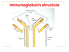
Name what the different boxes are referring to What is the bond that the black box is pointing to? What regions are represented by the purple and blue areas of the chains?

A
  • The green box is the antigen binding site
  • The blue box is the light chain
  • The black box is pointing to the disulphide bones that connect both heavy and light chains
  • The heavy chain is the backbone of the immunoglobulin
  • The yellow box refers to the Fc fragment
  • The red box refers to Fab fragement of the immunoglobulin

Purple = variable regions Blue = constant regions

9
Q

Antigens bind at the antigen binding site of the immunoglobulin Where do the Fc fragments bind?

A

The Fc fragments bind to Fc receptors which is a protein found on certain cells that antibodies can then join onto and await for antigens to bind to the binding sites

10
Q

B cell Initial production and development is in the bone marrow under control/influence of microenvironment What is the Ig variable element determined by?

A

The Ig variable element is determined by the V-D-J region recombination early in development of the cell

11
Q

After the VDJ region recombination, the Ig variable element is determined - thousands of combinations can be produced form this VDJ region recombination Immature B cells with (Ig) on their surface exit bone marrow ready to meet their target. Where do B cells mature?

A

The B cells mature in the lymph nodes

12
Q

The process of generating antibodies with increased binding affinities is called affinity maturation. Affinity maturation occurs in mature B cells after V(D)J recombination, and is dependent on help from helper T cells Where do the B cells travel in the lymph node to mature?

A

The B cells travel to the follicle germinal centre to mature The B cells are exposed to antigens - the ones that bind antigens well will survive, the ones that don’t bind as well will undergo aspoptosis

13
Q

Once the B cell is matured in the lymphoid follicle germinal centre, it may return to the bone marrow as what two cells?

A

The B cell may return to the bone marrow as a plasma cell or a memory B cell

14
Q

Plasma cells pump out antibodies What is the appearance of the plasma cell on blood film described as? What does the plentiful blue cytoplasm of the plasma cell contain? What is the open chromatin synthesising? On blood film, there may be a slight perinuclear pale area (perinuclear halo) - what is this?

A

The appearance of the plasma cell on blood film is described as a fried egg appearance Open chromatin is synthesising mRNA to help make proteins which are found in the plentiful blue cytoplasm There is also a perinuclear halo which is where the golgi apparatus lies

15
Q

Plasma cells are large lymphocytes with a considerable nucleus-to-cytoplasm ratio and a characteristic appearance on light microscopy. What is the normal number of plasma cells to find on a bone marrow aspirate?

A

Usually only about 2% of the bone marrow is found to have plasma cells but in conditions affecting the plasma cells, can see more than this 2%

16
Q

Because plasma cell malignancies arise from one cell, they are monoclonal It is monoclonal therefore giving the same one abnormal immunoglobulin When is there a polyclonal increase in immunoglobulins?

A

There can be a polyclonal increase in response to reactive things such as: * Infection * Autoimmune * Malignancy * Liver disease

17
Q

What is a monoclonal rise in immunoglobulins due to? Are the antiboides produced identical or not?

A

A monoclonal rise in immunoglobulins is due to clonal expansion of a single B-cell The antibodies are identical in structure and specificity (size and charge)

18
Q

Presence of a monoclonal immunoglobulin implies there is a clone of faulty B cells or plasma cells somewhere What is the name given to a monoclonal immunoglobulin?

A

The name given to a monoclonal immunoglobulin is a paraprotein

19
Q

Again, what is the marker of underlying clonal B-cell disorder?

A

This would be a monoclonal rise in immunoglobulins is a marker of an underlying clonal B-cell disorder

20
Q

If a patient presents ie with back pain that is not improving and systemic cancer symptoms WHat test can be carried out to measure for monoclonal protein bands?

A

Look for monoclonal protein bands in serum or in urine by using electropheresis

21
Q

The electropheresis separates the proteins in the serum into distinct bands(or zones) WHich protein lies nearest the positive charge of the electropheresis and why?

A

Albumin is very negatively charged and therefore lies nearest to positive charge on electrophereisis Gamma proteins are positively charged and therefore move furthest away form the positive charge on electropheresis

22
Q

It is in the gamma proteins in the blood where the immunoglobulins are found On electrophereisis, what is it that affects the rate at which proteins move?

A

Proteins move at different rates dependent on their size and charge

23
Q

On electropheresis the gamma band should be very fuzzy as there should be polyclonality and not a monoclonal immunoglobulin rise which would cause what?

A

A monoclonal rise in immunoglobulins would cause a distinct zone in the gamma band and this would warrant further investigation

24
Q

If an abnormal protein band is identified on serum or urine electropheresis, what is carried out to identify what type of paraprotein (monoclonal immunoglobulin) is present?

A

Serum immunofixation would be carried out - this enables the paraprotein present to be identified

25
Q

g - IgG a - IgA M - IgM K&L - kappa and lambda light chains respectively

A

Figure on left - can see that on electropheresis there was a distinct zone in the gamma protein band Matches up with IgGK (IgG Kappa is the paraprotein) Figure on the right - can see that on the electropheresis there was a dinstinct zone in the gamma protein band as well Matches up with IgAK (IgA Kappa is the paraprotein) * Use electrphersis to tell us there is a paraprotein * Use immunofixation to tell us the type of paraprotein

26
Q

After defining that there is a paraprotein present (electropheresis) and determining the type (immunofixation), have to quantify the ammount How is the amount quantified?

A

The serum electropheresis can be pltted telling us of the percentage that the gamma protein take up of the total electrophereisis after the immunofixation Means firstly -Serum protein electropheresis to find out if paraprotein is present, then immunofixation to determine the type of paraprotein then use the findigns from the electrophereisis to quantify the amount of the type of paraprotein that has been found

27
Q

If there is an increase in the immunoglobulin production due to an abnormal proliferation of a single clone of plasma cells, this will obviously increase the heavy and light chains Free light chain production by normal plasma cells is 0.5g/day Why is it that the heavy chains cannot leak into the urine where as the light chains can?

A

The heavy chains are too big too get through the glomeruli of the kidney whereas the light chains are able to leading to an increase light chain deposit in the urine

28
Q

What are these immunoglobulin light chains that are found in the kidney known as and how are they detected?

A

These immunoglobulin light chains are Bence Jones proteins - they are free Ig light chains of kappa or lamdba type They are diagnosed based on urine electrophoresis (The build up of bence jones is a paraprotein and the paraproetin in the blood can also be used to measure the myeloma)

29
Q

Usually there is a small excess light chain production and most gets reabsorbed in the renal tubules but over production can saturate the absorption and be found in the urine What is the free light chain production by normal plasma per day? Shape of KappavsLambda chains?

A

The free light chain production by normal plasma cells is 0.5g/day and it is usually reabsorbed by the kidney tubules In conditions where excess chains are being produced this can cause leakage into the urine as Bence jones proteins Kappa light chains are usually monomeric Lambda chains are usually dimeric

30
Q

Causes of paraproteinaemia (paraproteins (monoclonal immunoglobulins) in the blood) can be grouped under a term known as plasma cell dyscrasias What is a plasma cell dyscrasia?

A

Plasma cell dyscrasias is when there is an abnormal proliferation of a single clone of plasma cells or lymphoplasmacytic (ie lymphocytes - B cells or plasma cells) cells leading to the secretion of immunoglobin (Ig) causing the dysfucntion of many organs esp kidney . The Ig is seen as a monoclonal band -a paraprotein - on serum or urine electropheresis.

31
Q

What is the commonest plasma cell dyscrasia?

A

This would be MGUS - monoclonal gammopathies of undetermined significance - accounts for 56% of PCDs and is benign MGUS – benign condition where there is a small clone of plasma cells but the clone is not proliferating or taking over the plasma cells

32
Q

At what rate is the second most common cause of PCDs increased by due to monoclonal gammopathy of undertemrine significance (MGUS)?

A

Second most common cause - multiple myeloma Risk of progression from MGUS to multiple myeloma increases by 1% every year

33
Q

Myeloma is a plasma cell malignancy The clonal plasma cells can affect the body directly or indirectly via the paraproteins How does the tumour affect the body directly in myeloma? Talk about how it affects the bone

A

There is bone lesions (osetolytic bone lesions) leading to weakened bones and increased calcium in the blood The lytic bone lesions also result in bone pain The monoclonal plasma cells also replace the nomral cells in the marrow leading to bone marrow failure - pancytopenia

34
Q

Go over the direct tumour effects on bone on this card What effects does the paraprotein have on the body?

A

Direct tumour effects * Causes osteolytic bone lesions resulting in calcium release (hypercalcaemia has its own problems) * Also the weakened bones are in pain * The plasma cells can infiltrate the normal bone leading to marrow failure - pancytopneia (anaemia, infections, bleeds) Paraprotein effects- leads to renal failure, immune suppresion, hyperviscoisty due to increased cells in the blood and amyloid (rare)

35
Q

Although the plasma cells are malignant, they usually retain the ability to make antibody – therefore these cells are just chuingin out antibdoies resulting in massive overproduction of one type of antibody – the paraprotein resulting in different things Why can hypogammaglobulinaemia occur due to myleoma?

A

This is because the massive proliferation of one abnormal immunoglobulin - monoclonal rise in immunoglobulin is known as a paraprotein - results in the potential underproduction of other types of antibody

36
Q

Multiple myeloma patients can be classified on the basis of the type of monoclonal protein they produce. What is the main antibody that is produced in myeloma? What is the second most common produced? What is usually produced in the urine when there is monoclonal rise in immunoglobulins?

A

The main Ig produced is IgG - accounts for over 50% of all Myeloma cases The next most common Ig produced is IgA - accounts for almost a quarter Normally there is Bence-Jones proteins produced in conjunction with the rise in IgG or A but in 15% cases there is only a rise in Bence Jones proteins

37
Q

What is Bence Jones protein? WHat is the normal production of free light chains per day?

A

Bence Jones protein is a moncolonal immunoglobulin light chain found in the urine - it can be kappa (most of the time) - monomeric light chains or lambda - dimeric Normally there is excess light chains produced per day by the plasma cells compared to heavy chains- so there is free light chains prouced ~0.5g/day that are reabsorbed by the kdiney tubules IN myeloma there is increased proliferation so the tubules are saturated and the proteins are found in the urine

38
Q

What may the ostelytic bone lesions look like on Xray? Which bones are usually affected?

A

The osteolytic bone lesions look like punched out lesions on the xray - typically described as pepper-pot skull The lytic bone lesions can also make the bone more likely to fracture under stress and hypercalcaemia

39
Q

This shows a wedge fracture - usually results form spine degeneration or trauma - seen in osteoporosis as well How does myeloma cause the osteolytic bone disease?

A

Myeloma - activates the production of cytokines mainly IL-6 which causes most of the localised damage IL-6 causes suppresses osteoblast activity and promotes osteoclast activity resulting in ltyic bone lesions Increase in bone destruction releases Ca2+

40
Q

What are the symptoms of hypercalcaemia?

A

Abominal groans - nausea, dyspepsia Stones - kidney or biliary Bones - bone pain due to increased osteoclast activity Psyhicatry overtones - depression, anxiety confusion There is also polyuria leading to dehydratuin

41
Q

Hypercalcaemia can cause abo groans, renal stones, bones, and psychiatric overtones s What is the treatment of hypercalcaemia?

A

If patient presents with hypercalceaemia It is important to reyhdrate the patient -IV saline and IV biphosphontes to stop the breakdown of the bone

42
Q

In myeloma, 30% of patients have renal impairment at diagnosis The light chains that are produced in abundance in myeloma are toxic to the tubules What can the light chains deposit as in the kidneys when in excess? What else can affect the kidneys in myeloma?

A

The light chains can deposit in the kidneys leading to cast nephropathy * The hypercalcaemia can cause the patient to be deyhdrated and have kidney stones * If there is amyloid in myeloma - can cause kidney problems also * Taking NSAIDs to the back pain can also tip the patient into renal failure * Also due to infection thanks to the nuetropenia

43
Q

https://s3.amazonaws.com/classconnection/403/flashcards/11907403/png/paella-recipedocxpngjpgpngjpg-162247B0E010052EA53.png

A

The kidney produces Tamm-Horsfall proteins - in the thick asceding loop of henle - which combines with the free light chains to produced insoluble casts which can lead to acute renal failure

44
Q

Damage to the kidneys may be reversible if treated promptly What may be used to treat the cast nephropathy in the kidney?

A

Use steroids/cehmo - also treats the myeloma

45
Q

Measuring the levels of what globulin in the blood can be used as a prognostic factor in the staging of multiple myeloma? What else is measured in the blood? measures how fast blood will settle

A

The levels of B2-microglobulin is an important prognostic measure in myeloma - increasing levels means there is an increased stage of myeloma Erythrocyte sedimentation rate is aslo increased in myeloma

46
Q

Treatment for myeloma is focused on therapies that decrease the clonal plasma cell population and consequently decrease the signs and symptoms of disease. What is the usual age of diagnosis for myeloma? What are the symptoms?

A

Usual age is patient in their 60s for diagnosis Symptoms - CRAB HyperCalcaemia - leads to abdominal pain/nausea, renal calculi and dehydration, bone pain, psychiatric overtones Renal complications - stones, dehydration, cast nephropathy Anaemia - as well as neutropenia (infections) and thrombocytopenia (bleeding) Bone disease - lytic bones - increase risk of fractures & pain

47
Q

If a patient is symptomatic of myeloma what tests are carried out?

A

Serum or urine electrophereisis to identify the presence of a paraprotein Immunofixation to identify the type of paraprotein and then quantify the amount of paraprotein Plasma cells are increased on bone marrow biopsy as well Evidence of pancytopenia in bloods, hypercalcaemia and renal insufficiency ie increased creatinine Skeletal survery - xray of chest, spine, skull and pelvis

48
Q

What is the percentage of plasma cells seen on marrow biopsy?

A

There is a greater than 10% plasma cells seen on marrow biopsy - fried egg appearance, lots of cytoplasm and open nucleus, also perniculear halo

49
Q

Treat the hypercalcaemia and spinal cord compression of any Treat the hyperviscosity Also treat anny acute renal injury How is the hyperviscosity and spinal cord compression treatment?

A

Hyperviscosity - plasmapheresis to take blood out, remove the light chains, and transfuse the blood back in Spinal cord compression - Urgent MRI if suspected and treat with steroids / local radiotherapy

50
Q

What is used for treatment in myeloma? What can be given in the case of relapse or to imporve myeloma event free survival? What can be used in younger fitter patients as treatment?

A

Treatment would be corticosteroids (dexa or prednisolone) in combination with chemotherapy as treatment Can use thaldiomide to improve treatment prognostics or in the case of recurrence In younger fitter patients, can try giving the patient high dose chemo and an autologous stem cell transplant

51
Q

In myeloma, is the chemo (mephalan) + prednisolone curative?

A

This is not a cure to the disease but helps to treat the cancer and keep patients in a remissive state

52
Q

So - coritcosteroids + chemo therapy for treatment Can also use thalidomide to help with treatment prognostics High dose chemo and autologous stem cell transplant given in younger fitter patients WHat is used to monitor the response to treatment?

A

Measuring the paraproteins is used fo monitoring the response to therapy

53
Q

What is given for symptom control in myeloma?

A

Give: Opiate analgesics (Avoid NSAIDs as nephrotoxic) Biphosphonates - alendornic/zolendronic acid - corrects hypercalcaemia and bone pain Steroids and local radiotherapy if any spinal cord compression Can inject sterile cement into fractured erterba - vertebroplasty

54
Q

What is the most common type of plasma cell dyscrasia?

A

This would be MGUS - monoclonal gammopathy of uncertain significance The main causes of paraproteinaemia * MGUS * Myeloma * Primary amyloidosis * Waldenstrom’s macroglobulinaemia * Paraproteinaemia in lymphoma or lerrukamiea * Heavy chain disease

55
Q

How would MGUS be defined/diagnosed?

A

A paraprotein level of <30g/l in the bloods Less than 10% plasma cells on bone marrow aspiration No signs of myeloma end organ damage - normal calcium, normal renal function, normal Hb, no lytic lesions and no increase in infection MGUS is basically the situation where you have a paraprotein but you are well – usually the paraprotein is also present in less concentration than in myeloma

56
Q

What is the risk of progression of MGUS to myeloma?

A

Risk of progression is 1% increase in risk per year

57
Q

AL amyloidosis is a rare disorder causing paraproteinaemia What causes AL amylodisos? (primary) (AA amyloidosis is secondary to other causes)

A

AL amyloidosis is caused by a small plasma cell clone usually due to a mutation in the light chains leading to an altered structure that causes its deposition in different tissues

58
Q

In contrast to myeloma, amyloidosis usually is just a small clone of faulty plasma cells producing small number of paraprtoeins/light chains In amyloid the clonal light chains produced just happen to have the right structure/charge etc to stick together What do the cloncal light chains form when in the tissues?

A

The clonal light chains form insoluble pleated-beta-sheets which build up in the tissue causing tissue damage

59
Q

AL amyloidosis (immunoglobulin light chainamyloidosis) - Amyloid light-chain (AL) amyloidosis * Accumulation in tissues causes organ damage * Slowly progressive * Multisystem disease * Different protein to SAA amyloidosis (chronic inflammation) and familial amyloidosis What tissues does the abnormal amyloid light chains usually deposit in?

A

Deposit in kidney causing nephrotic syndrome Heart causing cardiomyopathy Liver Neuropathy - peripheral and autonomic GI tact causing malabsroption

60
Q

How is AL amylodisos confirmed? What abnromal light chains in the urine may be seen?

A

It is usually diagnosed by biopsy of an affected organ and staining under congo red staining and expecting to see apple green birefringence under polarised light May see bence jones proteins in the urine

61
Q

To stage the amyloidosis, also want to identify if any other organs are affected than the one from which the biopsy was taken How can this be done? What is the treatment for AL amyloidosis?

A

Echocardiogram for the heart Heavy proteinuria of kidneys Treat with chemo + steroids - same as myeloma

62
Q

What is the type of plasma cell dyscrasia in which IgM parprotein is produced? What is the neoplasm which produces the IgM cells? What is used to detect the IgM paraprotein?

A

This is Waldenstroms macroglobulinaemia It is a lymphoplasmocytic neoplasm - there is a clonal disoder between lymphocyte and plasma cells resulting in the prouction of a monoclonal IgM paraprotein IgM parapprotein detected on serum electropheresis

63
Q

Waldenstroms macroglobulinaemia also has direct tumour effects and indirect effects due to the paraportein What are these effects? What is the structure of the tumour in WM?

A

Tumour effects Splenomegaly Lymphadenopathy Marrow failure Paraprotein effects There is neuropathy and hyperviscosity in the blood WM has the IgM which is a pentamer immunoglobulin

64
Q

What are the clinical features of the hyperviscosity? Why is hyperviscosity much more common in Wladenstromas macroglobuinaemia than in normal myeloma? What is done to correct the hyperviscosity present in WM?

A

Fatigue, visual disturbance, confusion, coma and bleeding , can also cause cardiac failure Hyperviscosity is more common because WM features IgM as the overproduced monoclonal antibody - these are pentamers compared to IgG monomers and therefore are far bigger in the blood making it more viscous Treat with plasmapheresis to remove the paraprotein chains

65
Q

What is the treatment of waldenstoms macroglobilinaemia?

A

Treat with chemotherapy and plasmapheresis for the hyperviscosity Plasmapheresis is the removal, treatment, and return or exchange of blood plasma from and to the blood circulation. It is thus an extracorporeal therapy (a medical procedure performed outside the body).

66
Q
  1. An 83-year old female patient has complained of recurrent low back pain and tiredness. Recently she has fallen and fractured her T11 vertebra. She complains of feeling dizzy at the time of falling. In addition she has had some abdominal pain, which she thinks is unrelated. Urine protein electrophoresis shows Bence-Jones protein. A Acute lymphoblastic leukaemia B Burkitt’s lymphoma C Chronic lymphocytic leukaemia D Myeloma E Acute myeloid leukaemia F Hodgkin’s lymphoma G Chronic myeloid leukaemia H Non-hodgkin’s lymphoma
A

D - Myeloma Low back pain and tiredness with a fracture Feeling dizzy - anaemia Abdo pain - hypercalcaemia Light chains in urine