Week 1: Tuesday (1/12/16) Lumbar Anatomy & Arthrokinematics Prep Guide + Notes from class Flashcards

1
Q

What is the function of the curves in the spinal column (in other words, why specifically is a curved spine better than a straight spine)?

A
  • [Helps] Minimize compression force to reduce load through vertebral bodies (Mincer video)
  • The sagittal plane curvatures of spine provide strength and resilience to axial skeleton. A reciprocally curved vertebral column acts like an arch. Compression forces between vertebrae are partially shared by tension in stretched connective tissues and muscles located along the convex side of each curve. Similar to long bones like the femur, the strength and stability of the vertebral column are derived, in part, from its ability to “give” slightly under a load, rather than to support large compression forces statically. (pg 314 Neumann)
2
Q

What is the main function of the vertebral body?

What other structure helps with this function?

A
  • The drum-shaped vertebral body, designed to bear weight and withstand compression or loading. (knowyourback.org)
  • Intervertebral discs also serve this function.
3
Q

Besides bearing weight and withstanding compression or loading, is another important function of a vertebral body?

A

The upper and lower surfaces of the vertebra body give attachment to the intervertebral discs. (wikipedia)

4
Q

In general where is the axis of rotation for intervertebral movement?

Why is it helpful to know this?

A
  • The axis of rotation for intervertebral movement is near or through the region of the vertebral body.
  • Helpful to know because we can remember that With sagittal plane movement, any ligament located posterior to the vertebral body is stretched during flexion. Any ligament located anterior to the vertebral body is stretched during extension. >>>directional orientation to spine is done at the the vertebral body. (Neumann pg 318)
5
Q

What type of joint is a facet joint?

A

synovial

6
Q

How do synovial joints get nutrition?

A

Small blood vessels with capillaries penetrate the joint capsule, usually as deep as the junction of the fibrous layer of the joint capsule and the adjacent synovial membrane. ( Neumann pg 29)

7
Q

Can synovial joints be sources of pain?

A

Yes!

Sensory nerves also supply the external layer of the capsule and ligaments with receptors for pain and proprioception. (Neumann pg 29)

8
Q

How can synovial joints be sources of pain?

A

Sensory nerves also supply the external layer of the capsule and ligaments with receptors for pain and proprioception. (Neumann pg 29)

9
Q

In what way does the facet joint capsule act like a ligament?

A

Basically, the capsule is very similar to ligaments in structure and function

  • (Neumann, pg 316) Capsular ligaments of the apophyseal joints consist mostly of collagen fibers that attach along the rim of the facet surfaces ( see fig 9-11, A). Those apophyseal joints help interconnect and stabilize the intervertebral junction. Also have a unique role in guiding the specific direction of intervertebral movement. Sensory mechanoreceptors embedded within the capsule likely provide muscles info to assist with this guidance.
  • The capsular ligaments are relatively loose in the neutral position, but become taut as the joint approaches the extremes of all its movements. Passive tension is greatest in motions that create the largest translation or separation between joint surfaces.
10
Q

What structure primarily determines the amount of spinal motion (flex/ext, lateral flex, rotation) in the lumbar region?

A

The facet joints and the orientation of their surfaces.

11
Q

How do the facet joints of L1-L4 affect spinal motions (flex/ext, LF, rotation)?

A

L1-L4: The facet surfaces of most lumbar apophyseal joints are oriented nearly vertically (not great for horizontal plane rotation), with a moderate-to-strong sagittal plane bias (best for flex/ext).

  • The orientation of the superior articulating facet of L2, for example, is on average about 25° from the sagittal plane. This orientation favors sagittal plane motion at the expense of horizontal rotation [and probably frontal plane lateral felxion]. (Neumann pg 346)
12
Q

How do the facet joints of L5-S1 affect spinal motions (flex/ext, LF, rotation)?

A

L5-S1: The L5-S1 junction has an anterior interbody joint and a pair of posterior apophyseal joint (like any vertebral junction). However, the facet surfaces of the apophyseal joints have more of a frontal plane orientation than the other lumbar vertebrae. (Neumann pg 346)

  • This restricts saggital plane movement (flex/ext) and horizontal plane movement (rotation)
  • It allows frontal plane movements (lateral flexion).
  • It also resists anterior translation of L5 on S1.
13
Q

lumbar flexion/extion:

  • What is the normal range given in Neumann for each movement?
  • does L1-L4 or L5-S1 contribute to this motion the most?
A
  • Lumbar Flexion= 40°- 50° (pg 350)
  • Lumbar Extension= 15° - 20° (pg 350)

L1-4 contributes the most because of the almost saggital orientation of the facet joints (about 25 degrees from saggital plane).

14
Q

lumbar lateral flexion:

  • What is the normal range given in Neumann for this movement?
  • does L1-L4 or L5-S1 contribute to this motion the most?
A

Lateral Flexion= 20° (pg 357)

L5-S1 contributes the most to this motion because of the frontal plane orientation of the facet joints.

15
Q

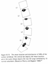
Gravity imposes a much higher degree of ________ force at L5-S1 than the rest of the lumbar spine.

A

Gravity imposes a much higher degree of anterior shear force (and posterior compressive) force at L5-S1 than the rest of the lumbar spine.(Neumann pg 348)

16
Q

The intervertebral foramen (IVF) is created by what bony structure(s)? (4)

A
  • IVF is the lateral opening between adjacent vertebrae
    1. The Superior Notch of the adjacent vertebra (inferior to IVF)
    2. The Inferior Notch of the vertebra superior to the IVF
    3. The body of the vertebral body
    4. Facet joints on the transverse process of the vertebra.(310 and wikipedia)
17
Q

What passes through the IVF? (6)

A
  1. root of each spinal nerve
  2. dorsal root ganglion
  3. the spinal artery of the segmental artery
  4. communicating veins between the internal and external plexuses
  5. recurrent meningeal (sinu-vertebral) nerves
  6. transforaminal ligaments. (wikipedia)
18
Q

True/False: Adult intervertebral discs are essentially avascular

A

True

19
Q

Adult discs are essentially avascular, so how does a disc receive nutrition? What is the effect of movement on this process?

A
  • (Neumann, pg 330) Only the outer, more peripheral rings of annulus fibrosus contain blood vessels. For this reason, most of the disc has a limited healing capacity. Essential nutrients, such as glucose and oxygen, must diffuse a great distance to reach the deeper cells that sustain the disc’s low but essential metabolism. The source of these nutrients is in the blood vessels located in the more superficial annulus and blood stored in the adjacent vertebral bodies. most of these nutrients must diffuse across the vertebral end-plate and through the disc’s extracellular matrix.
  • Movement causes a larger fluxuation in the water content of the disks and improves diffusion of nutrients into the disc (and waste out of the disk)
20
Q

How can age affect the ability of nutrients to reach the intervertebral disc?

How does this affect the ability of the disk to handle loading?

A

Aged discs show reduced permeability and increased calcification of the vertebral end plates, which reduces the flow of oxygen and nutrients into the disc. This age related process can inhibit cellular metabolism and synthesis of proteoglycans. Less proteoglycan content reduces the ability of the nucleus to attract and retain water, limiting its ability to effectively absorb and transfer loads.

21
Q

Describe the structure of the annulus

A

It is tough and strong, but subject to cracks and fisures as it ages.

  • The annulus (in the lumbar disc) consists of 15 to 25 concentric layers of rings or collagen fibers (like dough surrounding the jelly in a doughnut). Materials and cells are similar with the nucleus pulposus but the proportion is different. In the annulus collagen make up about 50 to 60% of the dry weight as compared to 15 to 20% in the nucleus. Elastin protein is abundantly interspersed in parallel with the rings of collagen adding circumferential elasticity to the annulus.
  • The outermost layer of annulus is primarily type I and type II collagen for strength and flexibility, as well as a mean of bonding the annulus to the ALL, PLL and to the adjacent rim of the vertebral bodies and end plates. Collagen fibers are arranged in multiple concentric layers, with fibers in every other layer running in identical directions (alternating) (fig. 9.34). The orientation of each collagen fiber is about 65 degrees from the vertical.
  • The outer layer contain the disc’s only sensory nerves.
  • The deeper internal layers contain less type I collagen and more water starting to gradually become similar in structure with the nucleus.
22
Q

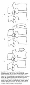
During flexion, what part of the annulus becomes taut? How many layers of this part are taut?

A
  • The posterior portion becomes taut to resist the migration of the nucleus pulposus during extreme flexion. (pg 350)
  • I believe all the fibers become taut.
    • I think since the fiber layers alternate in oblique directions (at about 65 degrees from vertical), when a person flexes forward, all fibers become taut. This is different from if someone performed lateral flexion, where half of the fibers (those oriented in the opposite direction of side flexion) would become loose, while the remaining half would get extra-taut. I think this holds true for rotation as well. This explains one reason the annulus is more at risk during movements that involve twisting (rotation) and flexion, whereas it is safer to perform flexion in a neutral position.
23
Q

What happens to the anterior annulus during flexion?

A

According to a book called Manual Physical Therapy, the anterior annular fibers become slack during flexion. (Not sure if this is what Mincer is looking for) - Sounds right to me (Sara)

  • The anterior aspect of the disc is compressed (pg. 350)
  • Nucleus moves slightly posterior
  • Annulus becomes taut posteriorly and the anterior fibers become slack and bulge anteriorly.
  • The nucleus pulposus of the disc is compressed anteriorly and pressure is relieved over the posterior surface.
24
Q

If the flexed position is maintained for a couple of hours, what structural change does the posterior part of the annulus undergo and how does this affect its strength?

A
  • Based on class discussion, I beleive she was asking us to identify the term “Creep”
    • something called “Creep” occurs, which is a slow deformation of the tissue over time when exposed to stress that is below the yield strength of the tissue. This is a mechanical property of soft tissue that is related to its viscoelastic properties
    • Creep reduces strength of the tissue.
25
Q

Say the flexed lumber position is maintained for a couple of hours and the person experiences “creep” in the posterior part of the annulus.

When this person returns to neutral, does this change reverse immediately?

A

It does not reverse immediately. Something called “set” occurs that takes about 15-20 min for it to get back to neutral.

26
Q

During rotation, what part of the annulus is taut?

A
  • The fibers in the annulus are configured in concentric rings. Every other ring has the fiber oriented 65° from vertical. The alternating rings are oriented in the opposite direction.
  • The reason this is important is because only collagen fiber oriented in the direction of the twist become taut; fibers in every other layer slacken (330)
27
Q

During rotation, what happens to intradiscal pressure and why?

A

–Vertebral bodies approximate, causing ­↑ intradiscal pressure (PP on D2L re:discs)

28
Q

What position(s) of the lumbar spine cause the least intradiscal pressure?

A

rest in the supine position

(From Dr. M’s PPt, from least to most intradiscal pressure):

  • Lying down < upright standing < sitting < sitting with more flexion <flexion></flexion>

</flexion>

Other things in reading guide, probalby from book:

Other positions that have relatively low pressure:

  • sustained full lumbar extension reduces pressure in the disc (allows water to be reabsorbed into the disc , reinflating it to its natural level)
  • lifting a load with the knees flexed
  • sitting erect
29
Q

What position(s) of the lumbar spine cause the most intradiscal pressure?

A

Flexion in Standing with load in hand

(From Dr. M’s PPt, from least to most intradiscal pressure):

  • Lying down < upright standing < sitting < sitting with more flexion <flexion></flexion>

</flexion>

Other things in reading guide, probalby from book:

  • excessive flexion, forward bending combined with vigorous muscle contraction
  • lifting a load with the knees straight
  • sitting in a forward slouched position
30
Q

Why is the ALL so much wider and stronger in the lumbar spine than the PLL?

A
  • to limit the degree of natural lordosis (pg. 316)
  • to prevent extreme extension and reduction of the IVF space which would determine encroachment onto the structures passing through the IVF.
31
Q

What is different about the ligamentum flavum from other ligaments and why does this matter?

A
  • The LF is located posterior to the spinal cord
  • Is characterized by a composition of approximately 80% elastin & 20% collagen signifying a very special function for this ligament
  • It has been proposed that this highly elastic structure, which is under pretension throughout all levels of flexion, acts as a barrier to material that could buckle and encroach on the cords in some regions of the ROM (especially during extension).
32
Q

Describe the location and function of the thoracolumbar fascia.

A

The thoracolumbar fascia (TLF) consists of both aponeurotic as well as fascial connective tissue. Its most important functions are providing a retinaculum for paraspinal musculature in the lumbar region and an insertion site for a substantial amount of trunk and extremity muscles. Through these attachments, muscles and passive tissues are able to exert moments and reaction forces on the TLF, essentially providing stability in several degrees of freedom of the lumbar spine.

(it also contributes to SI joint stability)

33
Q

What is the location of the transversus abdominus relative to the other abdominals and what is the functional advantage of its broad attachments?

A

(pg 390) The TA is the deepest of the abdominal muscles. The muscle is also known as the “corset muscle,” reflecting its role in compressing the abdomen as well as stabilizing the lower back through attachments into the thoracolumbar fascia. Of all the abdominal muscles, the TA has the most extensive and consistent attachments into the TFL, followed closely by the internal oblique muscle.

34
Q

An individual vertebral segment is sometimes conceptually divided into anterior elements and posterior elements. What bony/discal structures are included in each?

A

“Each vertebra is composed of a body anteriorly and a neural arch posteriorly” (according to Emory.edu and Neumann)

Anterior Element (body)

  • Vertebral body
  • Disc- Shock absorber or spacer
  • (Interbody joint - bonds disc & joint)

Posterior elements-

  • Spinous processes,
  • Transverse processes,
  • Articular facets,
  • Lamina

The Pedicle bridges the anterior & posterior elements

From Mincer’s video

35
Q

What is an alternate name for cortical bone?

A

Compact bone

36
Q

What are alternate nmes for Trabaecular bone?

A

Spongy

Cancellous

37
Q

Name the two types of bone in a vertebra and describe briefly the structural implications of each.

A
  • Cortical Bone Aka Compact Bone (surrounds cancellous bone & stiffer than spongy bone and could withstand greater stress)
  • Trabecular Bone Aka Spongy or Cancellous Bone - light but withstands compression. (hypertrophies in response to increased loading, becomes smaller in absence of loading thus less able to provide support, OA always affect this bone)
38
Q

Locate the pars interarticularis on a model of a lumbar vertebral segment. In standing or forward trunk flexion in the lumbar spine, what is the function of the pars interarticularis (it resists what)?

A

Pars interarticularis: is a region of the lamina located between the facet joints (midway between the superior and inferior articular processes) (pg. 349). The pars interarticularis can fracture, leading to spondylolisthesis.

It resist anterior slipping or sliding of one vertebra relative to another. (pg.349)

39
Q

Unilateral failure or lengthening of the pars interarticularis is called

A

Spondylolysis is actually a weakness or stress fracture in one of the bony bridges that connects the upper and lower facet joints. This fracture can happen at any level of the spine but usually occurs at the fourth (L4) or fifth (L5) lumbar vertebra.

40
Q

. Bilateral failure or lengthening of the pars interarticularis is called -

A

We put spondylolisthesis (Neumann pg. 349) in the google doc.

but I think this is actually just bilateral Spondylolysis

Checking . . .

41
Q

If the pars fails or lengthens bilaterally, what is likely to occur subsequently between the two adjacent segments? (name and describe the mechanics of what happens)

A
  • If the fracture gap at the pars widens and the vertebra shifts forward, then the condition is called spondylolisthesis.
  • Spondylolisthesis is the actual slipping forward of the vertebral body (the term “listhesis” means “to slip forward”). It occurs when the pars interarticularis separates and allows the vertebral body to move forward out of position causing pinched nerves and pain. Spondylolisthesis usually occurs between the fourth and fifth lumbar vertebra or at the last lumbar vertebra and the sacrum. This is where your spine curves into its most pronounced “S” shape and where the stress is heaviest.
  • Slippage is measured on a scale from grade 1 slippage (25%) to grade 4 (100%). The more the lower back curves in (swayback or lordosis), the steeper the grade.
42
Q

What are two main functions of the annulus fibrosis?

A
  • contain the nucleus pulposus
  • offer resistance against intervertebral distraction (vertical separation) shear (sliding) and torsion (twisting) (Neumann pg.330)
43
Q

How does the inner annulus differ structurally from the outer annulus?

A
  • The outermost layer of annulus is primarily type I and type II collagen for strength and flexibility, as well as a mean of bonding the annulus to the ALL, PLL and to the adjacent rim of the vertebral bodies and end plates. Collagen fibers are arranged in multiple concentric layers, with fibers in every other layer running in identical directions (alternating) (fig. 9.34). The orientation of each collagen fiber is about 65 degrees from the vertical. It also has some nerves and blood vessels.
  • The deeper internal layers contain less type I collagen and more water starting to gradually become similar in structure with the nucleus.
44
Q

What is the composition of the nucleus pulposus?

A
  • is a pulp-like gel located in the mid to posterior part of the disc. In youth the nucleus of the lumbar disc consists of 70-90% of water
  • The nucleus is thickened into a gel-like consistency by relatively large branching proteoglycans. Each proteoglycan is an aggregate of many water binding glycosaminoglycans linked to core proteins. Dispersed throughout the hydrated proteoglycan are thin type II collagen fibers, elastin fibers and other proteins, which help support the proteoglycan network. Very small number of chondrocytes and fibrocytes are interspersed throughout the nucleus responsible for the synthesis and regulation of the proteins and proteoglycans. (pg 328-329)
45
Q

What is the clinical significance of the nucleus being hydrophilic?

A
  • it allows the disc to function as a modified hydraulic shock absorption system, capable of dissipating and transferring loads across consecutive vertebrae. (pg 328)
  • When outside pressure is reduced (thereby reducing IVP) for a long period of time (like when you are sleeping), the nucleus attracts water and nutrients.
  • When outside pressure is increased (thereby increasing IVP) for a long time (like when standing all day), the nucleus looses water and waste products.
46
Q

Spine Biomechanics: What are 3 assumptions always made when describing movement?

A
  • Anatomical position, standing
  • Top segment moves on stationary bottom segment
  • Axis for most normal motions is just posterior to center of disc
47
Q

How many degrees of freedom are there for spine movement?

A

6

48
Q

What are the 6 degrees of freedom for spine movement?

A
  1. Along the longitudinal axis of the spine, i.e., under compression or disctraction effects
  2. Rotation in the transverse plane around the longitudinal axis of the spine
  3. Forward and backwards in the transverse plane, i.e., a degree of gliding or translation movement (“isolations” practiced in jazz dance class)
  4. Side bending to either side in the frontal plane around a sagittal axis
  5. Lateral gliding or translation in the transverse plane
  6. Forward and backward bending around the frontal axis, i.e., flexion and extension

**It must be recognized that spinal movement is complex and that normal physiological movement occurs through coupling of two or more of these possible movements simultaneously.

49
Q

Who is Fryette and why do we care?

A

Fryette, a researcher of spinal mechanics in the early 20th century, developed certain “laws” of spinal motion. Understanding these laws helps to correctly analyze spinal dysfunction and devise appropriate treatment strategies.

It looks like it is also a brand of sound equipment.

50
Q

Describe Fryette’s Law I:

A

Fryette’s Law I: If the segments are in neutral (or Easy Normal) without locking of the facets (erect standing posture), rotation is in the opposite direction of side bending. Simply stated, if the spine is side bent to the right, rotation of the spine occurs to the left. Some osteopathic literature refers to this as Type I motion of the spine. Type I motion is considered to be the normal adaptation or physiologic movement of the spine to changes in posture.

From http://www.passthenpte.com/2012/01/26/fryettes-law-of-spinal-motion-simplified/

FIRST LAW: When the spine is in neutral, side bending to one side will be accompanied by horizontal rotation to the opposite side.

TRANSLATION: When you’re standing in a neutral position (no bending or hyperextension) and you laterally bend/ flex or side bend to the RIGHT. Your spine will ROTATE to the LEFT (the opposite side).

51
Q

Describe Fryette’s Law II:

A

Describe Fryette’s Law II: If the segments are in full flexion or extension with the facets engaged (or locked), rotation and side bending occur to the same side. Thus, if one bends forward bends (flexes) and side bends to the right, rotation of the spine will occur to the right. In the lumbar and thoracic spine, this motion is considered to be non-physiologic. In the cervical spine, however, it is considered normal physiological motion. This is sometimes referred to as Type II motion and is a non-adapting spinal response to posture or external forces.

From http://www.passthenpte.com/2012/01/26/fryettes-law-of-spinal-motion-simplified/

SECOND LAW: When the spine is flexed or extended (non-neutral), sidebending to one side will be accompanied by rotation to the same side.

TRANSLATION: When you flex or hyperextend your spine and you laterally bend/ flex or side bend to the RIGHT. Your spine will ROTATE to the RIGHT (same side).

52
Q

Describe Fryette’s Law III

A

Fryette’s Law III: If motion is introduced into a segment in any plane, available motion in the other planes is reduced. This means that since vertebral movements are usually coupled (comcominant), movement into one plane lessens the range of movement available in the other two planes. Motion introduced into a segment in two planes further reduces the available movement in the remaining third plane.

From http://www.passthenpte.com/2012/01/26/fryettes-law-of-spinal-motion-simplified/

THIRD LAW: When motion is introduced in one plane it will modify (reduce) motion in the other two planes.

TRANSLATION: When you side bend or forward flex or you do any movement in one plane. Simply put, there won’t be as much motion in the other planes.

53
Q

What is the capsular pattern of the spine?

A

Capsular pattern: pattern of restriction of the joint when the capsule gets tight.

Capsular Pattern:

  • limited contralat (lateral) flexion,
  • Limited ipsilateral rotation;
  • will have ipsilateral deviation in flexion

Limited ipsilateral rotation (left facet cap pattern causes limited left rotation)

Limited contralateral flexion (left facet cap pattern causes limited right lateral flex)

Asymmetrical flexion - causes ipsilateral deviation in flexion (left facet cap pattern causes left deviation in flexion)

54
Q

explain the capsular pattern of the spine

A
  • Capsular pattern is indicative of disfunction (tightness) of an entire joint capsule
  • In the spine, the facet joints are the only synovial joints, therefore the only joints with capsules.

Limited ipsilateral roation:

  • When one rotates spine to the right, ultamately the left facet approximates and the right facet distracts some (opens like a door).
    • If the right capsule is tight, right rotation is limited because distraction is reduced

LImited contralateral flexion

  • When one performs lateral flexion, the contralateral facet must slide.
  • If the capsule of that facet is too tight it cannot slide, therefore reducing contralateral lateral flexion.

Asymetrical flexion - causes ipsilateral deviation in flexion

  • When one performs flexion, both facets must glide past each other equally to flex in midline.
  • If one facet capsule is too tight, it restricts this movement and causes the trunk to deviate towards that side during flexion.
55
Q

T/F: capsular pattern is very important in the spine

A

False

not as important in the spine because 1 facet joint is such a small joint in the overall scheme of things.

56
Q

What is a classic cause of capsular pattern?

A

OA

57
Q

What veiw do you need to see a “scotty dog” on a radiograph?

A

Angled radiograph needed

58
Q

Is a scotty dog normal?

A

Plain old scotty dog is normal

(but a scotty dog with a collar or who has been decapitated is not!)

59
Q

What is a scotty dog with a collar?

A

Scotty dog with a collar is spondylolysis (spine- broken)

  • pronounced: Spondo lolosis
60
Q

if you get a spondylolysis on one side, does it change your risk for spondylolysis on the other side?

A

if you get a spondylolysis on one side, the risk of spondylolysis on the other side is very increased.

61
Q

What is a scotty dog that is decapitated?

A

Scotty dog that is decapitated is Spondylolisthesis (spine- slipped) - pronounced normal

  • result of a fracture (could be traumatic; more likely a fatigue or stress fracture)
    • can also result from abnormal lengthening of the pars articularis.
      • but If she asks us about at spondylolisthesis, think of the classic fracture.
62
Q

Does spondylolisthesis always need a fracture/lengthening on both sides?

A

yes, in order for the shape of the central canal to change and get slippage.

63
Q

What is Spondylosis

A

Spondylosis - degeneration

  • usually means disks and facets are not so great anymore (but it is a poorly defined term)
64
Q

Is Spondylosis the same as DJD?

A

Not the same as DJD.

  • DJD is typically used to describe synovial joints (so just the facets in the spine).
  • Spondylolsis better describes degeneration of the disks and facets
65
Q

What did Dr. M say Fryette’s law was most clinically relavant for?

A

Most clinically relavent for maipulations

(so you can isolate movement in one direction?)

66
Q

Describe how spinal flexion effects the facet joints?

A

Spinal flexion causes facets to glide and then compression/approximation of the tips of the facets as the moment arm increases and puts more pressure on it. Also, the tiny contact area causes pressure to skyrocket! Kind of a recipe for disaster over time.

  • The large body weight forces during flexion is one reason why L5-S1 takes such a beating and also L4-5.
67
Q

What happnens if an intervertebral joint is compressed?

A

The inferior articular process of the facet of the upper vertebra impacts the laminae below, allowing weight to be transmitted through the inferior articular process.

68
Q

What does this picture represent and what is the significance?

A

The mean location oand distribution if IARs of the lumbar vertebrae. The central dot depicts the mean locaion, while the outer ellipse depicts the two SD range exhibited by 10 normal volunteers.

This is showing the axis of rotation of the spine in the sagittal plane. It helps to know where things are rotating in order to understand how the movment affects the tissues around the bones.

***IAR = Instantaneous Action of Rotation (rotation around the axis in general, not specific to plane)

69
Q

What is the angle of inclination (sacral angle)?

Why do we care?

A

It is the angle of the interbody joint of L5 on S1. The greater the angle, the more anterior shear force on the interbody joint and the more compression on the facets at L5-S1.

70
Q

If the flexed position is maintained for many hours of each day for years, what structural change does the posterior part of the annulus undergo?

A

From Dr. Mincer’s PPT:

  • Degeneration of the annulus (and therefore increased risk of disc protrusion) usually occurs after months or years of forward bending and lifting or sitting in a slumped position

Details From Reading Guide (probably from book):

  • Excessive or prolonged flexion of the lumbar region generates increased compression force on the anterior side of the disc, ultimately deforming the gel-like nucleus pulposus in the posterior direction. Sustained force of the nucleus pulposus posterior can cause a fissure in the annulus. This can lead to posterior migration of nucleus pulposus leading to a herniated or prolapsed disc which can impinge on nerve roots or the spinal cord. (350) The caveat was given in the book that this would not happen with a healthy spine. It happens when the disc is weak or cracked and become distended over time.
71
Q

If the flexed position is maintained for many hours of each day for years,

  • what structural change does the anterior part of the annulus undergo?
  • How would these changes affect one’s posture?
A

From Reading Guide: It can change the structure and shape of joints. As a result of prolonged flexion deformities can develop which are most easily seen in the elderly. Excessive prolonged flexion can resist the lungs and thus breathing. It can deprive the stomach and other organs on the support they need. Ultimately, the person will develop a bent and stooped posture (Treat Your Back p 24) - Not sure what she was looking for here

  • Maybe that it would tighten?
  • Maybe adaptive shortening or the annular fibers?