Week 1 Flashcards
What are Melanocytes? Where do they originate?
Pigment-producing dendritic cells, found in the basal layer and above.
Migrate from the neural crest to the epidermis in the first 3 months of foetal development
When describing a lesion’s morphology, what features should be noted?
1. Colour
- is it red?
- is it blanching?
- purpura is due to extravasation of blood, won’t blanche
- Erythema is due to vascular dilatation, will blanche
- are there pigmentation changes e.g. hypo or hyperpigmentation
- hypo = lack of melanin
- hyper = excess melanin, haemosiderin, staining
2. Size
3. Raised or flat
- Flat with localised colour change
- macule <1cm
- patch >1cm
- Raised
- papule <0.5cm
- nodule >0.5cm
- plaque - raised edge, flatter surface
- wheal
- fluid-filled vesicle (<0.5cm) or bulla (>0.5cm)
- cyst - contains semi-solid material
- pustile - contains pus
4. Border features
- well-defined/sharp - regular or irregular?
- poorly defined
5. Surface features
- Scale
- Crust
- Lichenified
- Scar
- Fissures
- Atrophy
- Erosion - superficial break in epidermis
- Ulcers - deeper break into dermis
What two substances is the skin important for regarding metabolism?
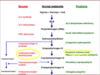
Vitamin D - UV light is taken up and results in the synthesis of vitamin D, which is stored in the liver as hydroxycholecalciferol and converted to 1,25-dihydroxycholecalciferol in the kidney
Thyroid hormone - T4 is converted into T3, partly (20%) in the thyroid gland but the majority is done in peripheral tissues, including the skin
What cells types are found in atopic eczema lesions?
Blocking what substance leads to a reduction in symptoms?
T cells (Th2), DCs, KCs, mast cells and macrophages are all found
Blocking IL-4 results in a reduction of symptoms
What are the two special nerve receptors found in the dermis? What do they sense?
Pacinian corpuscles - sense pressure
Meissner’s corpuscles - sense vibration

What enzyme is deficient in porphyria cutanea tarda?
Diagnosis is based mainly on clinical features, what might these be?
Uroporphyrinogen decarboxylase
Clinical features include blisters and fragility, but also hyperpigmentation, hypertrichosis, solar urticaria and morphoea
What enzyme is deficient in Acute Intermittent Porphyria?
What should be included in the list of differentials?
PBG deaminase, resulting in a build-up of Porphobillinogen (PBG)
Differentials
- acute abdomen
- mononeuritis multiplex
- Guillain-Barre syndrome
- Psychoses
Describe apocrine glands in terms of their distribution, function, sensitivity to hormones and what the produce
Distribution - axillae and perineum
Function - ?
Sensitivity to hormones - sensitive to androgens
Produce - oily fluid that has an odour following bacterial decomposition

What is the difference between disease and illness?
Disease - pathological condition of the body, can be measured and quantified. Illness - the experience of discomfort and suffering, subjective, hard to measure and quantify
Why is the choice of vehicle as important as the choice of drug when considering therapeutic agents in skin disease?
Concentration of the drug and the partition coefficient (“pushing force” of the drug) are highly dependent on the vehicle used
What enzyme is deficient in Erythropoetic protoporphyria?
How does this cause symptoms?
What investigation is best to diagnose this condition?
Ferrochelatase
Leads to a build up of Protoporphyrin IX which reacts with visible light and can damage the endothelium
Porphoryin Plasma Scan
Name some skin appendages
Melanocytes, glands (apocrine, eccrine, sebaceous), arector pili muscles and hair follicles, nails
What are Blashcko Lines?
Developmental growth pattern of skin. If a patient presents with something along these lines the cause is likely congenital
How does dose of a drug effect:
Immunological reactions
Non-immunological reactions?
Immunological - non-dose dependent
Non-immunological - dose dependent, usually resolves upon removal of drug (but half-life and tissue in which the drug has accumulated play a role as well)
CD4+ve cells are Cytotoxic/Helper cells. What are their subsets?
CD8+ve cells are Cytotoxic/Helper cells.
CD4+ve are helper
Th1 = activate macrophages to destroy pathogens
Th2 = Help B cells to make antibody
CD8+ve are cytotoxic and kill pathogens directly
List some of the presentations of Staph aureus infection seen in skin
Carbuncles
Impetigo
Scalded Skin Syndrome
Rash
Abscess
Folliculitis

What is the name of the junction between the dermis and the epidermis? Why is it important?
The dermo-epidermal junction
Plays a key role in epithelial-mesenchymal interactions:
- support, anchorage, adhesion, growth and differentiation of basal cells
- semi-permeable, acting as a barrier and filter
Describe the make-up of different populations of T cells within the epidermis and dermis
Epidermis - mainly CD8+ve T cells
Dermis - both CD4+ve and CD8+ve T cells, also other subsets
Briefly state what each of the following virulence factors do:
- Adhesin
- Invasin
- Impedin
- Aggressin
- Modulin
Adhesin - enables binding of organism to host tissue
Invasin - allows invasion of organism into host tissue
Impedin - allows organism to evade host defences
Aggressin - causes damage to the host directly
Modulin - induces damage to the host indirectly
What cell types make up the epidermis? What is their morphology?
Stratified squamous epithelium. 95% of epidermis is keratinocytes, containing structural keratins. Also melanocytes, Langerhans cells and Merkel cells
Describe theGranular Layer
2-3 layers of flatter cells Contains large keratohyalin granules made up of structural filaggrin and involucres proteins. Also contains Odland bodies (look like tennis rackets). High lipid content Cell nuclei are lost
What are the two important Toxinoses associated with Staph aureus to be aware of?
Toxic Shock
Scalded Skin Syndrome
Is the pilo-sebaceous unit located in the epidermis or the dermis?
Both! Has an epidermal component, and the papilla is located in the dermis

Describe the keratin layer
Made up of corneocytes - overlapping non-nucleated cell remnants Creates an insoluble cornified envelope, forming a tight waterproof barrier 80% keratin and filaggrin
Which Superantigen is particularly associated with Toxic Shock?
TSST-1
Causes a rapid progression (48 hours) of vomiting, diarrhoea, high fever, sore throat and muscle pain
Causes a cytokine storm and over-stimulation of the immune system
The Lancefield classification is used to serotype cell wall carbohydrates and further classify Strep strains.
What is Group A further subdivided according to? What are these divided into?
M protein antigens
M1 and M3 - major serotypes
M3 and M18 - severe invasive disease
Skin diseases are very common and everyone gets them! List some of the most common forms of skin disease
Eczema/dermatitis
Infections - viral/fungal/bacterial
Acne
Skin tumours
Psoriasis
What are the 3 phases of hair growth?
Anagen - growing (90% of hairs), 3-7 years with variable growth patterns
Catagen - involuting (10% of hairs), always 3-4 weeks
Telogen - resting, shedding phase

When describing a lesion’s configuration, what features should be noted?
Discrete? I.e. well demarcated with normal skin surrounding and a clear border
Confluent? Do the lesions join up?
Grouping - linear or annular?
What genetic defect causing problems with the DEJ leads to “mitten deformities’ and fragile skin in infants?
Epidermolysis bullosa
What causes virilism?
Excessive androgen, usually from a tumour
Seen in androgen receptor-controlled regions e.g. lip, chin etc.
What 2 main layers make up the skin, and where do they originate from? When does this happen?
Epidermis - outer layer made up of pseudostratified epithelium. Ectoderm cells form a single layer ‘periderm’, then there is a gradual increase in the layers of cells followed by casting off of the periderm cells. Dermis - inner layer made up of connective tissue. Formed from mesoderm Dermis and Epidermis are formed around day 8 during gastrulation (formation of the germ layers)
What other type of drug eruptions are there?
Urticarial (hives), usually an immediate IgE-mediated (type I) reaction
Pustular/Bullous - acne (e.g. due to steroids, androgens…) or rarely Acute Generalised Exanthematous Pustulosis (AGEP) caused by CCBs, anti-malarials, antibiotics
Fixed drug eruptions - usually well-demarcated round plaques, painful and red and found on the hands, lips, genitals… Associated with tetracycline/doxycycline, paracetamol, NSAIDs, carbamazepine.
What are some of the acute and chronic reactions seen in phototoxic drugs?
What drugs are associated with this?
Acute - skin toxicity, systemic toxicity, photodegradation
Chronic - Pigmentation, photoaging, photocarcinogenesis
Drugs:
- antibiotics (fluoroquinolones, tetracyclines)
- Thiazide diuretics
- Chlorpromazine
- NSAIDs
- Amiodarone
- Porphyrins
- BRAF inhibitors (vemurafenib)
- antifungals
- immunosuppressants
Name some of the important structural proteins found in the keratin layer a.k.a. the stratum corneum
Filaggrin
Involucrin
Keratin
What is alopecia areata?
hair loss due to an autoimmune cause
Briefly describe Langerhans cells
Of mesenchymal origin - created in the bone marrow
Found in the Prickle Cell Layer of the epidermis. Also found in the dermis and in lymph nodes.
Involved in the immune system of the skin. Acts as APCs, pick up antigen in the skin and circulate to lymph nodes.
Contain Birbeck Granules (tennis racket appearance)
What condition with effects seen on the skin develops if there is damage, via infection/trauma/old age etc., to lymphatic vessels?
Chronic lymphodoema
What are the two major organisms found on skin?
Staphylococcus aureus
Streptococcus spp.
(Others are important in IC/IS individuals)
What are the functions of melanocytes? What do they contain?
Melanocytes contain melanosomes and convert tyrosine to melanin pigment
- Eumelanin (brown or black)
- Phaeomelanin (red, yellow)
Melanin absorbs light, melanin caps protect nuclear DNA in basal cells
Name three skin conditions with an auto-immune component
Psoriasis
Vitiligo
SLE
What are the 4 types of hypersensitivity reaction?
Type I - allergic, IgE antibodies are produced by B cells in response to an antigen
Type II - auto-antibodies are produced by the immune system (B cells) due to recruitment of macrophages and dendritic cells
Type III - accumulation of immune complexes (antibody-antigen) that are not completely cleared by the innate response, resulting in inflammation and leucocyte recruitment
Type IV - delayed hypersensitivity, cell-mediated rather than antibody-mediated and involves CD4+ Th1 cells and IL-2 release
What skin infections are associated with Streptococcus pyogenes?
What kind of Strep is this?
Impetigo
Cellulitis
Necrotising fasciitis
Beta-haemolytic - Group A strep
What is the name of the autoimmune condition that occurs at the DEJ and results in the formation of large blisters (bullae)?
Bullous pemphigoid
Type II hypersensitivity reaction with the formation of auto-antibodies to hemi-desmosomes
What makes up a pilosebaceous unit?
Specialised keratins plus an adjacent sebaceous gland that produces sebum. Includes a dermal papilla.

What are Langerhans cells and what characteristic feature do they contain?
They are dendritic cells found within the keratin layer, and contain Birbeck granules, which process lipid antigen and microbial fragments and present them to effector T cells
What are the 3 types of skin glands?
Sebaceous - produces sebum to control moisture loss and protect against infection
Apocrine - sweat gland found in the axillary and genital regions. Androgen-dependent
Eccrine - sweat glands found throughout the body. Have a sympathetic cholinergic nerve supply
Briefly describe Merkel cells
Found in the basal layer between keratinocytes and nerve fibres.
A cancer of these is rare and caused by a viral infection. It has a high mortality rate, is extremely virulent and there is no cure.

Irritant contact dermatitis is an immunological/non-immunological process
Non-immunological
Contact with agents that abrade, irritate and traumatise the skin
Pattern is dependent on exposure
Pemphigus and Pemphigoid are Type _ hypersensitivity reactions
Type II
What cells and fibre types make up the dermis?
Cells - mainly fibroblasts, macrophages, mast cells, lymphocytes, Langerhan’s cells
Fibres - collagen (approx 80-90% of dermal tissue) and elastin
What is the most common form of drug eruption?
Exanthematous (90%)
T-cell mediated delayed type reaction (type IV)
Usually mild and self-limiting. Rash is widespread and symmetrical. Can potentially become very serious
Pruritus and mild fever are common
What severe adverse cutaneous reactions can occur in drug eruption?
- Steven-Johnson Syndrome (SJS)
- Toxic Epidermal Necrolysis (TEN)
- Drug Reaction with Eosinophilia and Systemic Symptoms (DRESS)
- Acute Generalised Exanthematous Pustulosis (AGEP)
Describe the Prickle Cell Layer
Larger polyhedral cells Lots of desomosomes connected to intermediate filaments
What is vitiligo?
Melanocytes are attacked by the immune system, resulting in a loss of melanocytes.
Treated with phototherapy (“melanotan”).
NB - this condition can be treated properly by replacing the melanocytes, but this is ethically tricky as this can lead to development of melanoma
How do keratinocytes function in the innate immune system?
Sense pathogens via cell surface receptors
Can be activated by UV light and sensitisers (e.g. in the case of allergic contact dermatitis)
Produce AMPs that can directly kill pathogens
Release cytokines and chemokines, including IFN gamma, which causes neighbouring cells to swell up
At week 4, the foetal skin is made up of the Periderm, Basal layer and Dermis. What does this change into? When?
Changes into the Keratin layer, Granular layer, Prickle cell Layer and Basal Layer (all epidermis), then the Dermis. Happens at week 16
What is Telogen Effluvium?
Lots of hairs enter the Telogen phase at the same time (usually this is staggered). Most common cause of hair loss, but is also reversible.
What is the name of the skin condition that develops due to overgrowth of blood vessels?
Angioma

Describe nail growth. What is included in the nail matrix and where is it located?
Nail matrix is located underneath the cuticle and contains interspersed melanocytes and stem cells.
Which layer of the skin is the single most important regarding absorption of drugs?
The keratin layer of the epidermis (the stratum corneum)
Describe sebaceous glands in terms of their distribution, function, sensitivity to hormones and what the produce
Distribution - found all over the body, with largest glands being on the face and chest
Function - control of moisture loss and protection from fungal infection
Sensitivity to hormones - yes, quiescent pre-puberty however
Produce - sebum, made up of squalene, wax esters, triglycerides and FFAs
Describe the Basal Layer
Small cuboidal cells, usually one layer thick. Lots of intermediate filaments (keratin) and highly metabolically active. Allows for continuous generation of cells which migrate upwards and differentiate.
What is the name of the rare genetic condition that results in overgrowth of nerve endings/neural tissue and appears as widespread nodules on the skin?
Neurofibromatosis

What Staph aureus toxin is specifically associated with severe skin infections (sepsis, necrotising faciitis) and CA-MRSA?
Panton-Valentine Leukocidin (PVL)
What are the 4 main groups of porphyria?
- Phototoxic skin porphyrias
- Blistering and fragile skin porphyrias
- Acute attack porphyrias
- Severe congenital porphyrias
When describing a lesion’s distribution, what features should be noted?
Sites affected
Symmetrical/unilateral
Local or widespread
What type(s) of T helper cell is associated with the following:
- atopic dermatitis
- psoriasis?
Atopic dermatitis - Th2 and Th17
Psoriasis - Th1 and Th17
What chromosome encodes for MHC?
What are the two types and how do they differ in presentation?
Chromosome 6
MHC I - found on almost all cells, presents endogenous antigen to CTLs
MHC II - found on APCs (B cells, macrophages etc.), presents exogenous antigen to T helper cells
What investigations can be performed if an allergy is suspected?
History - most important
RAST - specific IgE blood test, specificity and sensitivity 70-75%
Skin prick or prick-prick testing - Sens. and spec. 90+%, but small risk of anaphylaxis
Challenge test
Serum mast cell tryptase levels
What is Nelson’s Syndrome?
Melanin-stimulating hormone is produced in excess by the pituitary (presence of a benign tumour), leading to darker skin
What’s the difference between Incidence and Prevalence?
Incidence - the number of new cases of disease, defined as a % of the total population, over a fixed period of time
Prevalence - the total number of cases within a population at any one time
How is psoriasis triggered and blocking what substance leads to a reduction in symptoms?
How do keratinocytes function in psoriasis and what substances to they release?
Triggered by environmental factors in genetically susceptible individuals
Blockage of IL-17 results in imporvement of symptoms (mainly reduction of inflammation)
KCs are under stress, and this causes plasmacytoid DCs (pDCs) to produce IFN gamma, while KCs release IL-1/IL-6 and TNF alpha
When performing a skin examination, what are the 3 features that need to be noted?
Distribution - which areas are affected?
Configuration - how are lesions arranged?
Morphology - what do the lesions look and feel like?
How does albinism occur?
Genetic mutation affecting eumelanin production - absence of required genes leads to a partial loss of pigment production
Describe eccrine glands in terms of their distribution, function and how they are triggered
Distribution - whole skin surface
Function - cooling by evaporation, moistens palms/soles
Triggered by - sympathetic cholinergic nerve supply, activated by mental, thermal and gustatory stimulation
