Upper limb 1 Flashcards
Where is the axilla? And at what junction?
Below the glenohumeral joint - at the junction between the upper limb and thorax
What is so important about the axilla?
It is a passageway through which many neurovascular structures and muscular structures enter the upper limb
What are the 5 borders of the axilla?
- Apex (axillary inlet)
- Lateral
- Medial
- Anterior
- Posterior
What is the apex border?
- Lateral border of the first rib
- Superior border of the scapula
- Posterior border of clavicle
What is the lateral border?
The intertubercular groove of the humerus
What is the medial border?
- Serratus anterior
- Thoracic wall
What is the anterior border?
- Pec major
- Pec minor
- Subclavius
What is the posterior border?
- Subscapularis
- Teres major
- Lat dorsi
How does the size and shape of axilla vary with the degree of the abduction of the shoulder?
Apex decreases in size when arm is fully abducted, leading to risk of compression of contents of axilla
What are the contents of the axilla?
- Axillary artery and its branches
- Axillary vein and its tributaries
- Brachial plexus
- Axillary lymph nodes
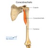
- Short head of biceps brachii and coracobrachialis (pass through the axilla after originating at coracoid process)
How many routes are there for structures to move through in the axilla?
3
What is the main route of passage in the axilla?
Inferolaterally, into the upper limb
What is the next passageway in the axilla?
Quadrangular space
What is the third passageway in the axilla?
Clavipectoral triangle
What is the quadrangular space?
The gap in the posterior wall of the axilla leading to the posterior arm and shoulder. It is made up of up:
- Teres minor
- Teres major
- Long head of biceps brachii
Nerves: axillary nerve
Artery: circumflex humeral artery
What is the clavipectoral triangle?
Opening in the anterior wall of axilla. Bounded by
- pec major
- deltoid
- clavicle
Special structures: medial and lateral pectoral nerves (leave the triangle)
-Cephalic nerve enters the triangle (along deltoid)
What is thoracic outlet syndrome? What are the common causes? What is the presentation
Compression of vessels and nerves between bones at the apex of the axilla.
- trauma
- repetitive movements
- cervical rib (from 7th vertebrae)
Presentation: tingling, muscle weakness and discolouration
What is the relevance of lymph node biopsy?
- 75% of lymph from the breast drains into axilla lymph nodes so can be biopsied for breast cancer
- remove axillary lymph nodes of cancer in detected (axillary clearance) - but can damage long thoracic nerve leading to winged scapula (paralysis of serratus anterior)
What are the anterior axillary folds, what are the posterior axillary folds? What is the midaxillary line?
- Anterior axillary fold = formed by lateral border of lat dorsi
- Posterior axillary fold = formed by lat dorsi and teres major
- Mid-axillary line = between the anterior and posterior axillary folds
What is the cubital fossa? What does it look like?
The transition from the arm to the forearm
-depression on the anterior surface of elbow joint
How many borders are there in the cubital fossa?
5
- Lateral
- Medial
- Superior
- Floor
- Roof
What is the lateral border of the cubital fossa?
Medial border of brachioradialis
What is the medial border of the cubital fossa?
Lateral border of pronator teres
What is the superior border of the cubital fossa?
A line between the two epicondyles of the humerus
What is the floor of the cubital fossa?
Brachialis and supinator
What is the roof of the cubital fossa?
- Skin
- Fascia
- Bicipital aponeurosis (aponeurosis of biceps tendon)
Where is the medial cubital vein within the cubital fossa?
Roof
What are the contents in the cubital fossa (lateral to medial)?
What is the mnemonic for it?
- Radial nerve (under brachioradialis, dividing into deep and superficial branches)
- Biceps tendon (runs through fossa and attaches to radial tuberosity)
- Brachial artery (bifurcates into radial and ulnar arteries)
- Median nerve (between the two heads of the pronator teres)
(also have radial and ulnar veins)
REALLY NEED BEER TO BE AT MY NICEST
How do you measure blood pressure?
Brachial artery –> brachial pulse. Medial to the biceps tendon
What is venepuncture?
Taking blood from median cubital vein (joining of basilic and cephalic vein)
What is a supracondylar fracture?
What can the damage cause?
What condition can it cause?
- Fracture to humerus
- Transverse fracture spanning both epicondyles
- May damage contents of cubital fossa
- The damage and swelling can interfere blood flow of the brachial artery
- It can cause Volkmann’s ischaemic contracture
What is Volksmann’s ischaemic contracture?
- Uncontrolled flexion of hand as flexor muscles become fibrotic and short
- Damage to median and radial nerve
What is the carpal tunnel?
What does it serve as?
The narrow passageway on the anterior part of the wrist.
It serves as an entrance to the palm for 9 tendons and the median nerve
What are the two layers of the carpal tunnel?
1) deep carpal arch
2) superficial flexor retinaculum
What is the deep carpal arch?
- It is concave and forms the base and sides of the carpal tunnel.
- Formed laterally by scaphoid and trapezium
- Formed medially by hook of hamate and pisiform
What is the superficial flexor retinaculum?
Thick connective tissue that forms the roof of the carpal tunnel
-Spans hook of hamate and pisiform to scaphoid and trapeziun
What are the 9 tendons in the carpal tunnel?
- Flexor pollicus longus
- Flexor digitorum profundus (x4)
- Flexor digitorum superficialis (x4)
What are the sheaths surrounding the tendons in carpal tunnel?
The 8 tendons are surrounded by a single synovial sheath
-Flexor pollicus longus has its own synovial sheath which allows free movement of the tendon
Tell me about the median nerve in the carpal tunnel
- Palmar cutaneous branch of the median nerve branches before the nerve enters carpal tunnel (so travels superficially). This nerve supplies central palm (sensory)
- After median nerve enters carpal tunnel, it gives rise to recurrent branches and palmar digital branches of the median nerve
What does the palmar digital branch of the median nerve supply?
- Sensory innervation to palmar skin (laterla 3.5 digits)
- sensory innervation to dorsal nail of lateral 3.5 digits
What do the recurrent branches of the median nerve supply?
Supplies the thenar muscles
What is carpal tunnel syndrome?
What is it caused by?
What does it cause on the thenar muscles?
What are the clinical features?
- Compression of median nerve within carpal tunnel
- mononeuropathy caused by thickened ligaments and tendon sheaths
- can cause weakness and atrophy of thenar muscles
- Clinical features: numbness, tingling, pain.
Worse in the mornings
How to test for carpal tunnel syndrome?
- Tinel’s sign (tapping the nap in the carpal tunnel to elicit pain)
- Phalen’s manoeuvre (holding the wrist in flexion for 30 seconds to elicit numbness/pain)
How do you treat carpal tunnel syndrome?
- Splint
- Corticosteroids to reduce swelling of connective tissue
- Surgical decompression in severe cases
What is the anatomical snuffbox (radial fossa)?
Where is it?
Triangular depression on the lateral aspect of the dorsum of the hand.
It is on the same level as the carpal bones
What are the borders of the anatomical snuffbox?
Ulnar = extensor pollicus longus
Radial = extensor pollicus brevis and abductor longus tendons
Proximal = radial styloid process
Floor = scaphoid and trapezium
Roof = skin
What muscle makes up the ulnar border of the anatomical snuffbox?
Extensor pollicus longus
What is the radial border of the anatomical snuffbox?
Extensor pollicus brevis
Abductor pollicus longus (closer to the thumb than extensor pollicus brevis)
What are the 3 contents of the anatomical snuffbox?
- Radial artery = travels between the heads of the adductor pollicus muscle
- Superficial branch of radial nerve = found in skin and subcutaneous tissue of anatomical snuffbox. innervates dorsal surface of 3.5 digits and the associated area on the back of the hand
- Cephalic vein = arises from dorsal venous network
What is a scaphoid fracture?
What is it caused by?
What are the clinical features?
- Fracture of scaphoid bone (most commonly fractured carpal bone)
- Normally caused by falling on an outstretched hand
- Clinical features = pain and tenderness of anatomical snuffbox
Why is the scaphoid at risk of avascular necrosis after fracture?
- It has a retrograde blood supply which means the blood flow enters from the distal end to the proximal end
- Hence, fracture to the middle/waist of this bone can interrupt blood flow to the proximal part of the scaphoid, rendering it avascular
What are the ligaments surrounding humerus and scapula?
- Coracoacromial liagment
- Coracohumeral ligament
- Glenohumeral ligament
- Transverse humeral ligament