Unit 2: Forelimb 1&2 Flashcards

1
Q

in most species, the _____ cartilage increases the surface area for attachment of the rhomboideus m. and part of serratus ventralis m.

A

in most species, the scapular cartilage increases the surface area for attachment of the rhomboideus m. and part of serratus ventralis m.

2
Q

in the horse, the _________ lies within the intertubercular groove of the humerus and stabilizes the biceps brachii m.

A

in the horse, the intermediate tubercle lies within the intertubercular groove of the humerus and stabilizes the biceps brachii m.

3
Q

two heads of the deltoideus m. in the ox

A
  • acromion head
  • spinous head
4
Q

in the horse, the thoracolumbar fascia continues cranially in the serratus ventralis m. as the ______

A

in the horse, the thoracolumbar fascia continues cranially in the serratus ventralis m.​ as the dorsoscapular ligament

5
Q

the ____ bursa is located over the caudal part of the greater tubercle of the humerus and doesnt communicate with the shoulder joint capsule

A

the infraspinatus bursa is located over the caudal part of the greater tubercle of the humerus and doesnt communicate with the shoulder joint capsule

6
Q

the _____/_____ bursa protects the proximal tendon of the biceps brachii m. and is separate from the joint capsule of the shoulder.

A

the intertubercular/bicipital bursa protects the proximal tendon of the biceps brachii m.​ and is separate from the joint capsule of the shoulder.

7
Q

list the three attachments of the biceps brachii m. in the horse (1 proximal and 2 distal)

A
  • supraglenoid tubercle of the scapula
  • radial tuberosity (on proximal, medial aspect of the radius)
  • metacarpal tuberosity (on cranial, proximal surface of 3rd metacarpal bone)
8
Q

______ is the long tendon of the biceps brachii m. and connects the fibrous core of the muscle to the CT of the extensor carpi radialis m.

A

lacertus fibrosus is the long tendon of the biceps brachii m. and connects the fibrous core of the muscle to the CT of the extensor carpi radialis m.

9
Q

in the horse, the _____ is entirely fibrous and also called “the long part of the medial collateral ligament”

A

pronator teres

10
Q

the ______ is the thick fibrous CT at the palmar aspect of the carpus that the carpal bones are embedded in. it prevents hyperexension.

A

palmar carpal ligament

11
Q

____ are remnants of carpal and tarsal pads in the horse

A

chestnuts

12
Q

distal attachements of the superficial digital flexor m.

ox:

horse:

A

distal attachements of the superficial digital flexor m.

ox: P2 only
horse: distal P1 and proximal P2

13
Q

attachements of the deep digital flexor m. in both ox and horse

A

P3

14
Q

the ____ of the metacarpophalangeal (fetlock) joint acts as a bursa for the common digital extensor

A

dorsal pouch

15
Q

____ is the remnant of the metacarpal pad in the horse

A

ergot

16
Q

at its distal border, the _____/_____ anchors the navicular bone to P3

A

distal navicular ligament/impar ligament

17
Q

the _______ is between the DDFT and navicular bone and is separate from the coffin joint capsule

A

navicular bursa

18
Q

the _____ are located between the axial surfaces of the proximal phalanges and at the distal interphalangeal joints and prevent splaying of the digits

A

interdigital ligaments

19
Q

Suprascapular n.

functional group:

muscles:

A

Suprascapular n.

functional group: lateral stabilizers of the shoulder

muscles: supraspinatus, infraspinatus

20
Q

subscapular n.

functional group:

muscles:

A

Subscapular n.

functional group: medial stabilizers of the shoulder

muscles: subscapularis

21
Q

Musculocutaneous n.

functional group:

muscles:

A

Musculocutaneous n.

functional group: flexors of the elbow joint

muscles: biceps brachii, brachialis, coracobrachialis

22
Q

Axillary n.

functional group:

muscles:

A

Axillary n.

functional group: flexors of the shoulder joint

muscles: deltoideus, teres major

23
Q

Thoracodorsal n.

functional group:

muscles:

A

Thoracodorsal n.

functional group: shoulder flexor

muscles: latissimus dorsi

24
Q

Radial n.

functional group:

muscles:

A

Radial n.

functional group: extensors of elbow, carpus, and digits

muscles: triceps, tensor fasciae antebrachii, ECLU

25
Q

Median n.

functional group:

muscles:

A

Median n.

functional group: flexors of carpus and digits

muscles: flexor carpi radialis, pronator teres (ox), SDF, DDF

26
Q

Ulnar n.

functional group:

muscles:

A

Ulnar n.

functional group: flexors of carpus and digits

muscles: flexor carpi ulnaris, DDF

27
Q

what muscle is essential for getting an animal up from a recumbent position and maintaining it during locomotion

A

triceps brachii

28
Q

list the seven components of the stay apparatus

A

1: triceps muscle
2: dorsoscapular ligament
3: biceps muscle
4: collateral ligaments of the elbow
5: palmar carpal ligament
6: SDF and DDF tendons and check ligaments
7: suspensory apparatus

29
Q

three components of the suspensory apparatus

A

1: interosseus (ox) or suspensory ligament (horse)
2: proximal sesamoids
3: distal sesamoidean ligaments

30
Q

what are the four distal sesamoidean ligaments

A
  • straight
  • oblique
  • cruciate
  • intersesamoidean
31
Q

list the four common nerve blocks of equine distal forelimb from proximal to distal

A
  • high 4-point nerve block
  • low 4-point nerve block
  • abaxial sesamoidean nerve block
  • palmar digital nerve block
32
Q

palmar digital nerve block (horses)

blocks:

doesnt block:

A

palmar digital nerve block (horses)

blocks: caudal 2/3 of the foot

doesnt block: the dorsal hoof wall or the entire coffin joint

33
Q

abaxial sesamoidean nerve block (horses)

blocks:

clinically considered:

A

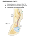
abaxial sesamoidean nerve block (horses)

blocks: entire foot and all of P2, distal sesamoidean ligaments

clinically considered: an imprecise block

34
Q

low 4-point nerve block (horse)

blocks:

clinically considered:

landmark:

A

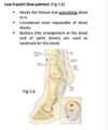
low 4-point nerve block (horse)

blocks: the fetlock and everything distal to it

clinically considered: the most repeatable of distal blocks

landmark: buttons

35
Q

high 4-point nerve block (horse)

blocks:

A

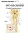
high 4-point nerve block (horse)

blocks: all palmar structures distal to the carpus (entire suspensory apparatus, splint bones, flexor tensons and sheath, distal check ligament)

36
Q

describe how to preform a Bier nerve block on an ox

A

first, apply a tourniquet above the fetlock. then, inject anesthetic (such as lidocaine) into a visible vein (usually the dorsal common digital vein III). the anesthetic diffuses into the region. the tourniquet stays in place during the procedure but must be removed by 90 minutes