Tooth Development Flashcards

1
Q

Growth

A
  1. An increase in size or number of cells in the whole or any part of the organism
    1. E.g. muscle tissue growth, fat tissue growth, bone growth
2
Q

Proliferation

A
  1. An increase in the number of cells as a result of cell division
3
Q

Differentiation

A
  1. The process of achieving a stable different phenotype
4
Q

Cytodifferentiation

A
  1. complex process by which a cell or cell line attains and expresses a stable phenotype
  2. Cytodifferentiation usually occurs over the course of several generations with cells expressing intermediate phenotypes
  3. Here, the differentiation is the increase in # of cells (of different types)
  4. Once they are committed, they go through the entire differentiation steps; along the way, some will go through apoptosis, a very natural & important events for proper development
  5. Mesenchymal cell divide very slowly because every division lead to mutation; the older the person, the more accumulation of these mutations –> potential for cancer
5
Q

Commitment

A
  1. The commitment of cells to specific cell fates and their capacity to differentiate into particular kinds of cells. Committed, or determined, cell develops along a certain pathway and is not susceptible to other influences.
6
Q

Potentiality

A
  1. Potentiality: a capacity within a cell that is not yet recognized. Differentiation leads to loss of potentiality
  2. Pluripotent: the capacity to differentiate along a variety of lines
  3. Totipotent: capacity to reproduce and differentiate into an entire multicellular organism (i.e. embryonic stem cells that can differentiate into anything).
7
Q

Competence

A
  1. the ability to differentiate along a certain line
8
Q

Modulation

A
  1. the process by which a cell becomes reversibly different in physical form. The cells are able to revert to their previous form i.e., modulat between forms
    1. e.g. ameloblasts modulate back and forth
9
Q

Expressed genes

A
  1. those genes which are actively transcribed
10
Q

Polygenic control

A
  1. aspects of embryogenesis that require multiple sequential gene function (e.g. limb development, tooth development, brain development, facial morphogenesis.
11
Q

Induction

A
  1. The action of physical or chemical factor which causes cell or tissue differentiation
  2. Differentiation of ectoderm which in turn signal mesenchymal cells go through a cycle between the two inducing each other into further development via signaling factor, growth factors, etc.
12
Q

Morphogenesis

A
  1. Morphogenesis: a change in shape or location of an organ or a tissue
  2. Morpho-differentiation: a change in shape of a developing organ due to morphogenetic movements or differential growth
  3. Morphogenetic movements: change in location of cells during development, e.g. gastrulation, neurulation, neural crest migration, etc.
13
Q

Patterning

A
  1. The establishment of a programmed subset of cells in proper relation to each other and to surrounding tissues, e.g. shaping of bones and muscles on limbs, positioning of specific tooth types
  2. Involves a number of induction sites spatially and temporarily localized
14
Q

Congenital malformations

A
  1. abnormalities resulting from errors arising during development
    1. e.g. cleft lip and pallet
    2. different types of oligodontia
15
Q

Ectomesenchymal cells

A
  1. Mesenchyme of the Neural Crest origin
  2. Ectomesenchymal cells give rise to:
    1. Fibroblasts
    2. Odontoblasts
    3. Cementoblasts
    4. Osteoblasts
    5. Chondroblasts
  3. Migation of the Neural Crest cells is critical for craniofacial development
    1. Maxillary and mandible all arise from the neural crest cells
16
Q

Signaling molecules (SP) and Transcription factors (TF)

A
  1. Signaling molecules/peptide:
    1. extracellular molecules that can modify cell metabolism, gene expression, structural organization and other parameters.
  2. Transcription factors:
    1. proteins that bind to specific DNA sequences, and regulate transcription of genetic information from DNA to mRNA
  3. Homeobox genes:
    1. a homeobox is a DNA sequence found within genes that are involved in the regulation of patterns of morphogenesis. Homeobox genes encode transcription factors that typically switch on cascades of other genes; complex of regulatory genes that have instructions for differentiation of particular cells
17
Q

Morphogenetic processes in odontogenesis

A
  1. Formation of primary epithelial bands and dental lamina
  2. Formation of dental placodes (regionalization of oral and dental ectoderm)
  3. Tooth type determination
  4. Tooth patterning
  5. These processes are controlled by cell signaling
18
Q

Animal models

A
  1. Information about tooth development is primarily obtained from animal models (mice, rats, rabbits); altough some data is generated from studies of human mutations
  2. Gene knockouts- allow to study functions of individual genes in morphogenesis
  3. Recombination experiments- recombination of different tissues (i.e. skin epithelium and dental mesenchyme) allow to reconstruct the spatial and temporal patterns of induction
19
Q

Epithelial-mesenchymal interactions

A
  1. Epithelial-mesenchymal interactions are essential for tooth development as it is required for proper series of reiterative induction events in odontogenesi
  2. Epithelial-mesenchymal interactions are not unique to tooth development as it is also found in the following:
    1. Lung
    2. Hair
    3. Kidney
    4. Mammary gland
  3. Epithelial and mesenchymal cells are very closely related and their interactions can be observed in many different development processes other than the tooth development; back and forth between epithelium and mesenchyme –> ectomesenchymal
  4. Tissue recombination experiments
    1. Provide a window into the induction processes during tooth formation
20
Q

Stages of tooth development in mouse

A
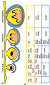
  1. Thickening/placode (E11.5)
  2. Bud (E13.5)
  3. Cap (E14.5)
  4. Bell (E18.5)
  5. Erupted tooth (P35)
  • Tooth development is a tightly controlled process with numerous factors contributing
21
Q

Reciprocal epithelial-mesenchymal interactions

A
  1. Epithelium at early stages provides instructions in epithelial-mesenchymal recombination:
    1. Epithelium at the dental lamina stage (E10 or slightly earlier in mice) induced tooth formation when combined with non-dental mesenchyme
    2. This experiment id not work if the epithelium was from E12 or later fetal mice
  2. Intraocular recombination of neural crest cells and dental epithelium
    1. Tooth formed from the combination of neural crest cells expanded from the neural folds and mandibular epthelium but NOT from combination with limb bud epithelium
    2. This indicates that tooth formation is initiated by factors residing in the oral epithelium
22
Q

Epithelial-mesenchymal interactions recombination experiments (past Day 12)

A
  1. Combination of molar epidermis with scalp mesoderm of papilum lead to the formation of hair
  2. Combination of scalp epidermis with molar mesodermal papilum lead to the formation of tooth
23
Q

Summary of recombination experiments

A
  1. The epithelium provides the first “set of instructions” at the stage prior to the presence of a recognizable bud (i.e., the placode or epithelial thickening stage).
  2. Mesenchyme is not predetermined at the early stages of tooth development.
  3. Mesenchyme directs later stages of tooth development
24
Q

Epithelial-mesenchyme interactions (cont.)

A
  1. Tooth buds can be micro-dissected into epithelial and mesenchymal components; enamel organ (epithelium) and the dental papila (mesenchyme)
  2. Prior to overt differentiation and when cultured separately, neither component will form dental structures by itself
  3. This indicates that cooperation of both tissues is essential for proper development
25
Q

Recombination of dental epithelium and ectomesenchyme at bud or cap stage (Day 13 or later)

A
  1. Incisor epithelium combined with molar papilla results in a molariform tooth
  2. Molar epithelium combined with incisor papilla results in an incisiform tooth
  3. Non-dental epithelium combined with a dental papilla results in tooth formation with the formation of an enamel organ and tooth type is determined by the mesenchyme (papilla)
    1. The opposite recombination does not form tooth
26
Q

Strategies for tooth regeneration

A
  1. Collect patient’s somatic cells
  2. Introduce reprogramming factors
  3. Patient specific iPS cell line
  4. Ectodermal epithelial cells & neural-crest derived mesenchymal cells
  5. Ameloblasts & Odontoblasts, cementoblasts, dental pulp cells, osteoblasts, etc.
  6. Recombination
  7. Transplantation
  8. Tooth regeneration
27
Q

Epithelial band

A
  1. Formation of primary epithelial band determines the region of the oral cavity where teeth will form
  2. Epithelial band forms by change in the direction of cell divisions, which lead to thickening of the epithelium; in the beginning it only divides horizontally but then later in various directions, leading to thickening
  3. The earliest stage of tooth development is recognized as an epithelial thickening followed by ectomesenchymal condensation
  4. 1st iteration: primary band signaling leads to the formation of dental lamina by recruitment of the mesenchymal cells
  5. Dental and Vestibular Lamina orginate from the primary band
  6. Pitx2 (TF) and Sonic Hedgehog (SP) are associated with the early differentiation of dental lamina
  7. Expression of Shh (SP) in the ectoderm induces expression of Ptc (receptor) and Gli (TF) in the underlying ectomesenchyme
  8. Reciprocal interactions between Shh (SP) and Wnt7b (SP) determine the position of dental lamina
    1. Shh is expressed where dental lamina wil form whereas Wnt7b is expressed everywhere except where dental lamina will form (i.e. inhibitory)
  9. Signals from dental lamina recruit ectomesenchymal cells which lead to formation of dental placode)
28
Q

Tooth bud

A
  1. Initiation of the tooth germ by interplay between FGF8 (SP) and BMP 2/4 (SP), via activation (FGF8) and inhibition (BMP 2/4) of Pax9 (TF) expression–> determination of the tooth bud positions.
    1. Example of polygenetic control; expression of Pax9 is ectomesenchymally determined where the tooth bud will form.
  2. Bud stage is when determination of tooth types occur
  3. The balance between stimulatory (FGFs, Wnts) and inhibitory signals (BMPs) is important in determining the site of placodes. Formation and growth of placodes is believed to involve the transcription factor p63, TNF, and ectodysplasin (Eda), among others.
  4. Smaller-than-normal placodes lead to missing and smaller teeth, whereas larger placodes induce supernumerary and larger teeth.
29
Q

Tooth Type Determination

A
  1. Tooth type determined by ectomesenchyme (which itself is induced by primary epithelial band
  2. Gradients of expression of different transcription factors and signaling molecules; these gradietns in the end determine which teeth form at where
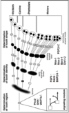
  3. Tooth type determination (field model) : interplay between Dlx1/2, Msx-1, Msx-2, and Barx-1 (all are TF’s)
    1. Differential expression of the transcription factors and signaling molecules along the mandible results in the formation of different tooth types at different locations
    2. Absence of Dlx-1/2 leads to absence of maxillary molars
    3. Overexpression of Barx-1 leads to too many molars
    4. Msx-1 & -2 are required for incisor development
  4. Expression of Barx-1 overlaps with Dlx-1 and Dlx-2,
    and corresponds closely to ectomesenchymal cells that will develop into molars in mice
  5. Two hypothetical models have been proposed to explain how these different shapes are determined, and evidence exists to support both.
    1. The first is the field model, which proposes that the factors responsible for tooth shape reside within the ectomesenchyme in distinct graded and overlapping fields for each tooth family
    2. The clone model proposes that each tooth class is derived from a clone of ectomesenchymal cells programmed by epithelium to produce teeth of a given pattern
30
Q

Dental and Vestibular Lamina

A
  1. Vestibular lamina splits to form the oral vestibule
  2. Primary teeth and permanent molars arise from the lateral dental lamina
  3. Permanent incisors, canines, and premolars arise from successional lamina
31
Q

Bud to Cap Transition

A
  1. Condensation of ectomesenchyme
    1. Cell proliferation without ECM synthesis; formation of dental papilla and dental follicle during cap stage
  2. Formation of the enamel organ
    1. A chamber at the end of the dental lamina; invagination enclosing forming tooth tissues
  3. Vestibular lamina also present during early cap stage of development
32
Q

Cap Stage

A
  1. Cells commit to major dental tissues at the cap stage i.e. histodifferentiation
  2. Inner enamel epithelium:
    1. Ameloblasts
    2. Stratum Intermedium
  3. Dental Papilla
    1. Dentin
    2. Pulp
  4. Dental Follicle:
    1. Tooth supporting tissues i.e. cementum
    2. Fibroblasts of PDL
  5. Stellate (star-like) Reticulum:
    1. A temporary structure containing stellate cells connected via long processes, with GAG molecules in between, later on disappears; keeping the shape up
    2. Glycosaminoglycans are hydrophilic and so pull water into the enamel organ. The increasing amount of fluid increases the volume of the extracellular compartment of the enamel organ, and the central cells are forced apart. Because they retain connections with each other through their desmosomal contacts, they become star-shaped
  6. Enamel knot is also seen at this stage
33
Q

Formation of the permanent tooth germs

A
  1. Occurs during the late cap stage
  2. The accessional lamina arises by posterior growth of the general dental lamina; it gives rise to the molar teeth
  3. Premolars arise from successional lamina of the primary molars (primary dentition)
34
Q

Bell Stage

A
  1. Deepening of the enamel organ due to the cell proliferation at the innter-outer enamel junction (cervical loop)
  2. Dental papilla determines the shape of the cervical loop?
  3. Cervical loop is an important “histological landmark” for distinguishing between cap and bell stages
  4. Stratum intermedium is a layer of epithelial cells between stellate reticulum and inner enamel epithelium, providing O2 and nutrition to ameloblasts during the matrix deposition phase
  5. Stratification of the enamel organ takes place in this stage
    1. Cells of the inner enamel epithelium polarize and adopt palisade morphology, with their nuclei moving into the apical portions away from the cells of dental papilla
    2. Basal lamina is formed between the inner enamel epithelium and dental papilla
    3. The stratum intermedium becomes apparent
    4. Differentiation of the outer cell layer of dental papilla into preodontoblasts takes place.
    5. The bulk of the dental papilla becomes pulp when the first calcified matrix appears at the cuspal tip of the bell stage tooth germ
35
Q

Enamel knot

A
  1. Organizing center for crown morphogenesis (patterning)
  2. Tooth shape is largely determined by a number and position of enamel knots (late cap and bell stages)
  3. FGFs are expressed in non-proliferating cells of the enamel knot, lacking FGF receptors, while other cells express FGF receptors
  4. Enamel knots differentiate at the cap stage
  5. Epithelial cells in enamel knots do NOT proliferate
  6. Cyclin-dependent kinase inhibitor p21 arrests cell proliferation in enamel knot
  7. p21 expression is induced by BMP4 expression in ectomesenchyme
  8. The expression pattern of the enamel knot is highly specific
    1. FGF4, SHH, and BMP2 is highly expressed on the entirety of the enamel knot
    2. cell proliferations occurs throughout except the enamel knot
  9. Secondary Enamel Knots
    1. appear over the tips of the developing cusps in the early bell stage of tooth development. They express growth factors and are organizing centers for cusp development
36
Q

Enamel knots (cont.)

A
  1. Primary and secondary enamel knots are associated with crown patterning
  2. Gradient of FGF leads to uneven cell proliferations; cells closest to the knot divide more actively
  3. Disruptors of the enamel knot formation Beta-Catenin
    1. Experiments with beta-catenin conditional KO show that lack of this protein causes changes in the enamel organ formation
  4. Supernumerary enamel knots form in the conditional B-catenin -/+ embryonic molars
  5. One molar tooth bud of a conditional B-cat -/+ embryo gives rise to dozens of misformed teeth
37
Q

Tooth fate determination

A
38
Q

Late Bell Stage

A
  1. The general dental and lateral laminae close thus sealing the tooth germ from oral epithelium
  2. The cells of cervical loop continue to proliferate, shaping the final crown pattern (outlined by the inner enamel epithelium cells
  3. The volume of stellate reticulum decreases
  4. The dental follicle becomes more defined
  5. At the end of the bell stage, appositional matrix deposition begins
39
Q

Bone-tooth interface

A
  1. Reciprocal signaling between forming tooth and bone is essential for the proper bone-tooth interface formation
  2. OPG, RANK, and RANKL in tooth development; coordination of odontogenesis and osteogenesis
40
Q

Appositional growth

A
  1. Appositional growth = matrix formation
  2. Basal membrane (lamina) is degraded before matrix secretion
  3. The next step in the development of the tooth is terminal differentiation of ameloblasts and odontoblasts and formation of the two principal hard tissues of the tooth: the dentin (that specialized hard connective tissue forming the bulk of the tooth) and the enamel, a process called histodifferentiation.
  4. At this stage we see:
    1. Preodontoblast & Odontoblast
    2. Preameloblast, Presecretory Ameloblast & Secretory Ameloblast
  5. Cells of the enamel organ and ental papilla move in opposite directions as matrix is deposited from odontoblast and secretory ameloblast. During this stage, the two dental matrices of the crown are being formed and the DEJ is established.
  6. Collagenous matrix of dentin is deposited FIRST and is mineralized at a distance from odontoblasts; predentin vs. dentin
  7. Dentin matrix deposition and mineralization precedes enamel matrix deposition and mineralization
  8. The primary dental lamina is loosing its connection with the oral epithelium. Apposition of mineralizaed matrices is taking place and a secondary (permanent) tooth is developing from the successional lamina
  9. Enamel organ, dental follicle and dental lamina collapse at the late stages of tooth development
    1. These structures play important roles in the eruption process
    2. As eruptive movement begins, the enamel of the
      crown still is covered by a layer of ameloblasts and remnants of the other three layers of the enamel organ. These are sometimes difficult to distinguish, and together the ameloblasts and adjacent cells form the reduced enamel epithelium
41
Q

Root formation

A
  1. Hertwig’s epithelial root sheath
    1. HERS is the organizing center for root formation
    2. HERS is the extension of the cervical loop, which continues to proliferate; however, the inner enamel epithelium cells do NOT differentiate into ameloblasts
    3. HERS induces differentiation of root odontoblasts from the cells of dental papilla and root dentin formation
  2. Cervical fold is the induction site; proliferation of cells and become epithelial rooth sheath and they become organizing structure of root formation
  3. Cells of the cervical loop continue to proliferate but do not differentiate
  4. HERS eventually fragments allowing mesenchymal cells of dental follicle to migrate toward dentin and form cementum and PDL; fragmented HERS are called epithelial rests of Malassez (ERM).
    1. PDL and cementum tissues originate from the dental follicle that slipped in
    2. ERM are remains of HERS, located directly adjacent to cementum
  5. Cell proliferation continues at the inner margin of the HERS aka epithelial diaphragm untill the full length of the root is completed, then it turns inward and forms apical foramen
  6. As the inner epithelial cells of the root sheath progressively enclose more and more of the expanding dental pulp, they initiate the differentiation of odontoblasts from ectomesenchymal cells at the periphery of the pulp, facing the root sheath. These cells eventually form the dentin of the root. In this way a single-rooted tooth is formed.
  7. Formation of multi-rooted teeth occurs by the fusion of HERS folds in the plane of the primary apical foramen
42
Q

Grand summary of tooth development

A
43
Q

Congenital disorders and tooth development

A
  1. Oligodontia caused by frameshift mutation in Pax9 gene
  2. In humans, heterozygous loss of function of either MSX1
    or PAX9 causes oligodontia
  3. They regulate reciprocal signals acting back on epithelium and regulating ename knot formation and epithelial proliferation. Knocking out any one of the three genes in mice arrests tooth development at the
    bud stage.