Thermal injury (all; including extravasation, XRT) Flashcards

1
Q

Outline principles for management of hand burns

A
  1. Initial assessment and management of total patient / total burn following ATLS/ABLS protocol including tetanus
  2. Proper evaluation of depth and extent of burn wound
  3. Consideration for prophylactic (therapeutic) escharotomies
  4. Prevention of infection with moist, anti-microbial dressings (ex: SSD)
  5. Early wound closure
    1. for superficial burns likely to close in 2-2.5 weeks, as above
    2. aim for excision and grafting for burns unlikely to heal in 2-2.5 weeks (all deep partial and full thickness, some intermediate thickness)
  6. Use sheet grafts when surgery is necessary
  7. Prevent stiffness and maintain/ regain ROM
    1. pre-operatively and immediately for non-operative
    2. at POD5 for patients having excision and grafting
    3. using “intrinsic plus” position of safety to prevent claw hand
  8. outpatient rehab including OT/PT and silicone therapy, pressure garments
  9. Reconstructive surgery for scars, contracture, claw hand, syndactyly @ 12 mos
2
Q

list criteria for prophylactic escharotomy

A
  1. circumferential or near-circumferential deep partial or full thickness burns
  2. extensive burns likely to require major fluid resuscitation and limb/digit at risk (at least partially, nearly circumferential)
  3. unable to evaluate patient (confounding factors like hypothermia)
3
Q

discuss how and where you will do an escharotomy on an upper extremity

A
  • Technical points
    • in tub room
    • use betadine prep
    • 15 blade or needle-tip cautery
    • just through dermis, just proximal and distal to extent of burn
  • Arm and forearm:
    • medial incision in antebrachial groove, ANTERIOR to elbow, along ulnar border
    • lateral incision lateral arm to radial border, be aware of sensory radial nerve branches
  • hand:
    • 2 dorsal interMC (btwn 4,5 and 2,3)
    • thenar (radial 1st MC) and hypothenar (ulnar 5th MC)
  • Digits
    • 1,5 radial border
    • 2,3,4 ulnar border
4
Q

what is approach to upper extremity fasciotomy?

A
  • most common to release is volar (Rowland’s)
    • curvilinear incision along volar forearm
    • leaves radially based flap to cover median nerve and brachial artery at AC fossa
    • leaves radially based flap to cover median nerve at wrist
    • release guyon’s canal and carpal tunnel
  • Dorsal incision is dorsal longitudeinal to release mobile wad and extensor compartment
  • Hand: incisions are same as for fasciotomy
    • 2 dorsal incisions: d/v i/o, adductor
    • thenar and hypothenar
5
Q

when would you consider fasciotomy in burn patient? what is another consideration against fasciotomy?

A
  • may want to consider fasciotomy when
    • late escharotomy
    • clinical suspision
      • pain out of proportion
      • pulseless limb despite adequate resuscitation
      • sensory deficit
      • non-resolving myoglobinuria or lactate
    • limb ischemia > 2 hrs
    • large volume resuscitation expected over short time
  • consideration against fasciotomy is taking a sterile wound bed and opening it, exposing it to risk of infection from open wounds, lungs,
6
Q

List common sequellae of hand burn

A
  • all suffer from edema, stiffness, pain
  • Hypertrophic scar / scar contracture
    • general hypertrophic scars
    • D5 abduction contracture
    • dorsal skin contracture (extension postures)
    • volar skin contract (flexion postures)
    • wrist, elbow, axillary contracture
  • Webspace contracture
    • syndactyly
    • thumb adduction contracture
    • webspace adduction contracture
  • Claw Hand
    • MCP hyper/extension
    • PIP (DIP) flexion
    • Boutonneire
    • “intrinsic minus” posture
    • extensor tendon adhesions
  • Other
    • Compression neuropathy (MN, UN, 10% of burns > 20% tbsa)
    • heterotopic ossification
    • gangrene, amputation
7
Q

List and discuss advantages / disadvantages of different flaps that can be used for burn scar reconstruction in upper extremity

A

Flap

Technical Considerations

Advantages

Disadvantages

Groin

  • SCIA
  • Large size, direct closure, hidden scar, reliable
  • 2 procedures
  • stiffness w/ immobilization

Radial Forearm

  • Harvest from other arm
  • Thin, supple, potentially sensate, can cover entire dorsum of hand/digits
  • Sacrifice major vessel. Donor site deformity

Reverse PIA

  • Posterior interosseous artery (pivot @ 3cm proximal to DRUJ)
  • Can cover dorsal/volar wrist, dorsal hand, 1st web
  • Does not compromise vascularity to hand

Dorsalis Pedis

  • Potentially sensate. Can cover dorsum & proximal digits. Can harvest extensor tendons, 2nd metatarsal for composite recon.
  • Cannot cover entire hand
  • Donor site deformity

Temporoparietal Fascia

  • Fascial flap then grafted with skin
  • Thin, pliable. Minimal donor morbidity (closed primarily). Can cover entire dorsum hand/digits. Potentially sensate (auricular N)
  • Variable venous outflow, makes it more tenuous
  • Short pedicle

Lateral arm

  • Fasciocutaneous
  • (PRCA)
  • Thin, supple. Potentially sensate (brachial cutaneous nerve). Minimal donor morbidity/primary closure of donor site
  • Smaller than RFF or tempoparietal

Local hand flaps

axial

  • Homodigital finger
  • Heterodigital finger
  • DMCA (antegrade, retrograde)
  • Local
  • Single stage
  • Easy and reliable
  • (homodigital – do digital allen’s test)
  • May not be available
  • STSG to defect

Local hand flaps

random

  • Z-plasty (2,4 flap)
  • Jumping man (5 flap)
  • V-M, V-Y
  • Reliable, effective, particularly for small or webspace contracture
  • Random flap in burned skin - ? increased risk of ischemia
8
Q

Draw a Z-plasty

what % increase do you anticipate?

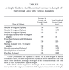
Draw a classic 4-flap z-plasty - what % increase do you anticipate?

A
  • 30’ – 25%
  • 45’ – 50%
  • 60’ – 75%
  • 75’ – 90%
  • 90’ – 120%
  • for a 4-flap Z plasty, expect 150% increase length
9
Q

draw a jumping man

how much % increase in length?

A

150%

10
Q

what is your approach to managing a flexed joint in claw hand?

A
  • first determine if the primary problem is skin scar/contracture or joint contracture (or both)
  • decide then if patient requires both soft tissue AND joint release
  • undertake appropriate choice of soft tissue procedure
  • undertake capsulotomy - volar plate / check-rein ligament release, capsule release, partial accessory liament release, +/- K-wire
  • Consider arthrodesis for refractory cases
  • Do not consider arthroplasty in this context, given poor soft tissue envelope, premorbid stiffness, risk of chronic pain and extrusion
11
Q

what is heterotopic ossification

A

bone formation in extra-skeletal soft tissue

12
Q

who is at risk for heterotopic ossification / what are the associations?

A
  • burn
  • critical care and immobilization
  • CNS injury, spinal cord injury
  • trauma; hip surgery
13
Q

what is the suspected etiology of HO?

A
  • BMP triggers dormant osteoprogenitor cells to differentiate into osteoblasts / osteoid
14
Q

treatment of HO

A
  1. prevention in high risk pts: NSAIDS, bisphosphonates (etidronate)
  2. treatment:
    1. non-operative
      1. PT/OT/SPLINTS
      2. NSAIDS, bisphosphonates,
    2. operative
      1. be cautious - high recurrence, low resolution of pain (what is the goal?)
      2. joint release and excision at > 12 mos
15
Q

what are the mechanisms of injury during extravasation injury?

A
  1. Pressure: vascular obstruction 2’ pressure in extra-cellular (intersititial) space occludes vessels - early necrosis
  2. Chemical: vascular obstruction 2’ inflammatory response to chemical - early necrosis
16
Q

Summarize injection material, typical treatment principle

A
  • high pressure injection injuries are variably defined, but generally administerd from a high pressure gun or hose of some sort
  • Toxic injection modalities include: grease (commonest), paint, paint thinner (worst), fuel
    • management includes history, physical (including for compartment syndrome), imaging (XR), tetanus administration, analgesia (via PO/IV not local), IV antibiotics, dress/splint/elevate, to OR urgently for decompression and debridement and second look
  • Non toxic injection injuries include air and clean water
    • management includes history/physical (for compartment syndrome), imaging (XR), tetanus, analgesia, IV antibiotics –> oral antibiotics, dress/splint/elevate
    • non-operative management as above with active surveillance (close fu)
17
Q

what do you wnat to know on history?

A
  • agent, toxicity, viscosity, concentration, estimate of volume? etc
  • pressure of unit
  • when it happened, what was done immediately
  • entry point (flexor = more dissemination)
  • symptoms now (pain, paresthesia, skin changes)
18
Q

describe principles of surgical management for high pressure injection injuries

A

· Removal of all material is often not feasible

· Surgical goals

o Neurovascular decompression

o Decontamination

o Debridement of non-viable tissue

o Prevention of infection

o Delayed reconstruction

· Anaesthesia: general or regional

· Tourniquet, but no exsanguination

19
Q

discuss management of normal pressure injection injuries

A
  • history:
    • agent: what was the agent, when injected, how injected, by whom, initial response, response over time, why injected
    • problems: what is the current problem (pustule, draining sinus, inflammation, pain, etc) and how has it been treated so far?
    • usual rest
  • Physical: signs of acute or delayed presentation:
    • Acute reaction –> chemical irritation, infection or pain, systemic illness/septicemia, embolization
    • Delayed presentation –> abscesses, scar, draining fistula/sinus, bleeding, extrusion, infection, granulomas, beading
  • Investigations:
    • CT or MRI to image cavity
    • tissue biopsy to rule out fungus, mycobacterium, other atpyical infection
  • Superficial localized granulomas may be improved with local or systemic steroids or imiquimod
  • Chronic inflammation, induration and sinus formation are usually treated by excision of involved tissue
  • Staged recon
20
Q

describe universal interventions for extravasation injuries

A

o Prevention

o Stop infusion immediately

o Aspiration via in-situ catheter (ie by RN prior to removal; often IV is removed by the time we’re called)

o Remove intravenous and re-start only at different site

o Elevation and splinting +/- cool compress

o Analgesia

o Serial examination

  • No prophylactic debridement; follow and treat w/ dsg (vs surg, below) as necessary once fully demarcated
  • *Adjunct: clysis with saline irrigation+/- hyaluronidase à not really done
21
Q

during an extravasation injury, describe agents that have an anti-dote

A

o Vasoconstricting agents (ie epi/norepi etc) –> phentolamine

o Cisplatin, nitrogen mustard (chemotherapeutics) –> sodium thiosulfate

o Doxorubicin (chemotherapeutic) –> DMSO

22
Q

Describe 5 factors determining the cellular response to radiation

A
  • Repair - ability of cell to recover from sublethal damage
  • Repopulation - cells ability to repopulate tumor
  • Reoxygenation - cell most sensituve to Rtx when oxygenated b/c ROS/free radicals formed
  • Reassortment - cell most sensitive to Rtx in mitosis/DNA replication phase
  • Radiosensitivity
    • sensitive if high oxygenation, reassortment
    • not sensitive if repair and repopulate
    • inherent cell sensitivty is based on frequency of cell division, time between division and life span
23
Q

How does radiation affect wound healing?

A

impairs all phases of wound healing

  • Inflammatory:
    • decreased macrophage function/GFs
  • Proliferative:
    • reduced fibroblast number, collagen production
    • impaired angiogenesis 2’ fibrosis and endoarteritis obliterans
    • delayd epitheliazation: abnormal and fewer keratinocytes
  • Maturation
    • impaired collagen turnover/ remodelling

RESULTS

  • Pre-op Rtx: delay surgery 1wk per wk of radiation prior to surgery - approx 6mths for breast OR
  • Post- op Rtx: Should delay rtx until wound healed - approx 3wks
24
Q

What are the phases of radiation dermatitis?

A
  • Acute - erythema phase (hrs-days)
  • Dry desqaumation phase (4-5thwk)
    • enough basal keratinocytes remain to repopulate epidermis
    • pruritus, scaling, hyperpigment which remains
  • Moist desquamation phase (4thwk) - at >40Gy doses
    • not enough basal keratinocytes remain
    • at lower Gy doses, re-epi occurs form appendages
    • at high Gy doses, re-epi occurs from edges
  • Late phase (1yr post-Rtx)
    • atrophic,fibrosis,scarring with latreed pigment, telangiectasia, risk of radiatio ulcer/necrosis

NB: most skin cancers require >40Gy= moist desqaum

<30Gy get dry desquamation

Gy = joule/kg of tissue, 1rad = 0.01 Gy - radiation absorbed dose

Conventional fractionation:

1.8-2Gy/day, 5day/wk, 5-6wks, total 50-70Gy

25
Q

What is your management and treatment options for a radiation ulcer?

A
  • Etiology: fibroblast and endothelial dysfx
  • Hx:
    • local trauma/injury, recent infections/systemic illness, Hx of Rx
  • PE: depth, surrounding itssue involvement
  • Investigation
    • BIOPSY
    • CBC (anemia) Alb,electrolytes (nutritional asx)

TREATMENT

  • wound care dressings w antimicrobials
  • correct systemic factors (malnutirtion Vit a,c Zn), anemia(Fe)
  • Hyperbaric Oxygeb (5x/wk) oxygen supplementation stimulates fibroblasts, halts progression of ulcer
  • Surgery if failed conservative
    • direct, skin graft, local not advised, regional/free
26
Q

What are geeral principles for reconstruction in a radiated field

A
  • confirm no residual/ de novo malingnacy
  • debridement: non -viable tissue
  • bone recon with vascularied BG
  • ST recon/coverage of prosthesis with non-radiated well vascularized tissue (regional/free)
  • Pedicle non-radiated and anastomosis not non-radiated
27
Q

What treatment options would you recommend for breast reconstruction in patient with

  • previous radiation
  • mastectomy and post-op radiation

NOT SURE ON THESE ANSWERS - check wi lipa

A
  • Previous radiation:
    • autologous tissue (pedicled flap - non radiated, or free tissue with anastomiss in non-radiated field)
    • NOT alloplastic b/c
      • poor wound healing, risk implant expousre
      • capsular contracture
      • poor cosmetic result
    • Timing - wait until late phase of radiation changes >6mths
  • Mastectomy + post-op Radiation
    • Delayed autologous recon preferred over immediate recon
    • less complications of fat necrosis, loss of flap volume
28
Q
A