The Red Blood Cell Structure and Function Flashcards
Describe the structure of RBCs.
- The RBC is unique amongst eukaryotic cells as it lacks a nucleus, mitochondria and ER, and its cytoplasm is essentially rich in haemoglobin.
- Mammalian RBCs are biconcave disc-shaped.
- They are 8um in diameter but are able to deform and pass through 3um capillaries or reticuloendothelial system without fragmentation.
- They have flexible membranes with a high surface-to-volume ratio.
“Reticuloendothelial system” - is an older term for the mononuclear phagocyte system, but it is used less commonly now, as it is understood that most endothelial cells are not macrophages.

Describe the main function of the RBCs, and what helps it achieve it.
- The primary function of RBCs is the transport of respiratory gases to and from the tissues.
- To achieve this: -
- The RBC should be capable of traversing the microvascular system without mechanical damage, and retain its shape.
- The red cell membrane should be extremely tough, yet highly flexible.
Traversing meaning - travel across or through.
Microvascular system meaning - relating to, or constituting the part of the circulatory system.
Describe the structure of the red blood cell membrane.
- It is a semipermeable lipid bilayer with proteins scattered throughout.
- It consists of: -
- An outer hydrophilic portion composed of glycolipids, glycoproteins and protein.
- A central hydrophobic layer containing proteins, cholesterol and phospholipids.
- An inner hydrophobic layer of mesh-like cytoskeletal proteins to support the lipid bilayer.

Describe the RBC membrane lipids.
- RBC membrane lipids make up about 40% of the membrane.
- There is an asymmetrical phospholipid distribution throughout the RBC membrane.
- There is unesterified free cholesterol between them.
- There are two types of phospholipids:
- UNCHARGED PHOSPHOLIPIDS IN THE OUTER LAYER
- Phosphatidylcholine (PC)
- Sphingomyelin (SM)
- CHARGED PHOSPHOLIPIDS IN THE INNER LAYER
- Phosphatidyl Ethanolamine (PE)
- Phosphatidyl Serine (PS)
- UNCHARGED PHOSPHOLIPIDS IN THE OUTER LAYER

Describe the membrane cholesterol.
- Membrane cholesterol exists in free equilibrium with plasma cholesterol.
- An increase in free plasma cholesterol results in an accumulation of cholesterol in the RBC membrane.
- RBCs with increased cholesterol levels appear distorted, resulting in acanthocytosis.
- An increase in cholesterol and phospholipid is a cause of target cell.
Acanthocytosis meaning - refers to a form of red blood cell that has a spiked cell membrane, due to abnormal thorny projections.
Target cell meaning - In optical microscopy these cells appear to have a dark center surrounded by a white ring, followed by dark outer second ring containing a band of hemoglobin.

Describe RBC membrane proteins (and it’s two categories).
- RBC membrane proteins make up about 50% of the membrane.
- They are split into two categories:-
-
INTEGRAL MEMBRANE PROTEINS
- They extend from the outer surface and transverse the entire membrane to the inner surface.
- Two major integral membrane proteins are Glycophorins (types we’ve identified are A, B and C) and Band 3 (an anion transporter)[It links lipid bilayer to underlying membrane cytoskeleton].
- Examples of other integral membrane proteins: - Na+/K+, ATPase, Aquaporin 1, surface receptors (eg. TfR).
-
PERIPHERAL PROTEINS:
- They’re limited to the cytoplasmic surface of the membrane and form the RBC cytoskeleton.
- Major peripheral proteins include: spectrin, ankyrin, protein 4.1, actin.
-
INTEGRAL MEMBRANE PROTEINS

Describe the function of the peripheral protein Spectrin.
- Spectrin is the most abundant peripheral protein.
- It is composed of α and β chains.
- It’s very important in RBC membrane integrity as it binds with other peripheral proteins to form the cytoskeletal network of microfilaments.
- It controls the biconcave shape and deformability of the cell.
Describe the function of the peripheral protein Ankyrin.
It primarily anchors the lipid bilayer to the membrane skeleton via interaction between spectrin and Band 3.
Describe the function of the peripheral protein Protein 4.1.
- It may link the cytoskeleton to the membrane by means of its associations with glycophorin.
- It also stabilises the interaction of spectrin with actin.
Describe the function of the peripheral protein Actin.
It is responsible for the contraction and relaxation of the membrane.
What are the functions of the RBC membrane?
-
SHAPE:
- provides the optimum surface area to volume ratio for respiratory exchange AND is essential to deformability
-
PROVIDES DEFORMABILITY, ELASTICITY:
- allows for passage through microvessels (capillaries)
- REGULATES INTRACELLULAR CATION CONCENTRATION.
What is the use of red cell metabolic pathways?
Metabolism provides energy required for: -
- Maintenance of cation pumps.
- Maintenance of Hb in its reduced state (reduction in oxidation state).
- Maintenance of reduced sulfhydryl groups in Hb and other proteins.
- Maintenance of RBC integrity and deformability.
Sulfhydryl meaning - A sulfhydryl is a functional group consisting of a sulfur bonded to a hydrogen atom.
List the key metabolic pathways.
- Glycolytic or Embden-Meyerhof Pathway
- Pentose Phosphate Pathway
- Methaemoglobin Reductase Pathway
- Luebering-Rapoport Shunt
What does the Glycolytic or Embden-Meyerhof Pathway do?
- It generates 90 - 95% of the energy needed by RBCs.
- In it, glucose is metabolised and generates two molecules of ATP.
- It functions in the maintenance of the RBC shape, flexibility and cation pumps.
What does the Pentose Phosphate Pathway do?
- RBCs need glutathione (GSH) to protect itself them from oxidative damage.
- The Pentose Phosphate Pathway provides the reducing power, NADPH.
- NADPH maintains glutathione in its reduced form (GSH).
(Glutathione is an antioxidant. It protects the RBCs from oxidative damage.)
(Antioxidants are molecules that prevent the oxidation of other molecules. Oxidation is a chemical reaction in which electrons are lost.)
What does the Methemoglobin Reductase Pathway do?
- It maintains ion in its ferrous state (Fe2+).
- In the absence of this enzyme, methemoglobin accumulates and cannot carry oxygen.
Methemoglobin meaning - is a form of hemoglobin, in which the iron in the heme group is in the Fe3+(ferric) state, not the Fe2+ (ferrous) of normal hemoglobin. Methemoglobin cannot bind oxygen, which means it cannot carry oxygen to tissues.
What does the Luebering-Rapoport Shunt do?
- It permits for the accumulation of 2,3-DPG (diphosphoglycerate), which is essential for maintaining normal oxygen tension, regulating haemoglobin affinity.
How long do RBCs live?
120 days.
What are two conditions caused by membrane abnormality?
Hereditary elliptocytosis - caused due to problems in ankyrin. It is an inherited blood disorder in which an abnormally large number of the patient’s erythrocytes (i.e. red blood cells) are elliptical rather than the typical biconcave disc shape.
Hereditary spherocytosis - caused due to problems in spectrin and ankyrin. The blood cells are spherical in shape. People with this condition typically experience a shortage of red blood cells (anemia).
What features allow RBC to withstand life without structural deterioration?
-
Geometry of cell; surface area to volume ratio.
- Facilitates deformation whilst maintaining constant surface area.
-
Membrane deformability
- Spectrin molucules undergo reversible change in conformation: some uncoiled and extended, others compressed and folded.
-
Cytoplasmic viscosity determined by MCHC (mean corpuscular hemoglobin concentration)
- as MCHC rises, viscosity rises exponentially.
Corpuscular meaning - an unattached cell, especially of a kind that floats freely, as a blood or lymph cell.
Describe the structure of haemoglobin.
- Haemoglobin is a globular haemoprotein,
- Haemoproteins are a group of speciallised proteins that contain haem as a tightly bound prosthetic group.
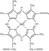
- Haem is a complex of protoporphyrin IX and ferrous iron (Fe2+).
- Iron is held in the centre of the haem molecule by bonds to the 4 nitrogen of a porphyrin ring.
- Haemoglobin is made up of 4 polypeptide subunits:-
- 2 alpha globin chains
- 2 beta globin chains

What is the composition of the RBC membrane?
It is :-
- 50% protein
- 40% lipids
- 10% carbohydrates


