Systems 2 - Integrated Physiology Flashcards

1
Q

Equivalents

A

= moles x valence

So 1 mol Na⁺ = 1 eq/L
1 mol Ca²⁺ = 2 eq/L

Moles are unit of quantity, 6 x 10²³

2
Q

PaCO₂

A

PaCO₂ (arterial) = Rate of CO₂ production / alveolar ventilation rate

3
Q

Quantity of moles

A

1 mole =
10³ mmol
10⁶ μmol
10⁹ nmol

4
Q

pH equations

A

pH = -log[H⁺]
So minor changes in pH -> major changes in [H⁺]

pH = pk + log[A⁻]/[HA]

5
Q

Importance of pH in body

A
  • enzyme activity/protein strucure affected
  • Ca²⁺ ions - 50% are free in blood, ionised, to stabilise nerve and muscle membranes. 50% are bound to albumin, which competes with H⁺ for binding. -> when decreased H⁺, less free Ca²⁺, so less of a membrane stabilising effect
  • –> so in hyperventilation, increased pH, hyperexcitable nerves
6
Q

Trousseau sign, Chvostek’s sign

A

Trousseau - hand cramped forward, claw
Chvostek - muscle twitch when tap facial nerve

-> indicate disturbance of plasma calcium, or acid/base balance disruption

7
Q

Buffers

A

Resist a change in pH by absorbing or releasing H⁺ when an acid or base is added

pH will still change slightly - buffer pair is weak acid and its conjugate base

8
Q

pk

A

= the pH where an acid is 50% dissociated, [A⁻]/[HA] = 1

Lower pk -> stronger acid

9
Q

Extracellular buffers

A

Bicarbonate
Haemoglobin
Phosphate
Plasma proteins

-> work together to resist change, isohydric principle

10
Q

Bicarbonate buffer system

A

pk = 6.1 CO₂ + H₂O = H₂CO₃ = H⁺ + HCO₃⁻

BUT rarely know [H₂CO₃], so use solubility coefficient of 0.03 - [H₂CO₃] = 0.03 x PCO₂

  • > pH ∝ [HCO₃⁻]/PaCO₂
  • > pH depends on the ratio of bicarbonate to carbon dioxide

IMPORTANT

  • high conc of buffer pair in plasma
  • PaCO₂ regulated by respiratory system
  • [HCO₃⁻] regulated by kidney
11
Q

Acid production in body

A

Body is net producer of acid

Kreb’s cycle makes CO₂
Metabolism makes H⁺
Gut below pylorus -> HCO₃⁻ to lumen in alkaline tide, H⁺ into blood

12
Q

Renal handling of bicarbonate

A

Reabsorption - of bicarbonate ions by glomerular filtration. If too high, exceeds tubular threshold and spills into urine

Regeneration - of bicarbonate lost in buffering, by secreting protons into nephron to be trapped and excreted by non-bicarbonate buffers, and by secreting ammonium

13
Q

-aemia

A

Acidaemia - acidic blood, pH less than 7.35

Alkalaemia - alkaline blood, pH more than 7.45

14
Q

-osis

A

Acidosis/alkalosis - processes that cause a change in pH of blood

Usually -> -aemia

‘osis-without-aemia’ when pH in normal range

15
Q

Compensation

A

Attempts to return pH to normal

Pathological chronic change in PCO₂ or HCO₃⁻ is compensated by homeostatic change in the other

Renal compensation (adjusting HCO₃⁻) is more effective than respiratory compensation (adjusting CO₂) but takes longer to get effect, days

-> if renal and lung disease, big problem

Change in same direction, if increase in HCO₃⁻, body will increase CO₂ to compensate

16
Q

Normal range of pH, PCO₂, HCO₃⁻

A

pH - 7.35-7.45

PCO₂ - 35-45

HCO₃⁻ - 21-29

17
Q

Alkalaemia

A

pH > 7.45

HCO₃⁻ raised, metabolic alkalosis

PCO₂ decreased, respiratory alkalosis

18
Q

Acidaemia

A

pH < 7.45

HCO₃⁻ decreased, metabolic acidosis

PCO₂ raised, respiratory acidosis
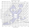
19
Q

Acid base map

A
20
Q

Electroneutrality

A

Total [cations] = total [anions] in body fluids, can’t have net charge

Anion gap in -ve ions, unsure where from
Normally 8-16mEq/L

Other anions are Cl⁻ and HCO₃⁻

21
Q

Hyperchloraemic metabolic acidosis with normal anion gap

A

As cations have increased, to fill in gap, Cl⁻ increases

Anion gap remains unchanged

Caused by too much bicarb out

22
Q

Increased anion gap metabolic acidosis

A

As bicarbonate has decreased, to fill in gap, anion gap increasases

Cl⁻ remains unchanged

Caused by too much acid in

23
Q

Causes of increased anion gap metabolic acidosis

A
  • more fixed acid production, eg lactic acidosis/ketoacidosis
  • ingestion of fixed acids, eg aspirin
  • inability to excrete fixed acids, eg in renal failure
24
Q

Causes of hyperchloraemic metabolic acidosis with normal anion gap

A
  • loss of bicarb from gut, eg diarrhoea, ileostomy
  • loss of bicarb via kidney, eg renal tubular acidosis
25
Causes of metabolic alkalosis
SALINE RESPONSIVE - vomiting, diuretic use, volume contraction - treated with saline to decrease RAAS activity SALINE UNRESPONSIVE - primary hyperaldosteronism
26
Causes of respiratory alkalosis
Decreased PCO₂, caused by: - mechanical ventilation, hyperventilation - stimulation of respiratory centre
27
Causes of respiratory acidosis
Increased PCO₂, caused by ALVEOLAR HYPOVENTILATION - defects in neuromuscular chain - work of breathing exceeds the strength of respiratory pump, eg in obstruction to airflow, restrictive lung diseases, decreased lung compliance, so increased CO₂ production
28
Constant body temperature
Homeotherms (birds and mammals) have physiological mechanisms to regulate temperature - allows them to inhabit physiological niches - enzyme reactions work in narrow range so good to keep constant Humans do vary by location in body in order to preserve core temperature (isotherms areas of equal heat), and also varies with time
29
BMR
Basal metabolic rate - without doing anything, we produce heat, ~100W
30
Clinical measurement of body temperature
Must be a representative site (into core) Must be easily accessible External auditory meatus most common now
31
Major routes for heat gain and loss equation
Metabolism - Evaporation ± Conduction ± Convection ± Radiation = 0, when heat gain = heat loss 20% loss by evaporation 40% loss by radiation 40% loss by convection
32
Thermoreceptors
SKIN (peripheral) - specific - sensitive to dynamic and absolute changes - small receptive fields - more cold receptors than warm CENTRAL (posterior hypothalamus) - more warm receptors than cold - also in midbrain, medulla, spinal cord
33
Central controller of temperature
Posterior hypothalamus Set point generated here ACh main transmitter Increase Na⁺, increase set point, increase body temp Increase Ca²⁺, decrease set point Peripheral and central input are integrated to fine tune response (eg in cool temps, there must be a greater core temp increase to stimulate sweating)
34
Adaptation/acclimatisation to heat
Lower sweating threshold Increased sweat rate Decreased electrolyte content of sweat Behavioural changes
35
Adaptation/acclimatisation to cold
Decreased skin blood flow Decreased shiver threshold Thyroid hormones Behavioural changes
36
Pyrexia
= fever Above 38 degrees is significant Infection -\> toxins -\> WBC reaction -\> pyrogens (IL family) -\> increased set point in hypothalamus --\> shivering, vasoconstriction, pyloerection -\> increase in core temperature
37
Temperature regulation in the newborn
Can't shiver, will develop at a few months old And small size, so large SA:volume ratio, lose heat easily - \> brown adipose tissue instead, on scapulae and around major arteries - abundant mitochondria here, for uncoupled oxidative phosphorylation
38
Ageing
= maturation and deterioration Maturation dominant until around 30 Maximum survival roughly constant, so intrinsic as well as extrinsic factors Senescence = post-reproductive decline in viability, accompanying biological age
39
Causes of ageing
Free radicals Apoptosis Genetics
40
Causes of ageing - free radicals
Occur in normal chemical reactions Normally involve molecular oxygen, making OH. and O₂. Production increased by environmental agents - UV, gamma, Xrays -MAINLY SMOKING -\> lipid peroxidation - breakdown products react with DNA -\> mutations -\> impaired protein function Enzymes can control: - superoxide dismutase, catalase - vitamins C and E are antioxidants, trap free radicals
41
Causes of ageing - apoptosis
Shrinkage, degrade nuclear DNA, breakdown mitochondria, break down cell, phagocytosis Useful in development, eg tissue remodelling In ageing - neurons, cardiac tissue, cells of immune system all die
42
Causes of ageing - genetics
DNA redundancy failure - non-error sequences will eventually be exhausted, so errors will be expressed Failure in chromosome replication - telomeres shorten with each division, if small enough cell will die - evidenced by progeria/Werner's syndrome - genetic condition -\> accelerated ageing
43
Homeostasis and ageing
Corrections to normal take longer to occur in older people - eg blood glucose, recovery from exertion, temperature acclimatisation - \> physiological effectiveness reduced
44
Structural cardiovascular changes in ageing
HEART Fewer cardiac myocytes - necrosis and apoptosis Increased collagen, so stiffer VASCULATURE Arteries - more collagen and smooth muscle, less elastic tissue, collagen cross linked, calcium deposited (so stiffer) -\> increased afterload on left ventricle Veins - fibrosis, intima thickening, loss of elastic tissues, so -\> varicosities under high pressure
45
Functional cardiovascular changes in ageing
- Reduced maximum heart rate, and reduced stroke volume -\> so reduced cardiac output - Increased bp, as stiffness in arteries increases resistance - Baroreflex sensitivity reduced, so postural hypotension
46
Renal changes in ageing
Decrease in kidney mass with age, loss of nephron units Reduced glomerular filtration rate Fewer glomerular capillary loops, change in vascular tone -\> respiratory adjustments needed to maintain pH, impaired ability by kidney to restore buffer systems
47
Thermoregulation and age
Reduced metabolic rate Reduced muscle mass Increased adipose tissue Reduced activity -\> so less heat generated by metabolism Also - less evaporation loss (sweat glands atrophy) - more radiation loss (less thermal insulation, thinner skin and subcutaneous fat) - less convection/conduction loss (reduced skin blood flow to compensate for reduced cardiac output) - \> so elderly are less able to cope with hot/cold challenges, though core temperature should not change - sweat later, and shiver later than would in young (though once established can do) Ideal temperature not changed, just precision lost
48
Hypoxia vs hypoxaemia
Hypoxia - low tissue O₂ content Hypoxaemia - low O₂ in blood ``` PᴀO₂ = alveolar pressure PaO₂ = arterial pressure ```
49
Types of hypoxia
Hypoxaemic - ventilatory defect, alveolar diffusion problems Anaemic - reduced O₂ carriage in blood Stagnant - cardiovascular shock, shunts Toxic - reduced extraction of O₂ by tissues
50
Symptoms of hypoxia
- dyspnoea (SOB), laboured breathing - fatigue, lethargy - confusion - tachycardia - deterioration of vision - headaches - peripheral cyanosis - euphoria, moodiness, dizziness - pins and needles - \> loss of consciousness
51
Ventilatory response to hypoxaemia
Gaseous exchange stops at PᴀO₂ of 40 Hypoxaemia -\> hyperventilation PᴀO₂ declines at a disproportionate rate, but more breathing also decreases CO₂ though, so more space for O₂ if CO₂ reduced - carotid bodies monitor PaO₂ (innervated by glossopharyngeal nerve), and direct to hyperventilate when gets too low
52
Limit to hyperventilation
Can't cope with very low PaO₂ - as blood-brain barrier is permeable to CO₂ - as PaCO₂ decreases, CO₂ also decreases in CSF - \> respiratory alkalosis, rise in pH - central chemoreceptor on medulla will inhibit drive to hyperventilation - contradicts with peripheral chemoreceptor (carotid bodies) drive -\> therefore hyperventilatory response not as high as should be Few days later, renal compensation will kick in, decreasing HCO₃⁻ to maintain pH another way - ACCLIMATISATION
53
Cardiovascular response to hypoxia
Transient rise in cardiac output Heart rate and stroke volume then decrease -\> can't adapt well Long term, better - increased capillarity to muscles - reduced muscle fibre diameter, so reduced O₂ diffusion distance - increased myoglobin content of muscle
54
Immediate response to hypoxia
(seconds) Increased ventilation Pulmonary vasoconstriction Tachycardia, increase in systemic bp
55
Intermediate response to hypoxia
(days) Increased ventilation, though with CSF compensation Renal compensation of respiratory alkalosis, HCO₃⁻ elimination O₂ dissociation curve shifts to right, reduced HbO₂ affinity, so easier to offload O₂ Increased urine loss
56
Long term response to hypoxia
(weeks-months) Polycythaemia - more RBCs, high haematocrit Increased muscle capillarity Increased muscle myoglobin/rate of anaerobic metabolism
57
Tissue hypoxia -\> erythropoiesis
Increased expression of hypoxia-inducible factor (HIF-1) - stimulates production of erythropoietin by kidney - increases liver production of transferrin - increases absorption of iron from intestine by regulation of hepatic mediators -\> more haemoglobin, more RBCs (BUT, increases blood viscosity, raised afterload on heart)
58
Increased urinary output at high altitude
Reduced aldosterone, so urine loss - \> risk of significant dehydration - blood volume may allow circulation to accomodate increase in RBCs -
59
Acute mountain sickness symptoms
- headache + one of: - dizziness - fatigue - sleep disturbance - GI disturbance Rarely progresses to pulmonary and cerebral oedema
60
Acute mountain sickness treatment
DESCENT Oxygen therapy Hyperventilation Acetazolamide
61
High altitude pulmonary oedema
- potentially fatal - around 3 days after ascent - hypoxaemia - \> pulmonary vasoconstriction - pulmonary pressures increase to maintain cardiac output - \> transudation of fluid into alveoli Symptoms - dyspnoea, fatigue, persistent dry cough (with pink frothy sputem)
62
High altitude cerebral oedema
- potentially fatal - even more dangerous but very rare - around 3 days after ascent - hypoxaemia - \> cerebral vasoconstriction - cerebral pressures increase to maintain cardiac output - \> transudation of fluid onto brain tissue Same symptoms as acute mountain sickness, + ataxia, altered consciousness/mental status, retinal haemmorhage
63
Stress response
Physiological changes that occur in response to stressors such as trauma, surgery, burns and sepsis, to aid survival and eventual repair - signalled via afferent neuronal impulses from site of injury, and release of cytokines by macrophages and monocytes in damaged tissues
64
Acute phase stress response
IL6 produced by macrophages - \> fever - \> increased granulocytes, especially neutrophils and platelets - \> liver increases production of proteins by CRP (which adheres to bacteria and promotes complement activation and phagocytosis)
65
Full blood count
To identify cause of infection TOTAL WHITE CELL COUNT Neutrophils raised -\> bacterial infection Lymphocytes raised -\> viral infection Eosinophils raised -\> parasitic infection/allergy
66
CRP test
To monitor degree of inflammation Measure CRP sequentially to see trend
67
Behavioural changes in stress response
As brain stimulated by neuronal and cytokine signals INCREASED - arousal - aggression - defence - vigilance DECREASED - sexual activity - feeding
68
Fight or flight response in stress response
Adrenaline released -\> α adrenoreceptor - relaxation of smooth muscle in GI tract - mydriasis (pupil dilation) - constriction of arterioles in skin and kidney (- hepatic gluconeogenesis and glycogenolysis) -\> β adrenoreceptor - relaxation of smooth muscle in GI tract - increase heart rate and contractility - bronchodilation - relaxation of detrusor in bladder - dilation of arterioles in skeletal muscle (- hepatic gluconeogenesis and glycogenolysis, lipolysis in adipose tissue, renin secretion in kidneys) -\> so blood diverted to skeletal muscle, away from gut and skin (-\> glucose production, salt retention, raised bp)
69
HPA axis activity in stress response
Hypothalmic-Pituitary-Adrenal axis Increases activity So hypothalamus releases CRH - corticotrophin releasing hormone To anterior pituitary, which releases ACTH - adrenocorticotrophic hormone To adrenal gland, which releases CORTISOL (will inhibit sequence now)
70
Cortisol effects
- increased gluconeogenesis - increased protein catabolism - increased lipolysis - \> survival during fasting - decreased immune response - decreased inflammatory response (steroids use!) - increased vascular response to catecholamines, as increases α1 adrenoreceptors - decreased histamine release from mast cells - \> treats anaphylactic shock
71
Adrenocortical insufficiency
Primary - addison's disease Secondary - secondary to exogenous steroid therapy
72
Salt and water metabolism in stress
SALT Sympathetic stimulation of kidney -\> RAAS activation, Na⁺ retention WATER ADH release from posterior pituitary -\> water retention by distal nephron -\> so post op (stress), dangerous to give hypotonic solutions, as retain lots of fluid -\> hyponatraemia, cerebral oedema, death
73
Insulin
For glucose -\> glycogen in liver, -\> fat in adipose, -\> protein in muscle Increases glucose uptake into cells, and reduces blood glucose
74
Metabolic changes to provide fuels in stress
- protein catabolism - stimulated by cytokines and cortisol - \> amino acids for gluconeogenesis in liver - gluconeogenesis and glycogenolysis - \> glucose and ketone bodies as fuel for heart and brain - lipolysis - stimulated by cortisol, catecholamines, insulin deficiency - \> free fatty acids and glycerol to liver
75
Hyperglycaemia
As catecholamines and cortisol stimulate hepatic glycogenolysis and gluconeogenesis So increased glucose in blood Decreased insulin production, insulin resistance (very high insulin concs reduce wound healing and increase infection)
76
Systemic Inflammatory Response Syndrome (SIRS)
Will become sepsis -\> severe sepsis -\> septic shock Triggered by surgery, trauma, infections, burns, haemmorhagic shock
77
Stress response a destructive force?
Intensive care uses drugs and machines to try to restore physiological values to normal - may not be good; high insulin always associated with death, increased fluids following traumatic haemmorhage give death -\> so consider different therapeutic end points
78
Na⁺/K⁺ levels
More Na⁺ extracellularly (blood tastes salty!) More K⁺ intracellularly