Surgical Recall - Ch. 66 Vascular Surgery Flashcards
Atherosclerosis
- What is it?
- How is it initiated?
- Risk factors?
- Common sites of plaque formation in arteries?
- What must be present for a successful arterial bypass operation?
- What is the major principle of safe vascular surgery?
- Which arteries supply the blood vessel itself?
- What is “ENDOVASCULAR” repair?
Atherosclerosis
- What is it? Diffuse disease process in arteries; atheromas containing cholesterol and lipid form within the intima and inner media, often accompanied by ulcerations and smooth muscle hyperplasia
- How is it initiated? Endothelial injury –> damaged vessel walls release thrombin, ADP, cytokines –> platelets adhere –> growth factors released –> smooth muscle hyperplasia/plaque deposition
- Risk factors? HTN, SMOKING***, DM, family hx, hypercholesterolemia, high LDL, obesity, sedentary lifestyle
- Common sites of plaque formation in arteries? Branch points (carotid bifurcation)
- What must be present for a successful arterial bypass operation?
- Inflow (e.g., patent aorta)
- Outflow (e.g., open distal popliteal a.)
- Run off (e.g., patent trifurcation vessels down to the foot)
- What must be present for a successful arterial bypass operation? Get proximal and distal control of the vessel to be worked on!
- Vaso vasorum
- Endovascular repair: placement of a catheter in artery and then deployment of a graft intraluminally

Peripheral Vascular Disease
- What is peripheral vascular disease?
- What is the most common site of arterial atherosclerotic occlusion in the lower extremities?
- What are the symptoms of PVD?
- What is intermittent claudication?
- What is rest pain?
- What classically resolves rest pain?
- What is Buerger’s Sign?
Peripheral Vascular Disease
- What is peripheral vascular disease? Occlusive atherosclerotic disease in the lower extremities
- What is the most common site of arterial atherosclerotic occlusion in the lower extremities? Occlusion of the SFA in Hunter’s Canal
- What are the symptoms of PVD?
- Intermittent claudication
- Rest pain
- Erectile dysfunction
- Sensorimotor impairment
- Tissue loss
- What is intermittent claudication?
- Pain, cramping… usually the calf muscle, after walking a specific distance; then pain resolves after stopping
- What is rest pain? Sign of advanced PAD… Pain in the foot, usually over the distal metatarsals; this pain arises at rest (classically at night, awakening the pt) –> LIMB-THREATENING ISCHEMIA**
- What classically resolves rest pain? Hanging the foot over the side of the bed or standing; gravity affords some extra flow to ischemic areas
- Buerger’s Sign: Physical exam sign of advanced chronic ischemia… the affected foot turns pale after it is elevated (usually for 1-2 min). Once the pt sits up and dangles the foot down, it becomes ruborous (like a cooked lobster) due to marked arteriolar dilation from chronic severe ischemia that causes a reactive hyperemia… such pts will have low ABI <0.4
Peripheral Vascular Disease
- What is the differential diagnosis of lower extremity claudication?
- What are the signs of PVD?
- What is the site of a PVD ulcer vs. a venous stasis ulcer?
Peripheral Vascular Disease
- What is the differential diagnosis of lower extremity claudication?
- Neurogenic (e.g., nerve entrapment/discs)
- Arthritis
- Coarctation of the aorta
- Popliteal artery syndrome
- Chronic compartment syndrome
- Neuromas
- Diabetic neuropathy pain
- Anemia
- What are the signs of PVD?
- Absent pulses
- Bruits
- Muscular atrophy
- Decreased hair growth
- Thick toenails
- Tissue necrosis/ulcers/infections
- What is the site of a PVD ulcer vs. a venous stasis ulcer?
- PVD arterial insufficiency ulcer–usually on TOES, FOOT
- Venous stasis ulcer–MEDIAL MALLEOLUS (ankle)
Peripheral Vascular Disease
- What is the ABI?
- What ABIs are associated with normals, claudicators, and rest pain?
- Who gets false ABI readings?
- What is the ABI? Ankle to Brachial Index (ABI); ratio of systolic BP at ankle to systolic BP at arm (brachial a.)… taken with doppler
- Normal ABI > 1.0; Claudicator ABI < 0.6; Rest pain ABI < 0.4
- Pts with calcified arteries, esp. those with diabetes
Peripheral Vascular Disease
- Prior to surgery for chronic PVD, what diagnostic test will every pt receive?
- What are the indications for surgical tx in PVD?
- What is the tx of claudication?
- A-gram (arteriogram: dye in vessel and x-rays) map disease and allows for best tx option (i.e., angioplasty vs surgical bypass vs. endarterectomy)… GOLD STANDARD FOR DX PVD
-
“STIR”
- Severe claudication refractory to conservative tx that affects quality of life/livelihood (e.g. can’t work b/c of claudication)
- Tissue necrosis
- Infection
- Rest Pain
-
Tx: for vast majority, conservative tx:
-
”PACE”
- Pentoxifylline (results in increased RBC deformity and flexibility)
- Aspirin (inhibits platelets aggregation)
- Cessation of smoking
- Exercise
-
”PACE”
Peripheral Vascular Disease
- What is the risk of limb loss with claudication?
- What is the risk of limb loss with rest pain?
- In the pt with PVD, what is the main post-op concern?
Peripheral Vascular Disease
- What is the risk of limb loss with claudication? 5% limb loss at 5 yrs (5 in 5, 10 in 10)
- What is the risk of limb loss with rest pain? >50% of pts will have amputation of limb at some point
- In the pt with PVD, what is the main post-op concern? Cardiac status b/c most pts with PVD have coronary artery disease (~20% have an AAA)
- MI = most common cause of post-op death after PVD operation
Peripheral Vascular Disease
- What is Leriche’s syndrome?
- What are the tx options for severe PVD?
Peripheral Vascular Disease
- What is Leriche’s syndrome?
-
“CIA”
- Claudication of butt (not rest pain b/c gradual… so time for collaterals to help)
- Impotence (erectile dysfxn… dec. blood flow to internal iliacs –> internal pudendals)
- Atrophy (from occlusive disease of the iliacs/distal aorta)
-
“CIA”
-
Tx options:
- Surgical graft bypass
- Angioplasty–balloon dilation
- Endarterectomy–remove diseased intima and media
- Surgical patch angioplasty (place patch over stenosis)
Peripheral Vascular Disease
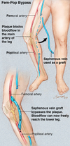
- What is a FEM-POP bypass?
- What is a FEM-DISTAL bypass?
- Dry vs. wet gangrene?
- What is blue toe syndrome?
- Bypass SFA occlusion with a graft from the femoral a. to the popliteal a.
- Bypass from femoral a. to distal a. (peroneal a., anterior tibial a., or posterior tibial a.)
- Dry: necrosis of tissue W/O signs of infection (“mummified tissue”)
- BTS: intermittent painful blue toes (or fingers) due to microemboli from a proximal arterial plaque
Acute Arterial Occlusion
- What is it?
- What are the classic signs & sx of AAO?
- What is the classic timing of pain with AAO from an embolus?
- What is the immediate pre-op mgmt?
- What are the sources of emboli?
- What is the most common cause of embolus from the heart?
- WHat is the most common site of arterial occlusion by an embolus?
- What diagnostic studies are in order?
- What is the tx?
- How is Fogarty catheter used? How many mm in diameter is a 12 French Fogarty catheter?
Acute Arterial Occlusion
- What is it? Acute occlusion of an artery, usually by embolization; other causes include acute thrombosis of an atheromatous lesion, vascular trauma
- What are the classic signs & sx of AAO?
- Pain
- Paralysis
- Pallor
- Paresthesia
- Polar (Poikilothermia)
- Pulselessness
- What is the classic timing of pain with AAO from an embolus? Acute onset; pt can tell you exactly when and where it happened
- What is the immediate pre-op mgmt?
- Anticoagulate w/ IV heparin (bolus followed by constant infusion)
- A-gram
- What are the sources of emboli?
- Heart–85% (e.g., clot from AFib, clot forming on dead muscle after MI, endocarditis, myxoma)
- Aneurysms
- Atheromatous plaque (atheroembolism)
- What is the most common cause of embolus from the heart? Afib
- What is the most common site of arterial occlusion by an embolus? Common femoral artery (SFA = most common site of arterial occlusion from atherosclerosis)
- What diagnostic studies are in order?
- A-gram
- ECG (looking for MI, Afib)
- Echo (looking for clot, MI, valve vegetation)
- What is the tx? Surgical embolectomy via cutdown and Fogarty balloon (bypass is reserved for embolectomy failure)
- Insinuate the catheter with balloon deflated past the embolus and then inflate the balloon and pull the catheter out; the balloon brings the embolus with it / To get mm from French measurements, divide the French number by pi (3.14); thus a 12 French catheter is 12/3 = 4 mm in diameter
Acute Arterial Occlusion
- What must be looked for post-op after reperfusion of a limb?
- What is compartment syndrome?
- What are the signs/sx of compartment syndrome?
- Can a patient have a pulse and compartment syndrome?
- Tx?
Acute Arterial Occlusion
- What must be looked for post-op after reperfusion of a limb?
- Compartment syndrome
- Hyperkalemia
- Renal failure from myoglobinuria
- MI
- What is compartment syndrome? Leg (claf) is separated into compartments by very unyielding fascia; tissue swelling from reperfusion can inc. intracompartmental pressure, resulting in dec. capillary flow, ischemia, and myonecrosis –> myonecrosis may occur after intracompartment P reaches only 30 mm Hg
- What are the signs/sx of compartment syndrome?
- Pain, especially after passive flexing/extension of foot
- Paralysis
- Paresthesias
- Pallor
- PULSES ARE PRESENT b/c systolic pressure is MUCH higher than minimal 30 mm Hg needed for syndrome
- Can a patient have a pulse and compartment syndrome? NO
- Tx: open compartments via bilateral calf-incision fasciotomies of all four compartments in the calf
AAA
- What is it?
- What is the M:F ratio and who is at the highest risk?
- Most common site?
- Classically, what do testicular pain and AAA signify?
- What are the signs of rupture?
- How can AAA typically be seen initially on imaging?
- What is the mean abdominal aortic diameter?
- What are the indications for surgical repair?
- Why is colonic ischemia a concern in the repair of AAAs?
- What are the signs of colonic ischemia?
- What is the study of choice to dx colonic ischemia?
- When is colonic ischemia seen post-op?
AAA
- What is it? Abnormal dilation of abdominal aorta (>1.5-2x normal) forming a true aneurysm
- What is the M:F ratio and who is at the highest risk? 6:1, white males
- Most common site? Infrarenal (95%)
- Classically, what do testicular pain and AAA signify? Retroperitoneal rupture with ureteral stretch and referred pain to testicle
- What are the signs of rupture? (prior to rupture.. typically asymptomatic)
- Abdominal pain
- Pulsatile abdominal mass
- Hypotension
- Abdominal plain x-ray - you will see the calcification outline… esp on lateral film so that calcification will not overlap with lumbar spine
- 2 cm
- >5.5 cm in diameter
- Often the IMA is sacrificed during surgery and collaterals can be inadequate
- BRBPR, diarrhea, abdominal pain
- Colonoscopy
- Usually in the first week

AAA
- What is the possible long-term complication that often presents with both upper and lower GI bleeding?
- What are other possible post-op complications?
- What is ASA?
- What artery is involved in anterior spinal cord syndrome?
- What are the most common bacteria involved in aortic graft infections?
- How is a graft infection with an aortoenteric fistula treated?
AAA
- Aortoenteric fistula (fistula between aorta and duodenum)
- Post-op complications:
- Erectile dysfunction (sympathetic plexus injury)
- Retrograde ejaculation
- Aortovenous fistula (to IVC)
- ANTERIOR SPINAL SYNDROME
-
Classically:
- Paraplegia
- Loss of bladder/bowel control
- Loss of P/T sensation below level of involvement
- Sparing of proprioception
- Artery of Adamkiewicz–supplies the anterior spinal cord
- Staph a. / Staph e. (usually late)
- Perform an extra-anatomic bypass with resection of the graft (axillofemoral bypass graft… graft goes from axillary a. to femoral a. and then from one femoral a. to the other (fem-fem bypass)

What is May Thurner Syndrome?

AAA
- Which vein crosses the neck of the AAA proximally?
- What part of the small bowel crosses in front of the AAA?
- Which large vein runs to the left of the AAA?
- Which artery comes off the middle of the AAA and runs to the left?
- Which vein runs behind the R common iliac a.?
- Which renal vein is longer?
AAA
- Which vein crosses the neck of the AAA proximally? Renal vein (left)
- What part of the small bowel crosses in front of the AAA? Duodenum
- Which large vein runs to the left of the AAA? IMV
- Which artery comes off the middle of the AAA and runs to the left? IMA
- Which vein runs behind the R common iliac a.? L common iliac vein
- Which renal vein is longer? L

Chronic Mesenteric Ischemia
- What is it?
- What are the sx?
- What is “intestinal angina”?
- What are the signs?
- How is dx made?
- What supplies blood to the gut?
- What is the classic finding on A-gram?
- What are the tx options?
Chronic Mesenteric Ischemia
- What is it? Chronic intestinal ischemia from long-term occlusion of the intestinal arteries; most commonly results from atherosclerosis; usually in two or more arteries b/c of extensive collaterals
- What are the sx? Weight loss, postprandial abdominal pain, anxiety/fear of food b/c of postprandial pain
- What is “intestinal angina”? Postprandial pain from gut ischemia
- What are the signs? Abdominal bruit
- How is dx made? A-gram, duplex, MRA
- What supplies blood to the gut?
- Celiac axis vessels
- SMA
- IMA
- What is the classic finding on A-gram?
- Two of the three mesenteric arteries are occluded, and there is atherosclerotic narrowing of the third patent artery
- What are the tx options?
- Bypass
- Endarterectomy
- Angioplasty
- Stenting
Acute Mesenteric Ischemia
- What is it?
- What are the causes?
- What are the causes of emboli from the heart?
- What drug has been associated with acute intestinal ischemia?
- To which intestinal a. do emboli preferentially go?
- What are the signs/symptoms of acute mesenteric ischemia?
- What is the classic triad of acute mesenteric ischemia?
- What is the gold standard diagnostic test?
- What is the treatment of mesenteric embolus?
- What is the treatment of acute thrombosis?
Acute Mesenteric Ischemia
- What is it? Acute onset of intestinal ischemia
- What are the causes?
- Emboli - to mesenteric vessel from heart
- Acute thrombosis - of longstanding atherosclerosis of mesenteric artery
- What are the causes of emboli from the heart?
- AFib**
- MI
- Cardiomyopathy
- Valve disease/endocarditis
- Mechanical heart valve
- What drug has been associated with acute intestinal ischemia? Digitalis
- To which intestinal a. do emboli preferentially go? SMA
- What are the signs/symptoms of acute mesenteric ischemia? Severe pain– “pain out of proportion to exam” … vomiting/diarrhea/hyperdefecation
- What is the classic triad of acute mesenteric ischemia?
- Acute onset of pain
- Vomiting, diarrhea, or both
- Hx of Afib or heart disease
- What is the gold standard diagnostic test? Mesenteric A-gram
- What is the treatment of mesenteric embolus? Perform Fogarty catheter embolectomy, resect obviously necrotic intestine, and leave marginal looking bowel until a “second look” laparotomy is performed 24 to 72 hours post-op
- What is the treatment of acute thrombosis? Papverin vasodilator via A-gram catheter until patient is in OR; then most surgeons would perform a supraceliac aorta graft to the involved intestinal artery or endarterectomy; intestinal resection/second look as needed
Median Arcuate Ligament Syndrome
- What is it?
- What is the median arcuate ligament comprised of?
- What are the symptoms?
- What are the signs?
- How is the dx made?
- What is the tx?
Median Arcuate Ligament Syndrome
- What is it? Mesenteric ischemia resulting from the narrowing of the cliac axis vessels by extrinsic compression by the median arcuate ligament
- What is the median arcuate ligament comprised of? Diaphragm hiatus fibers
- What are the symptoms? Postprandial pain, weight loss
- What are the signs? Abdominal bruit in almost all pts
- How is the dx made? A-gram
- What is the tx? Release arcuate ligament surgically

Carotid Vascular Disease
- What is the anatomy?
- 4 signs and symptoms?
- What is the risk of CVA in pts with TIA?
- What is the noninvasive method of evaluating carotid disease?
- What is the gold standard invasive method of evaluating carotid disease?
- What is the surgical tx of carotid stenosis?
- What are the indications for CEA in asymptomatic pt?
- What are the indications for CEA in symptomatic (CVA, TIA, RIND) pt?
- In bilateral high-grade carotid stenosis, on which side should the CEA be performed in the asymptomatic, R-handed pt?
- What is the most common cause of death during the early post-op period after CEA?
- Define “Hollenhorst plaque”

- ID: ICA, ECA, “Bulb,” Superior thyroid a., common carotid a.
- S&S:
- Amaurosis fugax: Temporary monocular blindness (“curtain coming down”): seen with microemboli to ophthalmic a. (first branch off the ICA in the brain) –> ipsilateral retinal ischemia lasting a few min; example of TIA
- TIA: Focal neurologic deficit with resolution of all sx within 24 hrs
- RIND: Reversible Ischemic Neurologic Deficit: transient neurologic impairment (w/o any lasting sequelae) lasting 24 to 72 hrs
- CVA: stroke: neurologic deficit with permanent brain damage
- ~10% a year
- Carotid U/S / Doppler: gives general location and degree of stenosis
- A-gram
- Carotid Endarterectomy (CEA): removal of diseased intima and media of carotid artery, often performed with a shunt in place
- Carotid artery stenosis >60% (greatest benefit in pts with >80% stenosis)
- Carotid stenosis >50%
- Left CEA first, to protect the dominant hemisphere and speech center
- MI
- “Hollenhorst plaque”: Typically seen with amaurosis fugax: Cholesterol microemboli to retinal arterioles seen as bright, yellow defects… highly suggestive of embolization from a plaque at the carotid bifurcation

Carotid Vascular Disease: classic CEA intra-op questions:
- What thin muscle is cut right under the skin in the neck?
- What are the extracranial branches of the ICA?
- Which vein crosses the carotid bifurcation?
- What is the first branch of the external carotid?
- Which muscle crosses the common carotid proximally?
- Which muscle crosses the carotid artery distally?
- Which nerve crosses approximately 1 cm distal to carotid bifurcation?
- Which nerve crosses the internal carotid near the ear?
- What is in the carotid sheath?
Carotid Vascular Disease: classic CEA intra-op questions:
- What thin muscle is cut right under the skin in the neck? Plastysma
- What are the extracranial branches of the ICA? None
- Which vein crosses the carotid bifurcation? Facial vein
- What is the first branch of the external carotid? SUperior thyroidal a.
- Which muscle crosses the common carotid proximally? Omohyoid m.
- Which muscle crosses the carotid artery distally? Digastric m.
- Which nerve crosses approximately 1 cm distal to carotid bifurcation? Hypoglossal nerve; cut it and the tongue will deiate toward the side of the injury
- Which nerve crosses the internal carotid near the ear? Facial nerve (marginal branch)
- What is in the carotid sheath?
- Carotid artery
- Internal jugular vein
- Vagus nerve
- Deep cervical lymph nodes

Subclavian Steal Syndrome
- What is it?
- Which artery is most commonly occluded?
- What are the symptoms?
- What are the signs?
- Treatment?
Subclavian Steal Syndrome
- What is it? Arm fatigue and vertebrobasilar insufficiency from obstruction of the left subclavian a. proximal to vertebral a. branch point; ipsilateral arm mvmt causes increased blood flow demand, which is met by retrograde flow from vertebral a., thereby “stealing” from vertebrobasilar arteries
- Which artery is most commonly occluded? L subclavian
- What are the symptoms?
- UE claudication
- Syncopal attacks
- Vertigo
- Confusion
- Dysarthria
- Blindness
- Ataxia
- What are the signs? UE BP discrepancy, bruit (above the clavicle), vertebrobasilar insufficiency
- Treatment? Surgical bypass or endovascular stent

Renal Artery Stenosis
- What is it?
- What is the etiology of the stenosis?
- Classic profile of a pt with renal artery stenosis from fibromuscular dysplasia?
- What antihypertensive medication is CONTRAINDICATED in pats with HTN from renovascular stenosis?
Renal Artery Stenosis
- What is it? Stenosis of renal artery, resulting in decreased perfusion of the JGA and subsequent activation of the RAAS (i.e., HTN from renal artery stenosis)
- What is the etiology of the stenosis? 66% result from atherosclerosis (men > women), 33% result from fibromuscular dysplasia (women > men)
- Classic profile of a pt with renal artery stenosis from fibromuscular dysplasia? Young woman w HTN
- ACEI (result in renal insufficiency)
Splenic Artery Aneurysm
- Causes? Women? Men?
- How is dx made?
- What is the risk factor for rupture?
- What is the tx?
- Women: medial dysplasia / Men: atherosclerosis
- Usually by abdominal pain –> U/S or CT scan, in teh OR after rupture, or incidentally by eggshell calcifications as seen on AXR
- Pregnancy
- Resection or percutaneous catheter embolization in high-risk (e.g., portal HTN) pts
Misc.
- “Milk leg”
- Phlegmasia cerulea dolens
- Raynaud’s phenomenon
- Takayasu’s arteritis
- Buerger’s disease
- “Paradoxical embolus”
- Behcet’s Disease
Misc.
- Milk leg: aka phlegmasia alba dolens (alba = white): often seen in pregnant women with occlusion of iliac vein resulting from extrinsic compression by the uterus (thus, the leg is “white” b/c of subcutaneous edema)
- Phlegmasia cerulea dolens: 2/2 to severe venous outflow obstruction –> cyanotic leg; extensive venous thrombosis results in arterial inflow impairment
- Raynaud’s: Vasospasm of digital arteries: white (spasm), then blue (cyanosis), then red (hyperemia)
- Takayasu’s arteritis: Arteritis of the aorta and aortic branches, resulting in stenosis/occlusion/aneurysms - seen mostly in women
- Buerger’s disease: aka thromboangiitis obliterans: occlusion of the small vessels of the hands and feet; seen in young men who smoke; often results in digital gangrene –> amputations
- ONLY TREATMENT = SMOKING CESSATION
- Paradoxical embolus: venous embolus gains access to L heart after going through an intracardiac defect, most commonly a PFO, and then lodges in a peripheral artery
- Behcet’s disease: Genetic disease with aneurysms from loss of vaso vasorum; seen with oral, oculuar, and genital ulcers/inflammation (inc. incidence in Japan, Mediterranean)

