Surgery Flashcards
What is a morton’s neuroma?
What symptoms does it usually present with?
treatment?
not a true neuroma! actually a mechanically-induced neuropathic degeneration that usually occurs in avid runners; sx include
- numbness and burning in the toes
- aching/burning that radiates from the distal forefoot to the 3rd/4th metatarsals
- when the 3rd/4th metatarsals are squeezed together, it reproduces the pain in the plantar surface and produces a clicking sensation (Mulder sign)
- sx worsened by walking on hard surfaces and wearing tight/high-heeled shoes
Treatment:
- metatarsal support (padded bilateral shoe inserts)
- surgical treatment if conservative management fails

What symptoms does a patient with morton’s neuroma usually present with?
treatment?
not a true neuroma! actually a mechanically induced neuropathic degeneration that usually occurs in avid runners; sx include
- numbness and burning in the toes
- aching/burning that radiates from the distal forefoot to the 3rd/4th metatarsals
- when the 3rd/4th metatarsals are squeezed together, it reproduces the pain in the plantar surface and produces a clicking sensation (Mulder sign)
- sx worsened by walking on hard surfaces and wearing tight/high-heeled shoes
Treatment:
- metatarsal support (padded shoe inserts)
- surgical treatment if conservative management fails

What symptoms do plantar fasciitis present with?
what is it usually caused by?
burning pain and point tenderness in the plantar aspect of the foot; worse with walking
common in runners with repeated microtrauma to the area

How do stress fractures usually pressent?
What are they normally caused by?
How are they usually diagnosed?
sharp and localized pain over a bony surface; made worse with palpation
caused by sudden increased in repeated tension/compression w/o adequate stress that eventually breaks the bone (avid runners/dancers or non-athletes who suddenly increase their activity)
diagnosed clinically, as x-rays are frequently normal but can sometimes reveal periosteal reaction in the site of the fracture
tarsal tunnel syndrome
what is it? how do patients usually present?
what is it usually caused by?
compression of the tibial nerve as it passes through the ankles under the flexor retinaculum -> burning/numbness/aching of the distal plantar surface of foot or toes +/- radiation to the calf
usually caused by ankle bone fractures

Tenosynovitis
what is it?
how do these patients usually present?
inflammation of the tendon and its synovial sheath, usually seen in the hand/wrist joints following a bite or puncture wound
pain/tenderness along a tendon sheath, esp with flexion + extension movements

elderly with multiple comorbidities presents with a hip fracture secondary to a fall. What is the next best step in management and why?
w/u for syncope - EKG, cardiac markers, CXR
surgery can be delayed for up to 72 hours to:
- address unstable medical comorbidities
- determine the etiology of his fall (which may be a possible syncope episode)
- assess preop risk prior to surgical intervention
how are femoral neck fractures classified and what are the risk associated with each?
intracapsular (femoral neck and head) - higher risk of avascular necrosis
extracapsular (intertrochanteric, subtrochancteric) - higher risk of implant devices (nails/rods)
pericardial tamponade
how does it present and what is the pathophysiology behind this? What can it lead to?
management?
hypotension that is unresponsive to IVF resuscitation
tachycardia
elevated JVD
acute bleed (>100-200cc blood) into a stiff pericardium (ø elasticity) results in a sudden increase in intrapericardial pressure that compresses the cardiac chambers, results in a compromised VR (elevated JVP) and CO (resulting in hypotension and compensatory sinus tachycardia)
can rapidly progress to pulseless electrical activity (PEA) with the ECG showing low voltage from the ensuing cardiac tamponade
emergent pericardiocentesis or surgical pericardiotomy to remove the blood and acutely reduce intrapericardial high pressure
pericardial tamponade
how does it usually present on a CXR?
normal cardiac silhouette due to the small amount of pericardial fluid
no evidence of tension pneumothorax
how do esophageal ruptures typically present?
severe retrosternal chest pain and mediastinal free air on CXR
prosthetic joint infection
when do these patients typically present?
what do the synovial fluid analysis usually show?
likely pathogen?
usually present months after surgery
synovial fluid - elevated WBC with PMN predominance
staph (usually s. epidermidis)
prosthetic joint infections
∆ btwn early- and delayed- onset of infection in terms of timing, presentation, and management?
early
- < 3 mo
- presents with wound drainage, erythema, swelling, +/- fever
- mgmt: removal/exchange of implant OR debridement and implant retention (keeping the implant)
delayed
- > 3 mo
- persistent joint pain, loosening of implant or sinus tract formation
- mgmt: removal/exchange of implant +/- debridement
there are two types (early vs delayed) of prosthetic joint infections. how do they differ in terms of pathogen?
early - s. aureus, GNR, anaerobes (SAG)
delayed - coag (-) staph, propionibacterium, enterococci (PECS)
Immediately after a prostetic joint replacement, your muscles will “<strong>SAG</strong>” because you are immobolized. After strength training, you will develop “<strong>PEC</strong>n<strong>S”</strong>
management for complicated diverticulitis with abscess formation?
CT-guided percutaneous drainage
surgery/laparotomy for drainage and debridement if standard treatment fails
acute diverticulitis
what are the two types and how do they present?
How are they managed?
uncomplicated - colonic diverticular inflammation resulting in LLQ pain, tenderness, fever, and leukocytosis. CT shows fat stranding + colonic wall thickening
- mgmt: bowel rest, oral abx, observation
complicated - diverticulitis with abscess, perforation, obstruction, or fistula formation;
- <3 cm - IV abx + obs; surgery if worsening symptoms
- >3 cm - CT-guided percutaneous drainage
- surgery for drainage and debridement only if drainage fails
when is surgery indicated for patients with acute diverticulitis?
- abscess - when CT-guided drainage does not control symptoms by the 5th day
- fistulas
- perforation with peritonitis
- obstruction
- recurrent attacks of diverticulitis
When is a tetanus immune globulin (TIG) ever indicated and why?
for dirty/severe wounds* in unimmunized, sub-immunized (<3 tetanus toxoid shots), or patients with ?immunization status, or those who are signficantly immunocompromised (HIV+)
(remember, TIG provides passive, temporary, but immediate immunity in these patients)
*dirty (contaminated with dirt, feces, saliva) or severe (puncture, avulsions, crush injuries, burns, frostbite) wounds are at higher risk for anaerobic growth environment favorable to Clostridium tetani
how does tetanus prophylaxis/management differ for dirty/major wounds in patients who have are fully immunized (> 3 tetanus doses) vs those who are unimmunized or sub-immunized (< 3 tetanus doses)?
fully immunized (> 3 tetanus doses) - tetanus toxoid vaccine if last booster given _>_5 years ago
unimmunized or sub-immunized (< 3 tetanus doses) - tetanus toxoid vaccine + tetanus immune globulin
how does tetanus prophylaxis/management differ for clean/minor wounds in patients who have are fully immunized (> 3 tetanus doses) vs those who are unimmunized or sub-immunized (< 3 tetanus doses)?
fully immunized (> 3 tetanus doses) - tetanus toxoid vaccine if last booster given >10 years ago
unimmunized or sub-immunized (< 3 tetanus doses) - tetanus toxoid vaccine
How does tetanus prophylaxis management differ in terms of clean/minor vs dirty/severe wounds in patients who are fully immunized (>3 tetanus doses)?
clean/minor - tetanus vaccine only if last dose >10 years ago
dirty/severe - tetanus vaccine only if last dose **>5 **years ago
How does tetanus prophylaxis management differ in terms of **clean/minor vs dirty/severe wounds **in patients who are unimmunized or sub-immunized (>3 tetanus doses)?
clean/minor - tetanus vaccine
dirty/severe - tetanus vaccine + TIG (tetanus immune globulin)
What is the equation for Aa gradient?
What is it a measure of?
What is a normal Aa gradient?
Aa = PAO2 - PaO2 = measure of O2 transfer from alveoli to blood
(PAO2 <strong>=</strong> FiO2 * (Patm - PH2O) - PaCO2/R <strong>=</strong> 0.21*(760-47) - PaCO2/0.8)
normal Aa is** <15**
T/F Aa gradient is normal in patients with reduced inspired O2 tension
true
T/F the Aa gradient is elevated in patients with hypoventilation
False - it is normal
T/F Aa gradient is elevated in the elderly compared to their youthful counterparts
True - it increases with age, but an Aa >30 is considered to be elevated regardless of age
In what scenarios is the Aa gradient elevated?
PE
atelectasis
pleural effusion/pulmonary edema
(all of these cause V/Q mismatch and subsequently elevations in Aa gradient)
definition of fever
>38C (100.4F)
Ddx of immediate post-op fever that occurs in the op or post-op period
prior infection or trauma
inflammation due to surgery
malignant hyperthermia
medications (anesthetics)
transfusion reaction/blood products given during or prior to surgery

Ddx of acute post-op fever that occurs within the first week after surgery
nosocomial infections (PE, UTI)
non-infectious (PE)

Ddx of sub-acute post-op fever that occurs >1 week after surgery
drug fever
surgical site infection
PE

Ddx of delayed post-op fever that occurs >1 month post-op
infection (viral infection from blood products, infective endocarditis)

5 major causes of post-op fever?

febrile non-hemolytic transfusion reaction
what causes it?
when does it usually occur?
how are these patients managed?
caused by **residual plasma or leukocytic cells **in PRBC taht release cytokines during storage; when transfused it can cause transient fevers, chills, and malaise (w/o hemolysis) within 1-6 hours of transfusion
mgmt: stop transfusion; r/o other causes of fever (acute hemolytic rxn) and anti-pyretics (aspirin)
when is laparoscopy vs laparotomy indicated for patients who suffered penetrating abdominal trauma?
laparoscopy = hemodynamically stable pts; assess injury to hollow viscus or other organs that cannot be readily determined clinically
laparotomy = hemodynamically unstable patients; used to diagnose and treat source of bleeding as well as any perforation of any abdominal viscus in an effort to prevent sepsis
when is peritoneal lavage indicated in cases of blunt abdominal trauma? 2
when US is not available for a FAST exam OR when a FAST exam is inconclusive
anterior dislocation of humeral head results in damage to which nerve?
what type of physical findings are notable on exam?
axillary n.
physical exam:
- prominent acromion with abnormal sub-acromial space (where humeral head normally resides)
- paralysis of deltoid and teres minor muscles
- loss of sensation over the lateral upper arm
classical presentation of patient with radial n. injury
wrist-drop
sensory loss on the posterior arm, forearm, and lateral dorsal hand
classical presentation of patient with ulnar n. injury
claw hand (secondary to paralysis of intrinsic muscles of hand)
sensory loss on the dorsal- and ventral- medial hand)
which part of the bladder wall is most susceptible to rupture when there is a sudden increase in intravesical pressure?
dome of bladder - this region is attenuated because it’s where the urachus atttaches to the bladder
most common cause of lower extremity edema
how does it classically present?
venous insufficiency (valvular incompetence)
edema that worsens throughout the day and resolves overnight when the patient is recumbent
femoral nerve
motor function
sensory areas?
innervates muscles of anterior compartment of thigh (quads, satorius, pectineus)
sensation to anterior thigh/medial leg via saphenous branch

obturator nerve
motor function?
sensory areas?
innervates medial compartment of thigh (gracilis, adductor longus/brevis/magnus)
sensation over medial thigh

superficial peroneal nerve
branch of?
motor function?
sensory area?
branch of common peroneal nerve aka fibular nerve

deep peroneal nerve
branch of?
motor function?
sensory area?
branch of common peroneal nerve aka fibular nerve

RLQ pain - indications for immediate appendectomy vs further imaging studies
immediate appendectomy - classic presentation (migratory pain, N, V, F, leuokcytosis, McBurney’s point, Rovsing sign); no further imaging before surgery is necessary
further imaging (US/CT) - atypical presentation or there are other potential causes of RLQ pain (diverticulitis, ileitis, IBD)
What is a Pilonidal Cyst?
What is it caused by?
How is it treated?
cyst or abscess near/on buttock cleft; often contains hair and skin debris
infection of hair follicles with sub-cu spread & abscess formation that then ruptures to form a pilonidal sinus tract; accumulation of hair/skin debris can subsequently cause infection/foreign body reactions, resulting in pain, swelling, and purulent discharge in the midline post-sacral intergluteal region
develops following chronic sweating/friction of skin overlying coccyx within the superior gluteal cleft -> promotes growth of anaerobic bacteria
Tx: I&D and excision of sinus tracts
perianal fistula
associated with what other disease?
where is it usually located?
Crohn’s disease
within 3 cm of the anal margin
Bowen’s disease
Squamous cell carcinoma in situ of the skin
typically presents as a gradually enlarging, well-demarcated erythematous plaque with irregular borders and crusting/scaling
Trauma patient presents with weakness and decreased pain sensation in both legs with proprioceptive sensation intact s/p MVC; AVSS, A&Ox4, IV lines in place. Next step?
bladder catheterization - in absence of obvious pelvic injury and blood at the urethral meatus, patients should have a urinary catheter placed to assess for urinary retention and prevent possible bladder injury from acute distension and damage
Children who present with supracondylar fracture of the humerus are at greatest risk of? 4
Why are these fractures so common in this population?

Complications (Barry’s Mnemonics broke his fall)
- median n. injury
- brachial a. injury
- cubitus varus deformity
- compartment syndrome/Volkmann ischemic contracture
Fractures are comon in children as the supracondylar area is thin/weak due to physiologic remodeling in childhood

Uncal herniations affect which 4 structures of the brain?
How do these patients usually present?
structures affected and presentation:
-
contralateral crus cerebri against tentorial edge
- ipsilateral hemiparesis
-
ipsilateral CN III (oculomotor)
- loss of parasympathetic innervation -> mydriasis (early)
- loss of motor innervation -> ptosis and down/out gaze of ipsilateral pupil due to unopposed CN4/6 activity (late)
-
ipsilateral posterior cerebral artery
- contralateral homonymous hemianopsia (due to ischemia of visual cortex)
-
reticular formation
- ∆ level of consciousness; coma

patient with head trauma develops HTN, bradycardia, and respiratory depression. Diagnosis? What do these patients also present with?
Cushing’s reflex - indicates increased ICP
Also present with ipsilateral oculomotor n. dysfunction (mydriasis, ptosis, down-and-out gaze) secondary to uncal herniation
management of patient with post-op pneumonia with fever, metabolic acidosis with compensatory tachypnea, hypotension with low UO
(2)
think septic shock, which is managed with
**IV NS (without vasopressor therapy) **to maintain intravascular pressure
**IV antibiotics **to correct underlying infection
(bicarb in treatment of lactic acidosis is controversial; only used in severe acute acidosis pH<7.2)
fever, limited neck ROM secondary to pain, esp with extension, trismus, dysphagia and odynophagia. XRays is as shown
dx? next step in management? (2)

retropharyngeal abscess
Get CT Neck to fully evaulate the extent of the infection
note: lateral X-rays of the neck show lordosis of cervical spine with gas + swelling in the retropharyngeal space
Trmt: IV broad spectrum abx + I&D of abscess to prevent spread into mediastinum
rapid associations: pseudomembrane pharyngitis
other symptoms associated with this?
diphtheria
low grade fever, unilateral nasal discharge, pharyngitis, cervical lymphadenopathy
rapid associations: herpangina
what other symptoms are associated with this disease?
coxsackie A virus
(vesicles on tonsils and soft palate, typically in children)
sore throat, fever, pain with swallowing
epigastric pain/tenderness with weight loss in the setting of non-specific systemic symptoms (weight loss, anorexia, fatigue) and a significant smoking history
malignancy affecting the upper GI or associated solid organs (liver, GB, pancreas
pancreatic adenocarcinoma risk factors
smoking
hereditary pancreatitis
non-hereditary chronic pancreatitis
obesity/lack of physical activity
duodenal ulcers
how do they typically present?
periodic pain that is relieved by food
PPI/H2 blockers provide some relief
in patients with suspected pancreatic cancer, diagnosis can be established with US or CT. How do you determine which test to use?
Easy!
If the patient is jaundiced (head tumors), get US
if the patient is not jaundiced (body + tail tumors), get CT
At what stage does pancreatic Ca usually present and what are the associated symptoms?
common symptoms: insidious onset of constant and gnawing epigastric pain, often worse at night, anorexia + weight loss, fatigue, migratory thrombophlebitis (Trousseau sign)
Other symptoms depends on the location of the tumor:
- head: painless jaundice (CBD obstruction), steatorrhea (inability to secrete fat-digesting enzymes/blockage of main pancreatic duct)
- **body/tail: jaundice **
don’t confuse with peptic duodenal ulcer, which typically causes periodic epigastric pain that is relieved by meals
first step in management in a patient who presents with acute variceal bleed
FIRST establish access wtih two large bore IV needles or central line
(don’t worry about management of the esophageal varices, as theyusually ceases bleeding eventually without further intervention;medical management(terlipressin, octreotide, or somatostatin) can be usedto control bleeding comes later). Can also useNG tube to minimize risk of aspiration)
common complication following surgical repair of AAA
how do these patients present (symptoms + radiological findings)
how does this occur?
colonic ischemia and infarction (usually distal L colon)
symptoms & radiological findings:
- LLQ pain with bloody diarrhea
- CT - thickness of bowel wall
- colonoscopy - discrete segment of cyanotic and ulcerated bowel
cause: inteference of blood flow to L distal colon secondary to:
- loss of collateral circulation
- mainpulation of vessels with surgical instruments
- prolonged aortic clamping and impaired blood flow through IMA
how does radiation proctitis classically present?
diarrhea
rectal bleeding
tenesmus
incontinence
later, strictures and fistulas may form
CT scan of the abdomen shows this in a 12 yo male who presented with direct blunt trauma to upper abdomen. What type of symptoms do you think he presented with? Management?

duodenal hematomas (forms in the submucosal and muscular layers of duodenum, thereby preventing gastric secretions from moving down the GI tract)
causes obstruction-like symptoms: epigastric pain with vomiting
NG tube with parenteral nutrition (most resolve in 1-2 weeks); surgery to evacuate the hematoma is considered if the more conservative method fails
management of patients with rib fractures and rationale behind this?
ensure appropriate pain control (opiates, NSAIDs, or intercostal nerve blocks) because rib pain can cause hypoventilation that may ultimately result in atelectasis and pneumonia
trauma patient presents wtih flat neck veins, significant bruising and abdominal distension
Diagnosis and pathophysiology?
hypovolemic shock
severe hemorrhage
- > decreased VR -> decreased EDV and CO
- > increased sympathetic activity -> constrict venous capacitance vessels -> improved VR
Why would someone who is undergoing hypovolemic shock (flat neck veins, abdominal distension) undergo cardiac arrest when put on mechanical ventilation?
What can you do to prevent this?
positive pressure mechanical ventilation
- > increases intrathoracic pressure during inspiration
- > increases RA pressure
-> decreases systemic VR to RA (preload) and
subsequently decreased pulmonary blood flow
-> acute circulatory failure and death
normally, (-) pressure created by inspiration assists with VR, and alleviates pressure on the pulmonary capillary system to encourage flow. On expiration, the intrathoracic pressure returns towards zero/atmospheric so that VR will increase.
prevent by IV fluid resuscitation BEFORE mechanical ventilation is attempted!

rapid association: pain worsened by passive extension of limb
compartment syndrome - soft tissue swelling caused by reperfusion following an arterio-occlusive ischemia, usually > 4-6 hrs (edema causes increased pressure within an enclosed fascial space, ultimately leads to muscle and nerve ischemia secondary to compromised blood flow)
other sx: pain out of proportion to injury, rapidly increasing and tense swelling, paresthesias
Tx: fasciotomy
patient presents with unilateral, increased lower extremity swelling. How would you differentiate btwn DVT vs compartment syndrome?
DVT: calf tenderness with vague, aching pain worsened by passive dorsiflexion of the calf with the knee extended (Homan)
Compartment: pain-out-of-proportion-to-injury increased on passive extension
patient develops jaundice on 2nd post-op days, with elevated Tbili and Alk Phos; mildly elevated AST/ALT, and normal amylase/lipase
diagnosis? pathophysiology? (3)
post-op cholestasis - usually develops after a major surgery characterized by hypotension, extensive blood loss into tissues, and massive blood replacement; pathophysiology involves
- increased pigment load caused by transfusion
- decreased liver function caused by hypotension
- decreased renal bilirubin excretion (caused by ATN secondary to hypotension)
when should patients with blunt abdominal trauma (BAT) undergo
fluid resuscitation?
fast scan?
DPL?
head CT?
Chest CT?
Abdominal CT?
- fluid resuscitation - first step always in a patient with hypotension/tachycardia
- fast scan - first step after IVF to determine if there is intraperitoneal free fluid or hemorrhage
- DPL - if FAST if unequivocal
-
CTs - done only in hemodynamically stable patients
- head CT - in patients with closed head injury coinciding with BAT
- Chest CT - in pts with multisystem injury ad suspected injury to aorta, CXR showing mediastinal abnormalities
- abdominal CT - in pts with negative findings and FAST to determine need for laparotomy
patient hears a whistling noise s/p rhinoplasty
nasal septal perforation (septum is poorly vascularlized and has poor regenerative capacity following trauma or surgery)
Patient on warfarin develops acute abdomen and requires emergent laparatomy, but the INR is currently 2.1. What is the next best step in management?
give FFP to restore the vitamin K dependent clotting factors
minimum platlet count that would provide adequate hemostasis for even the most invasive procedures?
50K
Patient with hemophilia A develops appendicitis and is required to go to the OR. What do you need to give him/her pre-operatively to prevent excess bleeding?
DDVAP - increases factor VIII by causing vWF release from endothelial cells
5 mechanisms for lowering ICP - what is the pathophysiology behind these mechanisms
- elevate head - increase venous outflow from the brain
- sedation - decreased metabolic demand and control HTN
- mannitol - osmotic diuresis (cannot cross BBB)
- hyperventilation - cerebral vasoconstriction
- CSF removal via therapeutic LP’s - reduces volume/pressure
43 yo F with acute onset of RUQ pain, fever, chills, and jaundice. US shows dilated CBD with stones in both the duct and GB. Her eyes are sunken in and she has dry mucous membranes. Started on IVF and abx, but continues to become increasingly confused and persistent fevers.
Diagnosis and next best step in management?
Reynold’s pentad (confusion + hypotension in addition to Charcot’s triad of fever, jaundice, RUQ pain)
get ERCP because urgent biliary decompression (either sphincterotomy w/ stone removal and/or stent placement) is imperative
when does fat emboli usually manifest s/p initial injury?
12-72 hours
patient presents with a femur fracture s/p unstrained MVC and complains of bilateral chest pain and SOB. How do you differentiate between pulmonary contusion vs fat emboli?
All in the presentation!
pulmonary contusion -> **hours **(cxr shows patchy, irregular infiltrates
fat emboli -> 12-72 hours
rapid association: marjolin ulcer
squamous cell carcinoma arising from burn wounds
Acute cholecystitis
definition?
classic presentation?
management?
inflammation + distension of the GB (usually due to obstruction of the cystic duct by a gallstone)
RUQ pain, fever, leukocytosis
cholecystectomy within 72 hours
Patients with acute cholecystitis has several contraindications to surgery. What other options are available for the patient?
percutaneous transhepatic gallbladder drainage

when is non-contrast CT of the brain useful?
detecting intracerebral hemorrhage
when is doppler examination of the carotid arteries useful?
when determining the etiology of TIA or strokes
patient with irregular heart beat presents with an acute onset of pain and tingling in her R hand but did not lose consciousness. Exam shows a cold hand and undetectable radial pulse. Diagnosis and management?
limb ischemia due to arterial thrombi occlusion secondary to afib - note the 5 P’s (pain, pallor, pulselessness, paresthesias, paralysis) in the question stem
get heparin infusion + vascular surgery consult for intra-arterial thrombolysis or surgical embolectomy
3 major contributors to defective bowel motility post-op ileus
increase splanchnic nerve sympathetic tone due to violation of the peritoneum
local release of inflammatory mediators
post-op narcotic (opiate) use, which causes disordered peristalsis
management of hemoptysis
depends on how much hemoptysis one is having, but bronchoscopy is the initial choice because it can localize the bleeding site, provide suctioning ability to improve visualization, and include therapeutic interventions, such as embolization, or resection when indicated
if mild/moderate –> work up (CXR, CBC, coag, CT +/- bronchoscopy)
if massive (>600cc/24h or 100cc/hr) -> secure ABCs; if bleeding continues, then bronchoscopy
management of hairline/stress fractures
rest and analgesics
why is it that atelectasis/pneumonia is a common post-op pulmonary complcation after abdominal and thoracoabdominal surgery?
pain and changes in lung compliance can cause:
- impaired cough -> small airway mucus plugging
- shallow breathing -> limit recruitment of alveoli at the basesza
patient presents with an acute onset of back pain, profound hypotension and hematuria
think AAA rupture and take to the OR
AAA can rupture into the retroperitoneum and create an aortocaval fistula with the IVC, leading to venous congestion in retroperitoneal structures (bladder). The fragile and distended veins in the bladder can rupture and cause gross hematuria
in patients without pre-existing intrinsic kidney disease, how is oliguria defined?
<400 cc/day
or
<6 cc/kg/day
normal BUN/Cr ratio
10
initial management of someone with an indwelling bladder cathether who presents with new onset oliguria (ie elevated BUN/Cr ratio)
first - change the foley to ensure it’s not clogged
next - if pre-renal azotemia is suspected, a fluid challenge (IVF bolus) is indicated
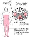
central cord syndrome
common causes? 2
signs and symptoms
Arnold Chiari malformations and prior spinal cord injuries (s/p MVA, whiplash, neck hyperflexion)
strength + sensory loss in upper extremities - damage involves the crossing fibers of the spinothalamic tract (pain/temp) and upper extremity motor fibers due to their medial locations within the corticospinal tra
patient’s is in mild respiratory distress s/p MVA; has breath sounds decreased at the L lung bases. CXR is as follows. What do you suspect and what is the next best step in management?

diaphragmatic injury
blunt abdominal trauma causes sudden increase in intraabdominal pressure that causes large radial tears in the diaphragm, allowing leakage of intra-abdominal contents into the chest, lung compression, and mediastinal deviation
(also shows a NG tube in the lung cavity)
Mgmt: surgical repair
why does a flail chest improve (ie chest movements become symmetrical) with positive pressure mechanical ventilation?
because the mechanical ventilator induces (+) intrapleural pressure (ie it replaces the normal (-) intraplaural pressure) during spontaneous ventilation, thereby causing the previously paradoxical moving flail segment of the thoracic cage to move out normally normally with the rib cage during inspiration
rapid association: intermittent bloody discharge from one nipple
intraductal papilloma
usually difficult to palpate because it is usually < 2mm and soft; US is usually negative as well
(US is best at detecting masses >1 cm)
rapid association: abdominal succussion splash
what type of symptoms are associated?
what are common causes of this?
gastric outlet obstruction caused by mechanical obstruction
post-prandial pain and vomiting with early satiety
causes: gastric malignancy, PUD, Crohns, pyloric stricture/stenosis, ingestion of caustic agents (acid, etc), and gastric bezoars
patient tries to commit suicide by ingesting acid, but regrets doing it after. what is he at risk for later on?
developing a pyloric stricture - acid causes fibrosis 6-12 weeks after the resolution of the acute injury.
when does diabetic gastroparesis tend to occur?
in patients who have had diabetes for >10 years
What doses of prednisone can result in HPA suppression? What are implications of this and how should these patients be managed?
20 mg daily for >3 weeks
patients may not respond appropriately to “stress” (surgery, infection, bleeding, MI) and will require “stress doses” of short-term steroids during the acute condition
oropharynx of a patient rescued from a burning building shows erythema and scattered blisters. What should you do?
endotracheal intubation because of the physical evidence of thermal damage to the upper airway.
Reason for early intubation is that progressive airway edema may preclude intubation later in the patient’s clinical course
patients with renal insufficiency who need to undergo CT with IV contrast cna be given this
N-acetylcysteine
What is the respiratory quotient (RQ)?
When is RQ important?
steady state ratio of CO2 produced to O2 consumed per unit time. Used to assess the body’s metabolism, as this ratio depends mainly upon the major fuel being oxidized for ATP production
RQ 1 = carbs is the major nutrient being oxidized
RQ 0.8 = proteins “ “
RQ 0.7 = fatty acids “ “
impt when trying to wean patients off mechanical ventilation since overfeeding (esp with carbs) can cause excess CO2 production and make weaning more challenging
what promotes atelctasis and a decrease in VC and FRC post-op?
post-op pain (promotes shallow, rapid breathing)
narcotics (decreases respiratory drive, deep inspirations, and coughing
abnormal mucociliary clearance
obesity + supine position
Rapid association: pulsatile groin mass
associated sx?
femoral artery aneurysm
associated with anterior thigh pain due to femoral n. compression that runs lateral to the artery
management of a trauma patient with tenderness in LUQ and US that shows fluid in the spleno-renal angle
depends on the hemodynamic status of the patient and their response to IV fluids:
- if hemodynamically unstable + unresponsive to fluid administration –> emergent exlap
- if hemodynamically unstable +** **responsive to fluid administration (SBP >100mmHg) –> CT abd
type of murmur normally heard in patients with aortic dissection
diastolic decrescendo murmur /aortic regurgitation - usually heard if the dissection involves the aortic root
greatest risk factor for aortic dissection?
HTN
patient on anticoagulation underwent cardiac catherization a few hours ago suddenly complains of back pain and is hypotensive and tachycardic
how do you make the diagnosis and how is this managed?
suspect bleeding/hematoma with retroperitoneal extension most form within 12 hours, esp if the arterial puncture site is above the inguinal ligament
diagnose with non-con CT or US
mgmt: supportive w. bed rest, intensive monitoring, IVF, and blood transfusion if necessary

best imaging modality for assessing soft tissue injuries of the knee
MRI - able to detect complete and partial tears, the exact site of the ligamentous injury and associated injuries to other ligaments/meniscus
patient is s/p partial gastrectomy for intractable PUD; comes in because of postprandial abdominal cramps, weakness, lightheadedness, and diaphoresis, which begins ~30 minutes after eating. Diagnosis? Management?
early dumping syndrome - rapid emptying of hypertonic gastric content into the duodenum/small intestines results in:
fluid shift from intravascular -> small intestines
release of vasoactive polypeptides
stimulation of autonomic reflexes
management: diet ∆s, octreotide for resistant cases, reconstructive surgery for intractable cases
how do you different between a psoas abscess vs appendicitis?
management of either one?
psoas abscess: fever + lower abdominal pain elicited with deep palpation; source of infection include hematagenous spread from the skin (ie furuncles) or contigious spread from nearby bone or bowel abscess
- mgmt: abx + drainage (percutaneous drainage tube, laparotomy, or laparoscopy)
appendicitis: vague periumbilical pain that localizes to RLQ
- mgmt: appendectomy
if you suspect one has an intraabdominal abscess, what imaging modality would you use to confirm?
CT scan
fracture of the midshaft of the humerus can result in what type of presentation?
damage to radial nerve (passes through the radial groover on the posterior side of teh humerus) results in a limitation of extension at the wrist, aka “wrist drop”
patient is anxious-appearing, hypertensive, and tachycardic s/p 30 feet fall from roof. CXR is as follows.
Management?

Note mediastinal widening on CXR, which is the most sensitive finding for aortic injury, which commonly ocurs after blunt deceleration trauma (MVA or fall >10 ft)
when is it safe to start anticoagulation in a hemodynamically stable patient after surgery without significantly increased risk of bleeding or other complications?
48-72 hours
acalculous cholecystitis tends to happen in which patient population?
what is it normally caused by?
how are tehse patients managed?
severely ill patients
(ex: ICU, multi-organ failure, severe trauma, surgery, burns, sepsis, prolonged TPN)
likely due to cholestasis and GB ischemia, which can lead to secondary infection by enteric organisms and resultant GB edema and necrosis. Can lead to sepsis and death if undetected
antibiotics + percutaneous cholecystostomy under radiologic guidance initially, followed by cholecystectomy when the medical condition stabilizes

in evaulating a patient with a shoulder injury, a positive arm drop sign suggests…

rotator cuff tear
falling on an outstretched hand commonly causes injury to the supraspinatous muscle, where the patient has severe shoulder pain and edema and is unable to abduct the arm past 90˚.
a clinical diagnosis of tension pneumothorax is made in the ED and a needle is handed to you for an emergent thoracotomy. Where should you stick your needle?
2nd intercostal space, midclavicular
how are amputated body parts managed?
wrapped in saline-moistened gauze, sealed in a plastic bag, placed on ice and brought to the ED with the patient
do not place directly on ice because this could result in frostbite injury to the amputated tissue and loss of viability
What is Leriiche Syndrome?
Who is it most common in?
arterial occlusion at the level of the aortic bifurcation into the common iliac arteries; characterized by the triad of
<strong>bilateral claudication of thehip, thigh, and buttock</strong>
<strong>impotence</strong>
<strong>symmetric atrophy of bilateral lower extremities due to chronic ischemia</strong>
most common in men with a predisposition for atherosclerosis, such as smokers
Diagnosis and management?

ruptured aorta with blood collection in the adventitial layer
if the patient is unstable –> OR for surgical repair
if the patient is stable –> CT
2 common causes of paralytic ileus
abdominal surgery
retroperitoneal hemorrhage associated with vertebral fracture
patient presents with colicky abdominal pain, vomiting, no bowel movement or passing gas, abdominal distension, diffuse tenderness. How do you know when to manage conservatively vs going to teh OR?
patients should proceed emergently for ex lap if
- they do not improve with conservative measures
- develop signs/symptoms of strangulation
- hemodynamically unstable
<u><strong>conservative measures:</strong></u> complete bowel rest, NG tube, pain control, fluid resuscitation, correction of metabolic derangements
<u><strong>sx of strangulation</strong></u><strong>:</strong>fever, tachycardia, leukocytosis, and metabolic acidosis, peritonitis (occurs only if there is frank bowel necrosis
patient is s/p CABG POD#3 develops fever, leukocytosis, and worsening retrosternal pain despite continuous analgesia Rx; PE cloudy fluid in the sternal wound drain and CXR shows widening of mediastinum. ECHO shows small amount of pericardial fluid.
Dx? Mgmt?
acute mediastinitis, likely secondary to intraoperative wound contamination (the cloudy fluid is actually pus in the mediastinum)
Mgmt: drainage, debridement with immediate closure, and prolonged antibiotic therapy
patient develops a-fib with RVR s/p CABG POD#3. Mgmt?
normal; self-limited and usually resolves in < 24 hrs.
anticoagulation +/- cardioversion is indicated if it doesn’t resolve within 24 hours
How do ligamentous injuries differ from meniscal injuries in terms of presentation? how are they the same?
ligamentous: rapid swelling due to hemarthrosis (ligaments have greater vascular supply than menisci, which rely on diffusion for nourishment)
meniscal: gradual joint swelling over 12-24 hr
BOTH are often associated with a popping sensation
patient has a pneumothorax + sub-cu emphysema s/p high speed MVC; CT placed in the ED. Few hours later, CXR shows an accumulation of air in the pleural space and pneumomediastinum. Ddx?
bronchial rupture - persistent pneumothorax despite CT placement + pneumoediastinum
(don’t confuse wtih esophageal rupture, which can also present with pneumomediastinum in addition to pleural effusions; also esophageal rupture secondary to blunt trauma is rare - usually it’s secondary to iatrogenic causes)
∆ btwn hernia, gastroschisis, and omphalocele
- hernia - covered by skin
- omphalocele - covered with peritoneum without overlying skin
- gastroschisis - no membrane
newborn infant has a large umbilical hernia - is surgery recommended?
yes, large hernias are less likely to close spontaneously, so surgery is recommended around age 5 for persistant hernias, or sooner.
first step in management of suspected urethral injury
retrograde urethrogram
how are urethral injuries treated?
urinary diversion via suprapubic cathether while the primary injury and associated hematomas are allowed to heal, after which assessment/repair of residual damage (ie strictures) is done.
patient presents with a broken clavicle; physical exam shows a palpable gap in the middle of the clavicle and auscultation shows a loud bruit beneath the clavicle. Next best step in management?
since a bruit is heard, get an angiogram to rule out injury to the underlying vessel
what are the 3 components of the GCS scale?
motor response
verbal response
eye opening

management of patient with a suspected scaphoid fracture
place thumb in a spica cast and repeat the x-ray in 7-10 days because the initial x-rays may not show anything if it is a minimally displaced or compressed fracture
if there is a non-displaced fracture –> wrist-immobolization for 6-10 weeks to minimize the risk of non-union since the scaphoid bone is at high risk of avascular necrosis

known complication of AAA repair
how can you prevent this?
bowel ischemia - results from inadequate colonic collateral arterial perfusion to the L and sigmoid colon after loss of the IMA during aortic graft placement
prevent by checking sigmoid colon perfusion following placement of the aortic graft
most common cause of SBO
adhesions secondary to inflammatory processes or abdominal operations (ie appendectomy)
what are black or tarry stools suggestive of?
what are the common causes of melena?
GI bleed that originates above the ligament of Treitz
PUD, gastritis, esophagitis, Mallory-Weiss tear
patient with grave’s disease s/p thyroidectomy 1 month ago has an EKG that shows NSR with QT prolongation.
hypOcalcemia
how does air emboli typically happen?
usually with blunt trauma with pulmonary injury that can result in communications between the airways and vessels - air emboli (esp when positive pressure mechanical ventilation is used) can form
how is bowel ischemia diagnosed and treated?
clinically: severe periumbilical pain that is out of proportion to exam findings, leukocytosis, elevated amylase, and metabolic acidosis (from increased serum lactate levels)
diagnosis: mesenteric angiography
trmt: supportive (IVF, abx, NG tube)
52 F presents with 3 episodes of bright red blood per rectum in the last 2 weeks. AVSS. Rectal eaxm shows internal hemorrhoids that prolapses and are manually reducible. Hg 13, WBC 6500. Next step in management?
get a colonoscopy
cancer until proven otherwise!
trauma patient has clinical signs of a large tension pneumothorax. what should you do?
urgent needle decompression in the second intercostal space, L mid-clavicular line
followed by
chest tube placement (tube thoracostomy) in the 5th intercostal space mid-axillary line
most frequently injured abdominal organs s/p blunt abdominal trauma (ie MVC)
liver and spleen
what is association with an abduction injury to the knee?
what test would help in the clinical diagnosis of this condition?
medial colalteral ligament injury
valgus stress test

what is association with an adduction injury to the knee?
what test would help in the clinical diagnosis of this condition?
lateral collateral ligament
varus stress test
patient who is s/p elective repair of a descending thoracic aneurysm develops flaccid paraplegia and loss of pain sensation in both lower extremities and urinary retention; vibratory sensation is intact. upper extremity exam is normal.
diagnosis? how did this happen?
anterior cord syndrome, likely due to spinal cord infarction
anterior spinal artery supplies the anterior 2/3 of the spinal cord, which includes the motor tracts (corticospinal) and sensory tracts (spinothalamic). it the ASA derives its blood supply from the radicular arteries that originate from the thoracic aorta.
thus thoracic aortic surgery can result in reduced blood flow through the radicular arteries (cross-clamping, systemic hypotension), and lead to anterior spinal cord infarction
signs and symptoms of anterior cord syndrome
spinal shock - abrupt onset of bilateral flaccid paralysis and loss of pain/temperature sensation below the level of the spinal injury
vibration and proprioception are preserved
ABGs of atelectasis
hypoxemia
hypocapnia
respiratory alkalosis
large areas of atelectasis cause VQ mismatch, leading to hypoxemia and increased work of breathing (dyspnea, tachypnea)
to compensate for the hypoxemia, patients usually ventilate and develop respiratory alkalosis and decreased PaCO2/hypocapnia
what is the management of patients with established aortic injury?
anti-HTN and immediate operative repair
differential for an anterior mediastinal mass 4
thymoma
teratoma
thyroid neoplasm
terrible lymphoma
∆ btwn seminomas and non-seminomatous germ cell tumors?
seminomas = usually just elevated ßhCG + normal AFP
non-seminomatous = elevated ßhCG + AFP
when do appendiceal abscesses typically present?
how are they managed?
typically >5 days after the onset of symptoms of appendicitis (when the appendix has perforated and an abscess has formed)
if they are clinically stable -> IV abx, bowel rest, and/or percutaneous drainage of the abscess
can return in 6-8 weeks for interval appendectomy
if FAST is inconclusive, what’s the next best step in the management of blunt abdominal trauma in hemodynamically unstable patients?
DPL
∆ btwn escharotomy and fasciotomy
escharotomy - incision of only the eschar layer
fasciotomy - incision through all fascial layers
(after escharotomy, patients are evaluated for clinical signs of adequate perfusion, and if there is no relief, a fasciotomy should be performed)
patient with hip pain and isolated alk phos elevation, normal serum calcium and phosphorus levels
dx?
associated symptom?
Paget’s disease of the bone (osteitis deformans)
disordered bone remodeling resulting in structurally inferior woven bone
may have hearing loss due to cochlear nerve compression from enlargement of temporal bone
SIRS criteria
T > 38.5˚C (101.3) or <35˚C
Pulse >90/min
RR > 20/min
WBC >12K, <4K, or >10% bands
What are some complications associated with severe burns?
hypovolemic shock
hypermetabolic response (hyperglycemia, muscle wasting, protein loss, hyperthermia, increase energy expenditure)
there is an increase in catecholamines+cortisol s/p burns, which can cause significant protein losses as muscle degradation is used for gluconeogenesis
infection (pneumonia/wound infections) leading to sepsis and septic shock
Patient with severe burns over his body - what are the indications that he is going into septic shock?
worsening hyperglycemia (due to worsening insulin resistance)
leukocytosis
thrombocytopenia
mild hypothermia (T <36˚)
Tachypnea
tachycardia
patients with penile fractures usually have injury to which structure?
management?
tearing of tunica albuginea, which invests the corpus cavernosum. This results in a rapidly-forming hematoma that causes bending of the shaft of the penis at the site of the fracture.
urgent urethrogram to assess for urethral injury and emergency surgery to evacuate the hematoma and to mend the torn tunica albuginea

rapid association: L lower fracture rib
management?
splenic injury
if the patient is stable -> get a CT with contrast, as this is the best diagnostic study to determine if there is a need for surgery
if patient is hemodynamically unstable despite IV resuscitation -> urgent laparatomy
patient with blunt abdominal trauma develops hypotension, LUQ pain + L shoulder pain
management?
splenic injury
(sometimes the stem will indicate L rib fractures as well)
if pt is stable -> get a CT w/ contrast, as this is the best diagnostic study to determine if there is a need for surgery
if pt is hemodynamically unstable despite IV resuscitation -> urgent laparatomy
how do meniscal injuries commonly caused by?
how do they often present?
best diagnostic test?
twisting injury with the foot in a fixed position
symptoms:
- GRADUAL swelling and pain usually within 12-24 hrs
- symptoms of popping, catching, knee giving out, and locking (inability to extend the knee) usually develops weeks later
MRI
best diagnostic test to assess for peripheral arterial disease in symptomatic patients
ankle-brachial index (=SBP in ankle / SBP in arm)
ABI <90 is diagnostic of occlusive peripheral artery disease
highly sensitive screening test for AAA
abdominal US
rapid association: 5 P’s (pain, pulselessness, pallor, paresthesias, paralysis)
arterial occlusion of an extremity
3 major causes of arterial occlusion in the lower extremity
thrombus
embolus
trauma
infection of which neck space carries the highest risk of mediastinal involvement?
retropharyngeal space
What is ludwig’s angina?
infection in the submandibular space
typically begins in the floor of the mouth and extends through the submandibular and sublingual space into tissues surrounding the airway

management of a non-displaced scaphoid fracture (fractures <2mm of displacement and no angulation)
justify answer
wrist-immobolization for 6-10 weeks to minimize the risk of non-union since the scaphoid bone is at high risk of avascular necrosis

A 77 YOM has a severe nosebleed not stopped by anterior packing and he spits clots out side his mouth. Most likely source of the bleed?
sphenopalatine artery
(MCC posterior nosebleeds)

52 F with history of T1DM presents with a 2 month history of R foot swelling w/o fever, pain, or trauma history. PE: swelling on R dorsal forefoot and medial and lateral ankles without ulcerations. The is a bony hypertrophy and small effusion over the ankle. Decreased sensation to light touch, proprioception, and pain below the ankles bilaterally. XRay shows osteopenia and disorganization of the mid-tarsal and tarsometatarsal joints. Cause of these findings?

lack of normal joint sensation (Charcot foot)
progressive degeneration of a weight-bearing joint, a process marked by bony destruction, resorption, and eventual defomity; commonly due to diabetic neuropathy or any condition resulting in decreased peripheral sensation, proprioception and fine motor control

24 hr after romoval of parathyroid adenoma for primary hyperparathyroidism, a 42 YOW has perioral numbness and tingling. He serum Ca is 6.8 (low). Normal albumin levels. Next step in management?
Low Ca levels can cause Prolonged QT -> give Ca gluconate
27 F, HIV + presents with 6 month history of nonbloody diarrhea, now with bloody diarrhea. She has a high fever, and a rigid abdomen. She ends up with an ileostomy for a perforated cecum and the path report shows nuclear inclusion bodies in colon. Most likely organism?
CMV colitis - inflammation of the colon

A 46 YOM with chronic alcoholism presents after 12 hours of N/V and mid-abdominal pain radiating to back. Pulse 120, RR are 20 and BP 110/60. Abdominal exam shows tenderness to palpation upper quadrants. WBC 24K, amylase 1842, albumin 4.1, Ca 7.7. Most important thing to give right now?
LR - needs to be fluid resuscitated
45 M has daily temps to 100.9 and 15 lb wt loss over 3 months. Physical exam is significant for pallor and a low pitched disatolic rumble at the apex that dissappears when he lies on his R side. Hb is 10. Most likely dx?
atrial myxoma - primary heart tumor derived from multipotential mesenchymal cells and may cause a ball valve-type obstruction; causes a mid-diastolic rumble (secondary to mitral stenosis caused by the tumor) that changes when the patient changes position
outine mammorgraphy on a 52 YOw shows six stippled microcalcification in a cluster in the upper outer quadrant of the left creast, not presen t 1 y ago. No lump. Next step?
needle localized open biopsy - performed when there is an abnormality seen on a mammogram that cannot be palpated.
Note that this is NOT a FNA!!
47 F with BMI 67 presents with chafed skin on inner thighs, under breasts. PE shows erythema and active fungal intertrigo, and there is extensive scarring of the axillae and submammary areas. Pelvic exam shows a thick, white, curdy vaginal discharge. Perineal exam shows poor hygiene. Over the last year, she has had two admissions for IV abx for panniculitis. Best long term management for this pt?
gastric bypass
Panniculitis is a group of diseases whose hallmark is inflammation of sub-cu adipose tissue.
52 nulligravid F presents with 2 months of progressive abdominal swelling and decreased appetitie. She has asthma treated with steroids and T2DM. PE shows markedly distended abdomen and (+) fluid wave. CT shows ascites, mulple pelvic masses and omental thickening. Most likely dx?
ovarian adenocarcinoma
As the cancer becomes more advanced, it can cause an accumulation of fluid in the abdomen
ovarian cancer risk factor: <strong>Not having children, </strong>obesity, HRT, increased age
OMENTAL THICKENING: typically caused by metastatic infiltration by tumors arising from the stomach, ovary, or colon (OT-SOC).
For 4 hours, a 55 YOM has acute intermittent pain that begins in right flank and radiates to right testicle. Most likely finding on UA?
microscopic hematuria (> 3/hpf)
A 42 YO is admitted to the hospital with a piece of meat lodged in the lower esophagus. With considerable difficulty, is it removed with esophagoscopy. That evening, the patient spikes a temp of 101˚F. Most apporopriate dx study?
water soluble contrast upper GI study to look for perforation
47yo s/p CABG POD#4 developed a sudden onset of severe pain in his L great toe. PE shows a tender, cyanotic L great tue with new ecchymoses over the trunk and upper/lower extremities. He was admitted to the hospital 10 days ago for chest pain with exertion, and received aspirin and heparin therapy for 3 days. Hct 37, WBC 12.2, Plt 8K PT/PTT WNL.
Diagnosis?
HIT - predisposes to thrombosis because IgG form a complex with heparin + PF4 and the tail of the antibody then binds to the FcγIIa receptor on platelets. This results in platelet activation and the formation of platelet microparticles, which initiate the formation of blood clots; the platelet count falls as a result, leading to thrombocytopenia.
reviously healthy 37 YOW has left groin and thigh pain for 3 d. Exam shows a non reducible mass in the groin. Operative exploration shows a hernia sac medial to the femoral vein. Dx?
femoral hernia
key words: female

24 M presents with burns covering 50% of his body; he weighs 75 kg. How much and what type of fluid does he need in the next 24 hours?
15 L of LR
calculate using Parkland Formula: V = 4* mass (kg) * % area burns
12 hours after rod stabilization of a femoral fracture, 27 homeless man has a sudden onset of combativeness and disorientation. HR 120, RR 24, BP 140/85. Exam shows petichiae over axila. Most likely cause?
Fat emboli - classically presents with
1) respiratory changes - first to present; usually dyspnea, tachypnea, and hypoxemia
2) neurological ∆s - early stages, ranges from confusion, drowsiness, and seizures
3) petechial rash - due to embolization of small dermal capillaries leading to extravasation of erythrocytes; most common in the axillary
NOT alcohol withdrawal, since these do not present with axillary petechiae, which is pathognomonic for fat emboli
A 68 YOM is broguht to the ED because of recurrent vomiting of bright red blood, and near syncope x 3 hours. He is afebrile, pulse 110, respirations are 16, BP 90/60 mm Hg and he has mild epigastric tenderness. Next step?
rapid infusion of IV saline - get the patient stabilized first, then figure out the bleeding source
∆ btwn bronchial rupture vs esophageal rupture?
bronchial rupture - persistent pneumothorax w/ chest tube, pneumomediastinum, sub-cu emphysema
esophageal rupture - pneumonmediastinum + pleural effusions; diagnose with gastrofin contrast esophagogram
how do patients usually present with anterior shoulder dislocations? posterior shoulder dislocations?
anterior = abducted + externally rotated (ant abd ex)
posterior = adducted + internally rotated (post add in)
which part of the bowel is most susceptible to rupture in blunt abdominal trauma and why?
2nd part of duodenum
it is retropertioneal and is the least mobile, perforation occurs when the bowel is compressed between the spine and external solid structure (ie steering wheel, seat belt)
how does osteosarcoma differ from ewing’s sarcoma in terms of clinical presentation and location?
osteosarcoma “osteo = bone pain”
- sites of rapid bone growth (metaphyses, proximal tibia/humeus)
- (+) persistent bone pain that is worse at night
ewing’s sarcoma “eww…symptoms”
- typically at diaphysis
- (+) systemic features (malaise, fevers, wt loss)

management of patients with gunshot wounds; rationale behind this?
how does this differ from stab wounds?
gun shot wound - to the abdomen (below the 4th intercostal space/nipple) requires ex lap to ensure that there is no life threatening injury. Why? because of the blast effect of the bullet
stab wounds - observed and treated conservatively in cases where the patient is hemodynamically stable
management of patient with blunt spinal cord trauma (ie anterior cord syndrome, central cord syndrome)
high dose IV steroids - should be given asap (ie administration should not be delayed for CT/MRIs)
(though its use is controversial)
management of a scrotal mass that is cystic and transilluminates with light in a newborn infant
reassurance + observation because hydroceles typically resolve within 12 months; those that do not resolve should be surgically removed due to increased risk of inguinal hernias
38 F complains of breast discomfort and recently starting having breast pain before menses. PE: smooth, soft, mobile mass in UOQ of breast with diffuse nodularity present bilaterally.
Diagnosis + management?
fibrocystic condition of the breast
- classically presents with breast pain that worsens around the time of menses
- cysts are tender + mobile
Get FNA to r/o malignancy - usually yields serous / thin, greenish, non-bloody fluid with resolution of the mass after aspiration.
a breast mass that produces a non-bloody aspirate and disappears completely w/ aspiration does not need further evaluation other than observation for recurrence
what is the pulmonary capillary wedge pressure a reflection of?
what are the normal values?
indirect measure of LA pressure
normally 6-12 mmHg
increased PWCP with persistent hypotension after a fluid bolus is indicative of …
LV failure/cardiogenic shock
best diagnostic study if an esophageal rupture is suspected
contrast study
avoid endoscopy because insufflation with CO2 may exacerbate the injury
where does appendicitis typically rupture into?
what would a typical physical exam show?
ruptures into rectovesical pouch
PE: tender fluctuating mass palpable with tip of fingers on rectal exam
management of pelvic abscesses
CT guided percutaneous drainage
recommendation if an xray shows:
intact scaphoid
radiolucent line across the R scaphoid bone?
displaced scaphoid fracture
intact scaphoid = analgesics + repeat xrays in 2 weeks
radiolucent line across the R scaphoid bone = cast immobolization
displaced scaphoid fracture = ORIF
angiography shows an intimal flap in the ICA just above the carotid bifurcation
neck exploration + repair of the flat
failure to do so will result in complete obstruction with emboli formation
incidental finding of an elevated alk phos in an elderly patient with hip pain and no other lab abnormalities
Paget’s disease/Osteitis deformans - disordered bone remodeling
patient who presented to the ED after a MVC remains hypotensive despite fluid resuscitation. FAST & DLP = negative
next step in management?
angiogram - consider a retroperitoneal bleed
What is the best imaging modality for a full-term infant is suspected to have developmental dysplasia of the hip? What if the patient was 5 years old?
< 4 yo = Ultrasound
> 4 yo = XRay
why is hematocrit a poor indicator of acute blood loss?
it remains the same/near normal blood levels in the time period immediately following hemorrhage
management of patient with mastitis
antibiotics, analgesics, and continue breast feeding
management of pancreatic pseudocyst
external drainage of the cystic lesion
why do patients develop hypotension and respiratory acidois /hypoxemia after an elective hernia repair?
replacement of the large herniated contents into the peritoneal cavity increases the pressure in the peritoneal cavity and results in:
- decreased VR to heart -> hypotension
- impaired motion of the diaphragm due -> hypotentilation -> decr. pH, incr PCo2, hypoxemia
of these, which ones have the longest duration of analgesia for wound management? shortest?
bupivacaine
chloroprocaine
lidocaine
prilocaine
bupivacaine
short acting (chloroprocaine, 45-90 minutes)
intermediate duration (lidocaine, mepivacaine, prilocaine 90-180 minutes)
long acting (bupivacaine, levobupivacaine, ropivacaine, 4-18 hours)
57M presents with 2 months of difficulty swallowing - food gets stuck when he swallows and he drinks more water to deal with this. Moderate discomfort when he has to make an effort to swallow. He has a 80 PY and drinking history. CXR normal. Next step?
CT chest
CT neck and chest
PPI
esophageal manometry
esophagogastroduodenoscopy
Esophagogastroduodenoscopy (EGD) - diagnostic endoscopicprocedure that visualizes the upper part of thegastrointestinal tract up to the duodenum; indicated when there is:
- Unexplained anemia (usually along with a colonoscopy)
- Upper GI bleed
- Persistent dyspepsia in patients > 45 years
- Heartburn and chronic acid reflux - this can lead to Barrett’s esophagus
- Persistent vomiting
- Dysphagia
- Odynophagia
- Persistent nausea
- IBD (Inflammatory Bowel Diseases)
best diagnostic test for carpal tunnel syndrome
nerve conduction studies - two electrodes are taped to your skin and a small shock is passed through the median nerve to see if electrical impulses are slowed in the carpal tunnel
Hidradenitis suppurativa is caused by what type of glands?
apocrine glands
sweat glands found in teh axillae, areola, nipples, ear canal, eyelids, nostril wings, perianal region, and external genitalia
what is celiotomy?
surgical procedure involving a large incision through theabdominal wall to gain access into theabdominal cavity. It is also known a laparotomy.
metabolic abnormalities associated with gastric cancer
serum IL-2 and TNF-a are associated with advanced gastric cancer and that these cytokines might be a useful tumor marker for gastric cancer, being associated with poor prognosis.
62 M presents with 5 months after noticing a mass in the right side of his neck. Hx of enlarged thymus treated with radiation duroing infancy. Exam shows 4 cm mass that moves with swallowing. TSH is normal. FNA shows follicular neoplasm. Next step?
thyroid function test
thyroid scan
treatment with radioactive iodine
repeat FNA
thyroidectomy
thyroidectomy - high suspicion for cancer in this gentleman given his history of neck radiation
30 wk infant has a harsh to-and-fro murmur consistent with PDA. Indomethacin is given and murmur stops. Explanation of effect?
COX inhibition with increased NE release
