Spinal Cord Flashcards

1
Q

How are spinal tumors classified?

A
  1. Extradural
    1. Fat suppresion is helpful both in pre- and post-contrast T1Wi
    2. Most lesions affecting the vertebral bone marrow then to have low signal on T1Wi
  2. Intradural-extramedullary
    1. Golf-tee sign: focal dilation of the subarachonoic space along the caudal and cranial margins of the intradular mass, best seen on T2Wi
    2. Dural tail sign: focal thickening/infiltration of the minienges adjacent to a meningeal mass (e.g. meningioma)
    3. Tend to enhnce significantly on post-conrtast
    4. Fat saturation may be useful to determina the extent
  3. Intramedullary
    1. Focal swelling of the spinal cord
    2. Circumferential attenuation of the bright CF signal +/- epidural fat on T2Wi
2
Q

In dogs, what category of spinal neoplasia is most common?

A

Extradural followed by intradural-extramedullary

3
Q

Spinal Cord Neoplasia (dogs)

  • Extra-dural spinal cord tumors

.

A
  1. Peripheral Nerve Sheath Tumors
  2. Osteosarcoma
  3. Synoval myxoma/myxosarcoma
  4. Plasma cell tumor
  5. Lymphoma
  6. Metastatic neoplasia (in particular from porstatic carcinoma or TCC)
  7. Solitary or disseminated hystiocytic sarcoma
4
Q

Spinal Cord Neoplasia (dogs)

  • Intradural-extramedullary spinal cord tumors
A
  1. Nerve Sheath Tumots
  2. Meningioma
  3. Nephroblastoma
5
Q

Spinal Cord Neoplasia (dogs)

  • Intramedullary spinal cord tumors
A
  1. Ependyoma
  2. Metastatic neoplasia (in partiacular TCC and hemangiosarcoma)

Less common:

  1. Asrtocytoma
  2. Nephroblastoma
  3. Chondroma
  4. Oligodendroglioma
6
Q

In cats what is the most common neoplasia and where is it usually located?

A
  • Lymphoma
  • Extradural and mixed extradural-ontramedullary are the most common location
7
Q

Spinal Cord Neopalsia (cats)

  • Extra-dural
A
  1. Lymphoma - most common spinal neoplasia overall
  2. Osteosarcoma - 2nd most common spinal neolasia in cats overall
  3. Fibrosarcoma
  4. Undifferentiated sarcoma
  5. Plasma cell tumors
8
Q

Spinal Cord Neopalsia (cats)

  • Intra-dural
A
  1. Meningioma
  2. Histiocytic neoplasia
9
Q

Spinal Cord Neopalsia (cats)

  • Intra-medullary
A
  1. Glial cell
  2. Primitive neuroendocrine tumors
10
Q

Are metastatic spinal cord tumors in dogs or cats?

A

Metastatic vertebral tumors are common in dogs while they are uncommon in cats.

11
Q

What are some benign spinal cord tumors?

A
12
Q

What are imaging characterits of chondrosarcomas on MRI?

A

The MRI appearance of specific vertebral tumors has been rarely reported. For example, chondrosarcoma tends to form:

  • lobulated masses
  • often in the dorsal vertebral compartment
  • markedly T2 hyperintense with few foci of hypointensity
  • iso to hypointense on T1W images with mild to marked peripheral or heterogeneous contrast enhancement
13
Q

What are Osteochondromas?

A
  • Osteochondroma is a cartilage-capped exostosis with a cortex and medulla that blend smoothly with those of the parent bone.
  • The lesion generally arises prior to skeletal maturity from the metaphyseal or juxta-epiphyseal regions of the axial and appendicular skeleton.
  • Although they can be clinically silent, signs associated with spinal osteochondromas may include pain and neurologic deficits as a result of spinal cord compression and impingement of adjacent nerves.
14
Q

What are locations of osteochondromas?

A
  • long bones
  • ribs
  • vertebrae
15
Q

Osteochorndromas can undergo malignant transformation into what kind of tumor?

A

Malignant transformation into osteosarcoma and chondrosarcoma is described with both isolated osteochondroma and multiple cartilaginous exostoses.

16
Q

What are MRI features of vertebral osteochondrfomas?

A
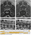
  • A well-defined mass arising from the surface of the affected vertebra and of similar signal intensity to the adjacent normal bone.
  • Presence of a thin cartilaginous cap recognized as:
    • a curvilinear T2 hyperintense, T1 iso- to hypointense structure conforming to the surface of the mass.
  • Variable degrees of spinal cord compression or foraminal impingement
  • There can be variable degrees of mineralization of the cap as the lesion matures, which can also alter the MRI signal intensity (T2W hypointense).
17
Q

In humans what risk factor has been identified to predict malignant transformation for vertebral osteochondromas?

A

In people, it was shown that increased thickness of the cartilaginous cap might be associated with malignant transformation, although this has not been demonstrated with dogs and cats.

18
Q

What are chondromas and what locations are common?

A
  • Chondromas are rare benign tumors characterized by the formation of mature cartilage, more common in flat bones and ribs.
  • In the spine, they may originate from the vertebral body, lamina, pedicles, or transverse or spinous processes, or could evolve from hyperplastic immature spinal cartilage or from metaplastic connective tissue in contact with the spine
19
Q

What are MRI features of synovial myxoma/myxosarcoma?

A
  • A lobulated mass
  • Associated with an articular process joint
  • Causing distortion/lysis of the adjacent articular processes and possibly widening of the joint space
  • The mass typically invades into the adjacent muscles (extends along the fascial planes) and into the vertebral canal, causing variable degrees of spinal cord compression
  • Markedly hyperintense on T2W images, with signal intensity similar to that of the CSF
  • On T1W images the mass is hypointense and there is moderate to strong enhancement on post-contrast images, which can be diffuse, patchy, or rim-like
  • Central non-enhancing areas are common, corresponding to pockets of mucinous substance
20
Q

What are MRI features of infitrative lipomas?

A
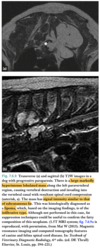
  • Hyperintense on T1W and T2W (turbo or fast spin echo) images with a signal intensity similar to that of normal subcutaneous fat.
  • Fat-suppression techniques are useful
  • A key feature is the local invasiveness into adjacent muscles and, depending on the case, into the vertebral canal, either through the intervertebral foramina and/or through direct invasion of the vertebrae
  • Areas of bony lysis/invasion are typically well marginated
  • There is typically no contrast enhancement, although mild enhancement of the muscles infiltrated by the mass may be present, presumably due to associated myositis
21
Q

What are myelolipomas?

What are common locations of them?

A
  • Myelolipoma is a benign tumor consisting of mature fat interspersed with myeloid and erythroid elements, resembling bone marrow tissue. These lesions are typically asymptomatic; however, large tumors may cause clinical signs as a result of the mass effect. When developing adjacent to the spinal cord, they can cause neurologic deficits and pain.
  • They are reported in dogs and cats in the:
    • spleen
    • adrenal glands
    • liver

Their etiology and classification into the neoplasia group are controversial, and some consider them as being ectopic proliferations, hamartomas, or choristomas

22
Q

What signalment is and location common for epidural myelolipomas?

A

There are a few reports of myelolipomas in the dog in an epidural location, causing spinal cord compression resulting in proprioceptive ataxia and spinal hyperesthesia.

All the dogs were:

  • older male
  • sled dogs (a Siberian Husky, an Alaskan Malamute, and a Husky-cross)

And all the lesions were in the:

  • thoracolumbar junction area
23
Q

What are MRI features of epidural myelolipomas?

A
  • Relatively well-marginated lesion in the epidural space, causing spinal cord compression
  • Mixed signal intensity with a bulk of T1 hyperintense and T2 hyperintense tissue compared with the spinal cord, interspersed with more hypointense areas, conferring overall a heterogeneous appearance
  • On fat suppression pulse sequences, suppression of the hyperintense signal is consistent with fatty components within the mass
  • No contrast enhancement
24
Q

What are peripheral primitive neuroectodermal tumors?

A

Primitive neuroectodermal tumors are rare embryonal undifferentiated tumors of neural crest origin, which in dogs and cats:

  • Most commonly develop in the central nervous system (CNS), in particular the cerebellum where they are referred to as ‘medulloblastomas’, which is mostly seen in young dogs and calves
  • The ‘peripheral’ form of the disease develops outside of the CNS; other terminology found in the literature includes ‘neuroblastoma’. It is derived from germinal neuroepithelial cells of the neural crest that are known to differentiate into autonomic ganglia, dorsal root gan- glia, adrenal medulla, skin melanoblasts, and parts of the peripheral nervous system.
25
Q

What are the two chategoties (based on location) of peripheral primitive neuroectodermal tumors?

A
  • CNS:
    • Medulloblastoma - cerebellum
  • Peripheral forms:
    • Neuroblastoma - these tumors can develop in various locations:
      • bone marrow
      • subcutaneous tissue with visceral metastases
      • cranial nerves
      • paraspinal location (adrenl medulla, autonomic ganglia, peripheral nerves) can cause spinal cord compression
26
Q

What are neuroendocrine tissues?

Given examples of neuroendocrine tumors.

A

Neuroendocrine tissues are tissues that have both nervous system and hormone-producing functions. These cells are located throughout various tissues throughout the body.

Pancreas:

  • Insulinoma
  • Gastrinoma
  • Glucanomas

Adrenal gland:

  • Pheochromocytoma - arise in the medulla of the adrenla gland

Chemoreceptor cells - detect very small changes in the carbon dioxide and oxygen content and pH of the blood, and they help regulate breathing and circulation.

  • Carotid body - neck
  • Aortic body - heart base

Thyroid

  • C-Cell Tumors (uncommon)
27
Q

What are paraganglionic cells?

A

A paraganglion (pl. paraganglia) is a group of non-neuronal cells derived of the neural crest. They are named for being generally in close proximity to sympathetic ganglia. They are essentially of two types:

  1. Chromaffin or sympathetic paraganglia made of chromaffin cells
    * Pheochromocytomas
  2. Non-chromaffin or parasympathetic ganglia made of glomus cells
    * Paraganglioma

The paraganglionic system is made up of the:

  • Adrenal medulla
  • Chemoreceptors
  • Vagal body
  • Small groups of cells associated with the thoracic, intra-abdominal, and retroperitoneal ganglia

Tumors of the paraganglionic tissues are known as paragangliomas, though this term tends to imply the nonchromaffin type, and can occur at a number of sites throughout the body.

Chromaffin paragangliomas are issued from chromaffin cells, and are known as pheochromocytomas.

28
Q

Give some examples of paragangliomas?

A
  • Pheochromocytomas - arise from chromafifn cells of the sympathetic nervous system
    • Adrenal gland
    • Extra-adrenal
  • Chemodectomas - arise from non-chromoffin type of cells of the parasympathetic nervous system that are located in various parts of the body
    • Aortic chemoreceptors - heart base
    • Carotid chemoreceptors - neck
29
Q

What are some imaging characteristics of parangangliomas that develop from paraganglion in the mediastinum or retroperitoneal space?

A

MRI apperance have not been reported:

  • May be locally invasive
  • May extend into the vertebral canal
  • Causing spinal cord compression

CT changes in a dog with invasive mediastinal paraganglioma causing:

  • Vertebral lysis
  • Vertebral canal invasion
  • Cord compression
30
Q

Differentials for soft tissue tumors arising from the paraspinal soft tissue causing secondary invasion of the vertebral column.

What are MRI features of these tumors?

A
  • Fibrosarcoma
  • Rhabdomyosarcoma
  • Other soft tissue sarcomas

MRI features:

  • Locally invasive mass in the paravertebral tissues
  • Extension into the vertebral canal
  • Vertebral destruction - disruption of the hypointense cortex, abnormal intravertebral signal
  • Spinal cord compression will be noted.
  • Signal intensity will be variable, but commonly the lesions are iso- to hypointense on T1W images, hyperintense on T2W images, and contrast enhancing
31
Q

What are the most common intradural-extramedullary spinal tumors in dogs? Less common?

A

Most common:

  • Meningioma
  • Peripheral Nerver Sheath Tumor

Less common:

  • Nephroblastoma
32
Q

What is the most common intradural-extramedullary tumor in cats?

A
  • Lymphoma, although most of these of have an extradural component
  • Meningioma - second most common spinal tumor after lymphoma
33
Q

What are Peripheral Nerve Sheath Tumor?

A

Peipehral nerve sheath tumors are spindle cell tumors that arise from:

  • Schwan cells
  • Perineural cells
  • Firbroblast cells
34
Q

Are PNST malignant or benign in dogs?

A

Most commonly malignant

35
Q

What are the common locations of the PNST?

A

They can arise anywhere from the nerve roots to the most distal portions of the nerves, and for this reason can be found in:

  • Intraspinal locations - most commonly affect of the nerve roots of:
    • Caudal cervical spine
    • Cranial thoracic spine
  • Paravertebral locations
    • Brachial plexus
  • Various regions of the body such as the limbs or the head
36
Q

What are MRI characteristics of PNST?

A

MRI features of intradural–extramedullary peripheral nerve sheath tumors include:

  • Well-marginated mass
  • Typically iso- to hypointense to the spinal cord on T1W images and hyperintense to the cord on T2W images, with moderate to marked contrast enhancement on T1W post-contrast images. Contrast enhancement may be homogeneous or heterogeneous.
  • The intradural component of the lesion causes focal expansion of the subarachnoid space; as described earlier, this is more easily seen on T2W images owing to the hyperintense signal of the CSF outlining the intradural mass. The focal expansion of the subarachnoid space along the cranial and/or caudal margins of the intradural component of the lesion can form a ‘golf-tee’ sign, similar to that observed on myelographic images, particularly on sagittal or dorsal plane images depending on the location of the lesion relative to the spinal cord.

The combined intradural–extradural location:

  • Following the proximal path of the nerve
  • Closely associated with the corresponding intervertebral foramen, is the more common presentation
  • The shape of the lesion varies but owing to its origin within the nerve sheath, an elongated, tubular, or fusiform shape is common
  • The classic appearance is that of a tubular mass extending from the intradural compartment across the intervertebral foramen and into the paravertebral soft tissues. The lesion may be mildly constricted at the level where the nerve traverses the dura mater or where it crosses the intervertebral foramen, which may confer on the lesion a dumbbell appearance.
37
Q

What is the most common type of primary spinal cord tumor (not metastatic) in a dog?

A

Meningioma

38
Q

What are the common localization of meningiomas? Less common?

A

Most commonly:

  • Intradural– extramedullary

Less commonly:

  • They occasionally can grow in the epidural space

Rarely:

  • Rarely, meningiomas arising from the leptomeninges can infiltrate and efface the underlying spinal cord parenchyma thereby giving the impression of an intramedullary mass
39
Q

What are common MRI features of Meningiomas?

A
  • A well-defined focal mass
  • Extradural–intradural compartment causing variable degrees of spinal cord compression
  • Broad-based dural margin seen on at least one of the imaging planes
  • Gradual expansion of the subarachnoid space cranial and caudal to the mass, which can be detected on T2W images, especially in the dorsal or sagittal plane, creating a ‘golf tee’ sign similar to that observed with conventional myelography or CT myelography; heavily T2W MR pulse sequences (‘T2-myelograms’) can be useful in recognizing this sign.
  • Extension of the lesion into the adjacent intervertebral foramen along with the nerve roots can occasionally be seen, and may mimic the appearance of a PNST
  • The mass is usually hyperintense to the spinal cord on T2W images or, less commonly, isointense; ill-defined areas of spinal cord parenchyma T2 hyperintensity around the margins of the mass are occasionally seen, and may represent edema.
  • Spontaneous hyperintensity on pre-contrast T1W images, ranging from mild to marked, has been reported quite frequently with meningiomas and may be a distinctive feature of this tumor in dogs; however, its cause has not been elucidated. The lesion can also be iso- to hypointense on T1W pre-contrast images.
  • Strong, homogeneous contrast enhancement, commonly with a ‘dural tail’ sign, seen as strong linear enhancement of the border of the tumor confluent with the adjacent meninges.
40
Q

What are uncommon MRI characteristics of spinal cord meningiomas?

A

Location in both the intradural and extradural compartments has been reported

Atypical presentations are possible:

  • Cyst-like mass lesion associated with the dorsal subarachnoid space in the cranial cervical spine, with signal intensity similar to that of CSF (T2 hyperintense, T1 hypointense, suppressed on T2-FLAIR, and non-enhancing) has been reported in one case of a spinal cystic meningioma. The myelographic and MRI appearance was similar to that of a spinal arachnoid diverticulum.
  • Very rarely, leptomeningeal meningiomas can infiltrate and efface the underlying spinal cord parenchyma and give the impression of an intra-medullary mass lesion. If adjacent meningeal enhancement is present, the appearance may be confused with other neoplasms such as histiocytic sarcoma.
41
Q

What are nephroblastomas and how are they formed? What are common locations?

A
  • Nephroblastoma is a rare embryonic tumor arising from the primitive metanephric blastema. During embryonic development, this tissue normally differentiates into epithelial and stromal components to form the nephrons and connective tissue of the kidneys.
  • Cells that do not differentiate comprise the ‘nephrogenic rest’ and eventually undergo apoptosis.
  • A nephroblastoma is thought to arise from neoplastic transformation of persistent nephrogenic rest that did not undergo apoptosis.

This can develop in the:

  • Kidney
  • Spine
    • When embryonic tissue becomes trapped in the dura during development.
    • The tumor is most commonly intradural–extramedullary and located consistently between T9 and L3
    • Spinal nephroblastomas typically have histomorphometric features of malignant lesions, although metastatic disease is rare.
42
Q

Do spinal nephroblastomas usually metastasize? What is the common signalment?

A
  • Spinal nephroblastomas typically have histomorphometric features of malignant lesions, although metastatic disease is rare
  • Spinal nephroblastoma occurs typically in young (less than 3 years old) dogs.
  • Some studies have suggested a predisposition in German Shepherd Dogs, although other series did not observe this predisposition.
43
Q

MRI features of spinal nephroblastomas?

A
  • A focal rounded, oval-shaped or lobulated mass
  • Intradural–extramedullary compartment or, less commonly, intramedullary location (rarely extradural).
  • The mass is typically isointense to the cord on T2W images or, occasionally, has a T2 heterogeneous signal or a T2 hyperintense peripheral rim.
  • Areas of T2 hyperintensity in the spinal cord parenchyma adjacent to the mass lesion may be present, and could represent edema or gliosis.
  • The mass is isointense to the cord on T1W images
  • Moderate to strong, often homogeneous, contrast enhancement after gadolinium injection.
  • Transverse and dorsal images typically show the peripheral location of the mass in the dural sac
  • Often with a broad base towards the meninges
  • Widening of the subarachnoid space cranial or caudal to the mass can be present and help in determining the intra-dural location of the mass; however, this sign is not consistently seen and it can be difficult to determine the intradural–extramedullary versus intramedullary origin of the mass, especially when the tumor focally invades the cord or arises from the pia mater.
44
Q

What MRI feature of nephroblastoma indicates poor prognosis?

A

The only MRI criterion that may be associated with prognosis is the location of the lesion:

  • intradural extramedullarylesions having a better prognosis than intramedullary lesions
45
Q

What is the most common type of primary intramedullary spinal cord tumors in dogs? Where are they commonly located?

A

In dogs, intramedullary tumors account for about 15% of all spinal tumors:

Most common primary (in order):

  • Ependymoma
  • Astrocytoma
  • Nephroblastoma, Chordoma, oligodendroglioma, and teratoma.
  • Gliomatosis cerebri is a diffuse glial CNS neoplasia where neoplastic cells reminiscent of astrocytes, oligodendrocytes, or cells with a transitional appearance insinuate among normal structures with minimal damage to neurons and axons. It is a widespread disease that frequently involves multiple divisions of the CNS, including the spinal cord
  • Hemangioblastoma
  • Hemangioma

In dogs, intramedullary tumors overall tend to be more common in the T3-L3 segment, although primary tumors tend to be more common in the cervical spine

46
Q

What are less common intradural spinal cord tumors in dogs?

A
  • Spinal metastatis from intracranial choroid plexus carcinomas
  • Intradural lumbosacral choiroid plexus papilloma (1 report in Shar-Pei)
  • Diffuse spinal meningeal oligodendrogliomatosis (2 boxers and 1 Stafford Bull Terrier)
47
Q

What is the most common type of metastatic intramedullary spinal cord tumors in dogs?

A

Most common:

  • Transitional cell carcinoma
  • Hemangiosarcoma

Less common:

  • Pheochromocytoma
  • Mammary/pancreatic/prostatic carcinoma
  • Sarcoma of unknown origin
48
Q

What is the most common location of intramedullary spinal cord tumors? What about primary intramedullary spinal cord tumors?

A
  • In dogs, intramedullary tumors overall tend to be more common in the T3-L3 segment
  • Although primary tumors tend to be more common in the cervical spine
49
Q

What are the most common types of intramedullary spinal cord neoplasia in cats? What location do they usually arise?

A

Intramedullary neoplasia is even less common in cats, and most are glial cell tumors. Most most commonly (in order):

  • Astrocytomas
  • Ependymomas
  • Oligodendrogliomas

They seem to be more common in the cervical spinal cord.

50
Q

What are general MRI features of intramedullary spinal cord tumors?

A
  • Thinning or attenuation of the subarachnoid space +/− epidural fat, best appreciated on T2W images
  • Swelling of the cord in all planes
  • Variable amounts of intraparenchymal contrast enhancement
51
Q

What are MRI features of spinal cord ependyomas?

A
  • Ependymomas can form fusiform or oval-shaped, focal, or multifocal lesions on sagittal images
  • On transverse images the lesions are centrally located (due to their association with the ependymal lining the central canal)
  • They are iso- or hypointense on T1W images, heterogeneously hyperintense on T2W images, with marked contrast enhancement,
  • Can contain cyst-like components (T2 hyperintense, T1 hypointense, non-enhancing, or ring-enhancing).
52
Q

What are MRI features of spinal cord astrocytomas and oligodendrogliomas?

A

Astrocytomas and oligodendrogliomas form:

  • Ovoid or elliptical mass lesions
  • Well marginated
  • Located eccentrically in the spinal cord on transverse images
  • tVariable degrees of spinal cord expansion
  • he lesions are iso- or hypointense on T1W images, hyperintense on T2W and STIR images, with moderate enhancement on T1W images post contrast injection
  • Mucinous oligodendroglioma may form extensive lesions replacing the normal cord parenchyma; this variant is formed of neoplastic cells separated by mucin causing a discontinuous patchy enhancement pattern
53
Q

What are MRI features of intramedullary spinal cord metastatic disease from TCC?

A
  • Intramedullary masses that are T1 isointense, T2 hyperintense, with mild uniform enhancement on T1W post-contrast image
  • Enlargement of the caudal sublumbar lymph nodes (such as the medial iliac lymph nodes), which appear T2 and STIR hyperintense secondary to metastatic infiltration
  • Concurrent lytic vertebral lesions may also be present.

The appearance of intramedullary metastatic disease is likely varia- ble depending on the nature of the primary tumor

54
Q

What are the MRI features of metastatic intramedullary spinal cord hemangiosarcoma?

A
  • Variable signal intensity on T1W images
  • Hyperintense on T2W images
  • Marked diffuse contrast enhancement or ring-like enhancement
  • Areas of low signal secondary to hemorrhagic foci may be present on T2W images and T2*W images
55
Q

What are the MRI features of spinal cord hemangioblastoma?

A

The appearance of spinal cord hemangioblastoma has been rarely reported.

  • A well-defined intramedullary mass is seen
  • T2 hypo- or hyperintense
  • Mildly hyperintense on T1W pre-contrast images
  • On T1W post-contrast images, there is either diffuse strong contrast enhancement or rim enhancement.
56
Q

What are MRI features of intramedullary spinal cord hemangioma?

A

A capillary hemangioma formed an:

  • Intramedullary mass
  • Heterogeneous but mostly hyperintense on T2W images
  • Mildly hyperintense on T1W images
  • Strong homogeneous contrast enhancement except for a central area.

A cavernous hemangioma formed a focal:

  • Intramedullary mass
  • Target-like appearance:
    • On T2W images there was a small isointense center surrounded by a large hyperintense area and a peripheral hypointense region
    • On T1W images there was a large hypointense center surrounded by a small hyperintense periphery and a peripheral hypointense region
  • No contrast enhancement
57
Q

What breeds are predisposed to hystiocytic sarcomas? What are the two forms of it and what are the common locations?

A

Breeds:

  • Bernese Mountain Dog
  • Rottweiler
  • Golden Retriever
  • Flat-coated Retriever
  • Labrador Retriever

Retrievers and Pembroke Welsh Corgis may be predisposed to CNS involvement with histiocytic sarcoma, with Corgis possibly predisposed to the primary CNS form.

Classification:

  • Localized:
    • Most commonly a soft tissue mass along the limbs
    • CNS
  • Disseminated:
    • Spleen
    • Liver
    • Lungs
    • Lymph nodes
    • Bone marrow
    • CNS
58
Q

What are MRI features of spinal cord hystiocytic sarcomas?

A
  • Focal or multifocal intramedullary and/or intraduralextramedullary mass lesion(s), most commonly isointense (less commonly hypo- or hyperintense) on T1W images, hyperintense or of mixed signal intensity on T2W images, hyperintense on STIR, with moderate to marked contrast enhancement after gadolinium injection
  • Ill-defined diffuse intramedullary T2 hyperintensity with loss of the epidural fat and gray/white matter definition, moderate parenchymal contrast enhancement after gadolinium injection, and mild diffuse meningeal enhancement.
  • Meningeal enhancement in the vicinity of focal mass lesions, sometimes with a dural tail sign.
  • Meningeal enhancement often extends over long distances from the mass lesion, and isolated areas of meningeal enhancement distant and separate from the mass lesion are also possible, suggestive of metastatic spread of the condition through the subarachnoid space.
  • In some cases, the only abnormality may be meningeal enhancement without a mass lesion (‘leptomeningeal histiocytic sarcoma’).
  • MRI features of spinal histiocytic sarcoma, such as focal mass lesions, meningeal thickening, and enhancement, can also be seen with other neoplasms such as lymphoma or rare tumors such as meningeal oligodendrogliomatosis, and are therefore non-specific.
  • Intradural–extramedullary masses with dural tail can also be seen with meningioma.
59
Q

What are the most common organs that are also affected in cats that have spinal cord lymphoma?

A
  • Bone marrow
  • Kidneys
  • In some reported case series, affected cats had lesions in multiple regions of the CNS including the brain, while in other series, single epidural lesions were more common
60
Q

What are MRI features of spinal cord lymphoma in dogs?

A
  • Multifocal disease more common than focal disease
  • Regional involvement of multiple compartments:
    • Vertebrae (none, one, several, or all vertebrae uinvolved) -with possible cortical involvement, seen as patchy T2W and STIR hyperintense signal with enhancement on T1W post-contrast images, disrupting the normal hypointense signal of the cortex.
    • Paraspinal soft tissues - often ill-defined
    • Epidural space - cause variable spinal cord compression
    • Leptomeningeal involvement with enhancement on post-contrast images is also reported, as well as involvement of the nerve roots and spinal cord parenchyma.
    • The vertebral canal and paraspinal components may be seen to communicate through the intervertebral foramina

Signal characteristics are similar for the lesions in the paraspinal and vertebral canal areas:

  • T1 iso- to hypointense, T2 and STIR hyperintense, moderately to strongly contrast enhancing.
  • In some cases, the lesions can be hyperintense on T1W pre-contrast images.
  • Heterogeneous signal on T2W images, with hypo- and hyperintense areas, is also reported, as well as isointensity to surrounding muscles.

Additional lesions that can be seen in the FOV include:

  • Lymphadenopathy
  • Hepatomegaly
  • Splenomegaly
  • Splenic nodules
61
Q
A