Skin Terminology/Diseases Flashcards
Papule
- a small pimple or swelling on the skin, often forming part of a rash
- can be a primary lesion resulting from a flea bite - developing as a direct consequence of disease process
- solid palpable skin elevation less than 1cm in diameter
EX: Canine FAD (crusted papules)
secondary lesion
- evolve from a primary lesion ot are caused by the patient (aka self trauma)
- selt trauma in veterinary patients may obscure the primary lesions
- secondary bacterial infection may present with primary lesions
Panniculus Adiposus
- vascular and nerve supply under the layer of the dermis
(generally find sweat glands in this area as well)
- The panniculus adiposus is the fatty layer of the subcutaneous tissues, superficial to a deeper vestigial layer of muscle, the panniculus carnosus. It includes structures that are considered fascia by some sources but not by others
the Epidermal Layer
(5 layers)
stratified squamous keratinizing epithelium with proliferation, differentiation, desquamation
happens in hair follicle as well - down to the insertion of the sebaceous gland duct - can get notable disruptions
shows there may be a foliclur keratinization or cornification (final stage of keratinization) defect going on

Cutaneous Basophil Hypersensitivity
- Cutaneous basophil hypersensitivity is a distinct form of hypersensitivity reaction with a delayed-time course that is different from both the classic delayed-type hypersensitivity reaction and immediate hypersensitivity reaction.
- It occurs in humans, guinea pigs, and other animals.
- It may be induced by sensitization with a variety of antigens (viruses, allografts, parasites, fungal antigens, etc.) in incomplete Freund’s adjuvant and elicited by skin testing 7 days later with the specific antigens.
- The cutaneous reaction in basophil hypersensitivity is characterized clinically by less indurated (hardened) erythema than in classic delayed hypersensitivity, and microscopically by numerous basophils in the papillary dermis.
- The reaction is mediated by both T- and B-lymphocytes.
Basophils

- Basophils appear in many specific kinds of inflammatory reactions, particularly those that cause allergic symptoms.
- Basophils contain anticoagulant heparin, which prevents blood from clotting too quickly.
- They also contain the vasodilator histamine, which promotes blood flow to tissues.
Primary lesion
If Foot and Mouth replicates in horse epidermis–> may lead to primary lesion in pigs and ruminants
- staphylococcal infection creating pustular lesion in the hair follicle
- a secondary lesion can have its own primary infections - staphylococcus taking advantage of allergy
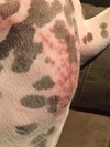
Wheals
- you know as they “pit” upon the application of pressure- may get some with traumatic pericarditis
- so it is different to firm nodules or plaques
- YOU GET WHEALS FROM PENICILLIN = hives (urticaria)
- Urticaria (hives) is a vascular reaction of the skin marked by the transient appearance of smooth, slightly elevated papules or plaques (wheals) that are erythematous and that are often attended by severe pruritus. Individual lesions resolve without scarring in several hours
- Horses–> present with wheals more commonly as a species (dogs as well) –> horses are not generally pruritic!
- A skin wheal is a patch on the skin that is elevated, discolored, changes shape, and often itches (+/-). It can be due to an insect bite, an adverse reaction to something that touched your skin, eczema, or another small puncture wound you experienced. You may have also heard a skin wheal referred to as a ‘welt’ or a ‘hive.
- In some animals, serum oozes from the wheals matting the hair coat

Angioedema
- extensive type wheal
- angioedema is a localized or generalized area of extensive deep dermal and subcutaneous oedema
- affecting the whole body region –> vaccine reactions or maybe (may get dogs swelling up around the head after routine vaccine)
- in comparison, the oedema involves the deep dermis and subcutis where in urticaria it involves only the superficial dermis
- both involve type I and type III hypersensitivities
- pruritis is not always present, especially in the horse
Urticaria
- Urticaria, also known as hives, is an outbreak of swollen, pale red bumps or plaques (wheals) on the skin that appear suddenly (involving the superficial dermis) – either as a result of the body’s reaction to certain allergens, or for unknown reasons.
- Hives usually cause itching, but may also burn or sting
- a unique form has been described in Jersey and Guernsey cattle bc of a type I hypersensitivity to casein (slow digesting protein) in their milk
- Urticarial lesions are wheals that typically arise suddenly and remain a few hours although chronic urticaria has been noted (lasting weeks to months)
macule
an area of skin discoloration
pustules
- A pustule is a bulging patch of skin that’s full of a yellowish fluid called pus
- discrete elevation of the epidermis containing PUS (within or just below epidermis)
- like a plaque with pus
- a mini abscess located near or in the epidermis
- Where as an ABSCESS: is a demarcated dermal/subcutaneous accumulation of pus

Vesicle/Bullae
Bullae: (Blisters)
Vesicles are circumscribed epidermal elevations in the skin containing clear fluid and less than ½ cm. in diameter. If the lesion has a diameter of greater than ½ cm, it is called a bulla. Vesicles and bullae arise from a cleavage at various levels of the skin
Sebaceous Adenitis

- Sebaceous adenitis in an uncommon skin disease found in some breeds of dog, and more rarely in cats, rabbits and horses. characterised by an inflammatory response against the dog’s sebaceous glands, which can lead to the destruction of the gland
- targets sebaceous glands and results in alopecia and epidermal and follicular hyperkeratosis
- useful to take a biopsy sample in suspected cases and then have disease confirmed by histopath
- In standard poodles, sebaceous adenitis is definitely a recessive genetic trait
Hyperkeratosis
abnormal thickening of the outer layer of the skin
- increase in the stratum corneum layer


- Hair follicles destroyed by fibrosis - where there should be a hair follicle is just collagen
- Could see cytotoxic lymphocytes are now seen attacking the hair follicles in this
- Can now think of drugs that would block lymphocytes mediated autoimmune disease - cyclosporine
- But some owner refuse due to the $$ and dog not suffering from itchy


- Hair follicles on normal histopathology
Examples of diseases that involve different areas of the skin:
- Dermis
- Panniculus (subcutis, hypodermis)
- Epidermis
- Hair Follicles
**note: panniculitis, not hepatitis
also in hair follicles: Demodecosis

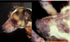
Puppy Strangles
(juvenile cellulitis)

- DERMAL DISEASE
- Puppy strangles, or juvenile cellulitis, is a nodular and pustular skin disorder that affects puppies. It usually occurs between the ages of three weeks and four months, and is rarely seen in adult dogs.
- The face, pinnae (outer part of the ear), and salivary lymph nodes are the most common sites to be affected. The cause of this condition is unknown, but there are breeds that have been shown to be predisposed to it, including golden retrievers, dachshunds, and Gordon setters
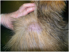
Clinical Signs
- Acutely (sudden and severe) swollen face – especially the eyelids, lips, and muzzle
- Salivary gland lymphadenopathy: a disease process affecting a lymph node or multiple lymph nodes
- Marked pustular and oozing skin disease, which frequently fistulates (develops into a hollow passage); develops within 24–48 hours
- Pustular ear infection
- Lesions often become crusted
Sterile Nodular Palliculitis

- Panniculitis is a rare condition in which the layer of fat under the skin, which provides warmth, protection and energy to the body, becomes inflamed.
- While it may be caused by an infection with bacteria, fungi or other organisms, sterile nodular panniculitis is a descriptive term for an inflammation of the fat cells that does not involve infectious agents. In most cases, the exact cause is not known.
- The inflammation results in bumps on the skin surface that can be soft or firm, and are sometimes painful.
- The bumps can rupture, releasing an oily discharge that may be clear, yellow-brown or bloody.
- Most pets with sterile nodular panniculitis are treated with drugs designed to modify the immune system, such as steroids, but Vitamin E may also be helpful
Patterns of Inflammation in the Skin
(5)

Hyperepidermis
- increase in the stratum spinosum layer
- epidermal living layers thicken
- may be a protective response by the body to a noxious stimulant from outside
- also as an accidental consequence of a dermal disease triggering cytokines and inflammatory mediators trigerring the basal keratinocytes to increase the proliferative rate
- may see both hyperkeratosis and hyperepidermis in a chronically allergic animal


- hyperplasia more regular
- hyperkeratosis is quite marked
- CLINICAL EQUIVOLENT: SCALING
- scale = an accumulation of loose fragments in the stratum corneum
- Migration of lymphocytes into the epidermis (lymphocyte exocytosis) is common - as seen here
- can also see in this periodic acid–Schiff (PAS) stain that there is some fungal hyphae - Dermatophytosis (ringworm)
Scaling
- accumulation of loose cornified fragments from the stratum corneum