S3) Innate Immune System Flashcards
What is the immune system?
The immune system is a network of cells and organs that contribute to immune defences against infectious and non-infectious conditions (self vs non-self)
Identify 4 roles of the immune system
- Pathogen recognition: cell surface and soluble receptors
- Containing/eliminating the infection: killing and clearance mechanisms
- Regulating itself: minimum damage to host (resolution)
- Remembering pathogens: preventing disease from recurring
Compare and contrast the innate and adaptive immunity
- Innate (immediate protection): fast (seconds), lack of specificity and memory, no change in intensity
- Adaptive (long lasting protection): slow (days), specificity, immunologic memory, changes in intensity
What is an infectious disease?
An infectious disease is when a pathogen succeeds in evading and/or overwhelming the host’s immune defences
Identify and describe the 3 mechanisms by which micro-organisms trigger the inflammatory cascade
- Pilus enhances attachment
- Lipopolysaccharide endotoxins triggers inflammation
- Polysaccharide capsule promotes adherence and prevents phagocytosis
The inflammatory cascade is triggered when an endotoxin binds to macrophages.
Outline its following phases:
- Local
- Systemic
- Sepsis
- Local: cytokines, TNFs and interleukins promote wound repair and recruit the reticuloendothelial system
- Systemic: cytokines released into circulation and stimulate GF, macrophages & platelets (aims to control infection)
- Sepsis: cytokines lead to activation of humoral cascades, RE System, circulatory insult (infection is not controlled)
Explain the relationship between sepsis and coagulation
- Cytokines released in sepsis initiates production of thrombin
- Thrombin promotes coagulation and cytokines also inhibit fibrinolysis
- Coagulation cascade leads to microvascular thrombosis → organ ischaemia, dysfunction and failure (sepsis)
What are the three factors determining the outcome of the host-pathogen relationship?
- Infectivity
- Host’s immune response
- Virulence
The first line of defence consists of factors that prevent entry and limit growth of pathogens.
Identify the different innate barriers to infection
- Physical barriers
- Physiological barriers
- Chemical barriers
- Biological barriers
Identify the 3 different physical barriers to infection in the innate immune system
- Skin
- Mucous membranes: mouth, resp tract, GI tract, urinary tract
- Bronchial cilia
Identify and describe the 4 physiological barriers to infection in the innate immune system
- Diarrhoea – food poisoning
- Coughing – pneumonia
- Sneezing – sinusitis
- Vomiting – food poisoning, hepatitis, meningitis
Identify and describe the 2 different chemical barriers to infection in the innate immune system
- Low pH – skin (5.5), stomach (1-3), vagina (4.4)
- Antimicrobial molecules:
I. IgA (tears, saliva, mucous membranes)
II. Lysozyme (sebum, perspiration, urine)
III. Mucus (mucous membranes)
IV. Gastric acid + pepsin
Identify and describe the biological barrier to infection in the innate immune system
Normal flora:
- Non pathogenic microbes
- Found in nasopharyngeal, mouth/throat, skin, GI tract, vagina
- Absent in internal organs/tissues
What are the benefits of normal flora as a biological barrier in the innate immune system?
- Compete with pathogens for attachment sites and resources
- Produce antimicrobial chemicals
- Synthesise vitamins (K, B12, other B vitamins)
Describe how the different innate barriers work together to maximise the response against microbes
Innate barriers trigger the second lines of defence:
- Phagocytes
- Chemicals
What does the second line of defence in the innate immune system do?
- Causes inflammation
- Contain and clear the infection
Provide some examples of opsonins
- Complement proteins: C3b, C4b
- Antibodies: IgG, IgM
- Acute phase proteins: CRP, MBL
Opsonis are essential in clearing encapsulated bacteria.
Identify three examples of this type of bacteria
- Neisseria meningitidis
- Streptococcus pneumoniae
- Haemophilus influenzae b
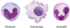
Identify the 3 main types of phagocytes
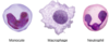
State the location and functions of macrophages

- Location: present in all organs
- Functions:
I. Ingest and destroy microbes by phagocytosis
II. Present microbial antigens to T cells
III. Produce cytokines/chemokines
State the location and function of monocytes

- Location: present in the blood (5-7%)
- Function: recruited at the infection site and differentiate into macrophages
State the location and functions of neutrophils
- Location: present in the blood (60% of blood leukocytes)
- Functions:
I. Increased during infection
II. Recruited by chemokines to the site of infection
III. Ingest and destroy progenitor bacteria e.g. Staph aureus
State the function of the following key cells in the innate immunity:
- Basophils/mast cells
- Eosinophils
- Natural Killer cells
- Dendritic cells
- Basophils/ mast cells: early actors of inflammation (vasomodulation) in allergic responses
- Eosinophils: defence against multicellular parasites (worms)
- NK cells: kills all abnormal host cells (viral infected/malignant)
- Dendritic cells: present microbial antigens to T cells (acquired immunity)

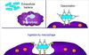
What is opsonisation?
Opsonisation is the process that involves coating microbial surfaces with opsonin proteins leading to enhanced attachment of phagocytes and clearance of microbes
Identify the structures involved in pathogen recognition
- Microbial structures: pathogen-associated molecular patterns (PAMPS) – carbohydrates, lipids, proteins, nucleic acids
- Phagocytes: pathogen recogniton receptors (PRRs)

In 7 steps, outline the process of phagocytosis
⇒ Recognition (PAMPS and opsonins)
⇒ Chemotaxis and adherence of the microbe to the phagocyte
⇒ Ingestion of the microbe by the phagocytes
⇒ Formation of phagosome
⇒ Fusion with lysosome to form a phagolysosome
⇒ Digestion of ingested microbe by enzymes
⇒ Discharge of waste materials
Identify and describe the 2 different phagocytic intracellular killing mechanisms
- Oxygen-dependent pathway (respiratory burst): toxic O2 products for pathogens – peroxide, hydroxyl radical, nitric oxide
- Oxygen-independent pathways: lactoferrin and transferrin, proteolytic and hydrolytic enzymes
The complement pathway consist of 20 serum proteins.
Identify and describe the 2 activating pathways
- Alternate pathway – initiated by cell surface microbial constituents e.g. endotoxins on E.coli
- MBL pathway – initiated when MBL binds to mannose containing residues of proteins found on many microbes e.g. Candida albicans
Describe the antimicrobial actions in the alternate pathway of the complement system
- C3a and C5a: recruitment of phagocytes
- C3b-C4b: opsonisation of pathogens
- C5-C9: killing of pathogens, membrane attack complex
Provide 4 examples of normal flora that inhabit the skin
- Staphylococcus aureus
- Staphylococcus epidermidis
- Streptococcus pyogenes
- Candida albicans
Provide 3 examples of the normal flora which live in the nasopharynx
- Streptococcus pneumoniae
- Neisseria meningitidis
- Haemophilus species
