S1B5 - Orbit (with CN III, IV & VI) Flashcards
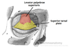
What is the action of the superior tarsal muscle? What is its innervation?
Superior tarsal muscle: The superior tarsal muscle attaches to the tarsus of the upper eyelid.
- Action: Elevation of the eyelid
- Innervation: Sympathetic nerves (superior cervical ganglion)
Clinical Correlate: Sympathetic innervation of the superior tarsal muscle explains the ptosis seen in Horner’s Syndrome

What is the action of the inferior oblique muscle on the eye? What is its innervation?
Inferior oblique:
The inferior oblique muscle is located inferiorly at the floor of the orbit.
- Action: Elevates, abducts, and externally rotates (extorts) the eye
- Innervation: CN III

What is formed by dense connective tissue plates that forms the walls of the eyelids?
Tarsus: The tarsus is formed by dense connective tissue plates that forms the walls of the eyelids.
What bones form the roof of the orbit?
Roof – orbital plate of frontal bone and lesser wing of sphenoid.

What bones form the floor of the orbit?
Floor – maxilla, zygomatic and small part of palatine (Note: the palatine bone forms the floor and lateral wall of the nasal cavity, the roof of the mouth, and the floor of the orbit).

What are the arteries of the orbit mainly derived from?
The arteries of the orbit are mainly derived from the ophthalmic artery, a branch of the internal carotid artery.

What is the action of the lateral rectus muscle on the eye? What is its innervation?
Lateral rectus:
- Action: Abducts the eye
- Innervation: CN VI

What are the 2 actions of the superior rectus muscle on the eye? What is its innervation?
Superior rectus:
The superior rectus muscle originates from the superior part of the common annular tendon.
- Action: Elevates and medially rotates (intorts) the eye
- Innervation: CN III

Describe the innervation of the muscles of extraocular movement.
Mnemonic: Remember the innervation of the muscles of EOM:
“SO4 LR6, and all the rest by 3”
- SO4: Superior oblique — CN IV (trochlear nerve)
- LR6: Lateral rectus — CN VI (abducens nerve)

What protects the orbit within the ocular cavity and is contiuous with the pericranium?
Orbital septum: The orbital septum protects the orbit within the ocular cavity; it is continuous with the pericranium.
What is the action of the medial rectus muscle on the eye? What is its innervation?
Medial rectus:
- Action: Adducts the eye
- Innervation: CN III

The following clinical findings are suggestive of what diagnosis and etiology?
- Ptosis - drooping eyelid
- Miosis - constricted pupil
- Anhydrosis - loss of hemifacial sweating
Clinical aside: Horner’s syndrome - damage to superior cervical ganglion or sympathetic pathway causing: A triad:
- Ptosis – drooping eyelid
- Miosis – constricted pupil
- Anhydrosis – loss of hemifacial sweating
Parasympathetic fibers involved with pupillary reflexes such as the lightreflex (direct, and consensual) and accommodation.
The following is a description of injury to what nerve?
- Weakness of downward eye movement
- vertical diplopia (double vision)
- Patient tilts the head forward in order to bring the fields back together
- Weakness of intorsion resulting in torsional diplopia
Injury to the trochlear nerve:
- Weakness of downward eye movement
- vertical diplopia (double vision)
- Patient tilts the head forward in order to bring the fields back together
- Weakness of intorsion resulting in torsional diplopia
The oculomotor nerve carries preganglionic parasympathetic fibers to what ganglion?
The oculomotor nerve (CN III) also gives off the preganglionic oculomotor root of the ciliary ganglion, which synapses with the ciliary ganglion.
The short ciliary nerves carry postganglionic parasympathetic fibers to the ciliary muscle and the pupillary sphincter muscle, which act on the lens and pupil. The ciliary muscle is responsible for accommodation and the pupillary muscle is responsible for miosis.
What bones form the lateral wall of the orbit?
Lateral Wall – zygomatic and greater wing of sphenoid

What is the action of the postganglionic parasympathetic fibers which leave the ciliary ganglion?
The oculomotor nerve (CN III) also gives off the preganglionic oculomotor root of the ciliary ganglion, which synapses with the ciliary ganglion.
The short ciliary nerves carry postganglionic parasympathetic fibers to the ciliary muscle and the pupillary sphincter muscle, which act on the lens and pupil. The ciliary muscle is responsible for accommodation and the pupillary muscle is responsible for miosis.

Through which opening do the nerves of extraocular movement enter the orbit?
The nerves of the orbit enter the orbit via the superior orbital fissure.
The following findings are from dysfunction of what nerve?
- Affected eye pointing inferolaterally during rest
- Ptosis (eyelid drooping)
- Mydriasis (pupil dilation)
Clinical aside: Oculomotor palsy – ‘down and out’
Paralysis of the medial rectus, etc. with unopposed superior oblique, and lateral rectus. The affected individual will also have a ptosis, or drooping of the eyelid, and mydriasis (pupil dilation).
Which ophthalmic vein communicates with the “triangle of danger”?
Superior ophthalmic vein communicates with supraorbital and angular veins - (triangle of danger)
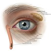
Describe fluid drainage from the lacrimal gland.
The lacrimal punctum a tiny opening which drains lacrimal fluid via the lacrimal canaliculus.
The lacrimal canaliculus drains to the lacrimal sac, which communicates inferiorly with the nasal cavity via the nasolacrimal duct.
Note: This is why you sniffle when you cry!
Where is the lacrimal groove located?
Lacrimal groove – between the lacrimal and maxillary bones for the lacrimal sac.

What is the direction of eye deviation in oculomotor palsy?
Clinical aside: Oculomotor palsy – ‘down and out’

What do the ophthalmic veins drain into?
Ophthalmic veins: Ophthalmic veins drain to the cavernous sinus.
The following findings indicate dysfunction of what nerve?
- esotropia, a convergent squint on a distant object
- to compensate, patients turn their face toward the affected eye
Abducens nerve (CN VI)
What is the action of the superior oblique muscle on the eye? What is its innervation?
Superior oblique:
The superior oblique muscle originates on the medial side of the orbit and passes through the trochlea to insert onto the sclera.
- Action: Depresses, abducts, and medially rotates (intorts) the eye
- Innervation: CN IV

What is the action of the levator palpebrae superioris? What is its innervation?
Levator palpebrae superioris:
- Action: Elevates the upper eyelid
- Innervation: CN III
What are the 2 actions of the inferior rectus muscle on the eye? What is its innervation?
Inferior rectus:
- Action: Depresses and externally rotates (extorts) the eye
- Innervation: CN III

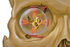
What artery is found at the center of the optic nerve and supplies the retina?
Central retinal artery: The central retinal artery is found at the center of the optic nerve and supplies the retina.
Clinical Correlate: Cholesterol emboli from the internal carotid artery can occlude the central retinal artery and cause central retinal artery occlusion (CRAO). CRAO often presents with amaurosis fugax, the sudden loss of vision in one eye lasting up to a few hours.
Aneurysms of the internal carotid artery can damage what nerve to the orbit?
Aneurysms of the internal carotid artery can damage the abducens nerve (CN VI).
What bones form the medial wall of the orbit?
Medial wall – orbital plate of ethmoid, lacrimal, frontal process of maxilla and a small part of sphenoid

Which 3 cranial nerves innervate the muscles of extraocular movement?
The muscles of extraocular movement (EOM) function to move the eyeball within the bony orbital cavity.
- Innervation: CN III, IV and VI
The common annular tendon is the attachment for the 4 rectus muscles.

