S11) Cancers of the Reproductive Tracts Flashcards
Where can gynaecological tumours arise?
- Vulva
- Cervix (neck of uterus)
- Endometrium (lining of uterus)
- Myometrium (body of uterus)
- Ovary
What are the clinical features of vulval tumours?
- Uncommon
- Approx. 2/3rds occur > 60 years of age
- Usually squamous cell carcinoma
How many vulval squamous neoplastic lesions are related to HPV infection?
- 30% HPV-related (6th decade) – risk factors the same as for cervical carcinoma
- 70% HPV-related (8th decade) – often occur in longstanding inflammatory and hyperplastic conditions of the vulva e.g. lichen sclerosis
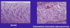
What is vulvar intraepithelial neoplasia?
- Vulvar intraepithelial neoplasia involves atypical squamous cells within the epidermis (no invasion)
- It is an in situ precursor of vulval squamous cell carcinoma

How does vulval squamous cell carcinoma spread?
- Spreads initially to inguinal, pelvic, iliac and para-aortic lymph nodes
- Thereafter spreads to lungs and liver
Almost all cases of CIN and cervical carcinoma are related to high risk HPVs.
How does an HPV infection lead to these conditions?
⇒ Infects immature metaplastic squamous cells in transformation zone
⇒ Produces viral proteins E6 & E7 which interfere with activity of TSGs to cause inability to repair damaged DNA and increase cell proliferation

What are the risk factors for CIN and cervical carcinoma?
- Early first sexual intercourse
- Early first marriage/pregnancy
- Multiple births
- Sexual promiscuity
- Immunosuppression (cannot clear HPV infection)
Why is cervical screening successful?
- Cervix accessible to visual examination (colposcopy) and sampling
- Slow progression from precursor lesions → invasive cancers (years)
- Pap test detects precursor lesions and low stage cancers
- Allows early diagnosis and curative therapy
What does cervical screening involve?
- Cells from the transformation zone are scraped off
- Cells are stained with Pap stain
- Cells are examined microscopically
- Cervical cells can be tested for HPV DNA
In cervical screening, abnormalities are referred for colposcopy and biopsy.
What sort of abnormalities could be seen?
- Increased nuclear:cytoplasmic
- Irregular nuclear outlines
- Hyperchromatic nuclei

What are the advantages of vaccinating men against HPV too?
- Reduce risk of oral and penile cancer
- Reduce risk of transmission of HPV
- Protect girls who cannot be vaccinated (herd immunity)
What is Cervical Intraepithelial Neoplasia?
- CIN is a dysplasia of squamous cells within the cervical epithelium, induced by infection with high risk HPVs
- Three stages: CN I mostly regresses spontaneously, some progress to CN II (in situ carcinoma) and 10% may progress to an invasive carcinoma (CN III – 2-10 years)
What is the treatment for CIN?
- CIN I – follow-up or cryotherapy
- CIN II & CIN III – superficial excision (LLETZ – large loop excision of transformation zone)
What are the different types of invasive cervical carcinomas?
- 80% – squamous cell carcinomas
- 15% – adenocarcinomas (also caused by high risk HPVs)
Which age group is usually affected by invasive cervical carcinoma?
Average age = 45 years
What do invasive cervical carcinomas look like?
Exophytic (external) or infiltrative (stromal invasion through basement membrane)
Identify the three ways in which invasive cervical carcinomas spread
- Locally to para-cervical soft tissues, bladder, ureters, rectum, vagina
- Lymphatic system to para-cervical, pelvic, para-aortic nodes
- Distally
How does cervical carcinoma present?
- Screening abnormality
- Postcoital, intermenstrual or postmenopausal vaginal bleeding
How are cervical carcinomas treated?
- Microinvasive carcinomas: cervical cone excision
- Invasive carcinomas: hysterectomy, lymph node dissection and radiation and chemotherapy (if advanced)
Describe the structure and location of the endometrium
- Location: lines internal cavity of uterus
- Structure: glands are within a cellular stroma
Why is endometrial hyperplasia a frequent precursor to endometrial carcinoma?
- Increased gland:stroma ratio
- Associated with prolonged oestrogenic stimulation:
I. Annovulation
II. Increased oestrogen from endogenous sources (e.g. adipose tissue)
III. Exogenous oestrogen
What are the clinical features of endometrial adenocarcinoma?
- Most common invasive cancer of the female genital tract
- Usual age: 55-75 years
- Presents with irregular or postmenopausal vaginal bleeding
What do endometrial adenocarcinomas look like?
Polypoid or infiltrative
Identify the two types of endometrial adenocarcinoma
- Endometrioid endometrial adenocarcinoma
- Serous carcinoma
What are the clinical features of endometrioid endometrial adenocarcinoma?
- More common
- Mimics proliferative glands
- Arises due to endometrial hyperplasia
- Spreads by myometrial invasion to local lymph nodes and distant sites
- Associated with unopposed oestrogen and obesity
How do endometrioid endometrial adenocarcinoma look?

What are the clinical features of serous carcinoma (endometrial adenocarcinoma)?
- Poorly differentiated
- Aggressive
- Exfoliates, travels through oviducts and implants on peritoneal surfaces
What is the commonest tumour of the myometrium?
- Leiomyoma – benign tumour of myometrium (fibroid)
- Probably most common tumour in women
What are the clinical features of a leiomyoma?
- Often multiple
- Range from tiny → massive
- Asymptomatic or heavy/painful periods, urinary frequency, infertility
- Malignant transformation rare
What does a uterine leiomyoma look like?
- Well circumscribed, round, firm and whitish in colour
- Bundles of smooth muscle (resembles normal myometrium)

Describe the clinical features of the malignant tumour of the myometrium
Uterine leiomyosarcoma:
- Uncommon
- 40-60 years
- Doesn’t arise from leiomyomas
- Metastasises to lungs
What are the clinical features of ovarian tumours?
- 80% are benign – 20-45 years
- 20% are malignant – 45-65 years
- Many are bilateral
Why do ovarian cancers have such a poor prognosis (70% 1 year survival)?
Ovarian cancers have often spread beyond the ovary by the time of presentation and therefore the prognosis is often poor
How do ovarian tumours present?
- Most non-functional – produce symptoms when large, invasive or metastasise
- Mass effects – abdominal pain and distension (GI & urinary symptoms)
- Ascites
- Hormonal problems – menstrual disturbances and inappropriate sex hormones
What are the clinical features of malignant ovarian tumours?
- Approx 50% spread to other ovary
- Metastasise to regional nodes and elsewhere
- Some associated with BRCA mutations
Which tumour marker is used in the diagnosis and monitoring of ovarian carcinoma recurrence and progression?
Serum CA-125
How do we classify ovarian tumours?
Dependent on the tissue from which they have arisen:
- Müllerian epithelium (including endometriosis)
- Germ cells (pluripotent)
- Sex cord-stromal cells (form the endocrine apparatus of the ovary)
- Metastases
What are the three main histological types of ovarian epithelial tumours?
- Serous
- Mucinous
- Endometrioid
How can one classify ovarian epithelial tumours?
- Benign
- Borderline
- Malignant
What are the risk factors for ovarian epithelial tumours?
- Nulliparity / low parity
- Oral contraceptive pill (protective)
- Heritable mutations e.g. BRCA1 and BRCA2
- Smoking
- Endometriosis
How do serous ovarian tumours present?
Often spread to peritoneal surfaces and omentum, therefore commonly associated with ascites

How do mucinous ovarian tumours present?
- Large, cystic masses – can be >25kg
- Filled with sticky, thick fluid
- Usually benign/borderline

What is pseudomyxoma peritonei?
- Pseudomyxoma peritonei is a condition caused by cancer cells (mucinous adenocarcinoma) which produce extensive mucinous ascites due to epithelial implants on peritoneal surfaces
- There’s frequent involvement of ovaries which can cause intestinal obstruction
How do endometrioid ovarian tumours present?
- Contain tubular glands resembling endometrial glands
- Can arise in endometriosis (15-20%)
- Associated with endometrial endometrioid adenocarcinoma (15-30%)
What are the clinical features of germ cell ovarian tumours?
- Most are teratomas
- Usually benign
Identify some malignant germ cell ovarian tumours
- Dysgerminoma (resembles seminoma of testes)
- Yolk sac tumour
- Choriocarcinoma
- Embryonal carcinoma
Identify and describe the three types of ovarian teratomas
- Mature (benign) – most common
- Immature (malignant) – rare, composed of tissues that resemble immature foetal tissue
- Monodermal (highly specialised)
What are the clinical features of ovarian mature teratomas?
- Most are cystic
- Almost always contain skin-like structures, usually contains hair, sebaceous material and tooth structures
- Usually occur in young women
- 10-15% bilateral

The most common types of monodermal ovarian teratomas is the struma ovarii.
Describe its clinical features
- Benign
- Composed entirely of mature thyroid tissue
- May be functional and cause hyperthyroidism
Describe the clinical basis of ovarian sex cord-stromal tumours
- Derived from ovarian stroma (which is derived from sex cords)
- Sex cord produces Sertoli & Leydig cells (testes) and granulosa and theca cells (ovaries)
- Tumours resembling all of these four cell types can be found in the ovary and can be feminising or masculinising
What are the clinical features of granulosa cell tumours?
- Most occur in post-menopausal women
- May produce large amounts of oestrogen → precocious puberty in pre-pubertal girls
- Associated with endometrial hyperplasia, endometrial carcinoma and breast disease in adults
What are the clinical features of ovarian Sertoli-Leydig cell tumours?
- Blocks normal female sexual development (in children – functional)
- Causes defeminisation and masculinisation (in women – functional): breast atrophy, amenorrhoea, sterility, hair loss
- Peak incidence in teens/ twenties
Metastases to the ovaries are most commonly due to Mϋllerian tumours.
Identify the structures involved
- Uterus
- Fallopian tubes
- Contralateral ovary
- Pelvic peritoneum
Metastases to the ovaries are most commonly due to Mϋllerian tumours.
Identify some other tumours which metastasise to the ovaries
- GI tumours (colon, stomach, biliary tract, pancreas, appendix)
- Breast tumour
- Krukenberg tumour
What is a Krukenberg tumour?
- A Krukenberg tumour is a metastatic gastrointestinal tumour within the ovaries
- It is often bilateral and usually from stomach
Identify three tumours which occur in the testes
- Germ cell tumours
- Sex cord-stromal tumours
- Lymphomas
What are the two different types of germ cell tumours?
- Seminomas
- Non-seminomatous germ cell tumours (NSGCTs)
What are the two types of sex cord-stromal tumours?
- Sertoli cell tumours
- Leydig cell tumours
Identify four types of non-seminomatous germ cell tumours (NSGCTs)
- Yolk sac tumours
- Embryonal carcinomas
- Choriocarcinomas
- Teratomas