S1) CVS Anatomy Flashcards

1
Q

State the structure and function of the pericardium

A
  • Structure: a fibroserous sac
  • Function: encloses the heart and roots of the great vessels
2
Q

Identify the 2 layers of the pericardium

A
  • Outer fibrous layer
  • Inner serosal layer
3
Q

Identify and describe the 2 components of the inner serosal layer in ther pericardium

A
  • Visceral pericardium adheres to the external wall of the heart
  • Parietal pericardium lines the outer fibrous layer and formed when the visceral pericardium reflects back on itself
4
Q

What is the pericardial cavity and what does it do?

A
  • The pericardial cavity is the space between the visceral and parietal layers in the pericardium
  • It consists of pericardial fluid which reduces friction
5
Q

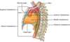
Describe the anatomical position of the pericardium

A

The pericardium is attached to the sternum and mediastinal portions of the right and left pleura

6
Q

Identify the blood vessels which emanate from the pericardium superiorly and inferiorly

A
  • Superiorly: aorta, pulmonary artery, superior vena cava
  • Inferiorly: inferior vena cava
7
Q

Describe the structures forming the different surfaces of the heart:

  • Apex
  • Base/Posterior surface
  • Anterior surface
  • Inferior surface
A
  • Apex - formed by the tip of the left ventricle
  • Posterior surface - formed by the atria (mainly the left)
  • Anterior surface - formed by the right atrium and ventricle
  • Inferior surface - formed by both ventricles (mainly the left)
8
Q

What is another name for the inferior surface of the heart?

A

Diaphragmatic surface - lies along the diaphragm

9
Q

Identify 2 functions of the heart valves

A
  • Direct blood flow in a forward direction
  • Prevent backward leakage
10
Q

Identify the four major valves in the normal heart

A
  • Atrioventricular valves:

I. Tricuspid valve

II. Mitral valve

- Semilunar valves:

I. Aortic valve

II. Pulmonary valve

11
Q

What is the endocardium?

A

The endocardium is the single layer of endothelial cells that lines the surface of the heart valves and interior surface of the chambers

12
Q

Identify 5 components of subendocardial tissue

A
  • Fibroblasts
  • Elastic and collagenous fibres
  • Veins
  • Nerves
  • Branches of the conducting system
13
Q

What is the myocardium?

A

The myocardium is the thickest layer of the heart and consists of bundles of cardiac muscle cells

14
Q

What is the epicardium?

A

The epicardium is the outermost layer of the heart and is identical to, and just another term for, the visceral pericardium

15
Q

What is external to the myocardium?

A

External to the myocardium is a layer of connective tissue and adipose tissue through which pass the larger blood vessels and nerves that supply the heart muscle

16
Q

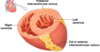
Identify the 3 vascular structures which open into the right atrium

A
  • Superior vena cava
  • Inferior venae cava
  • Coronary sinus
17
Q

Describe the purpose of the vascular structures which open up into the right atrium

A
  • Venae cavae return deoxygenated blood from the systemic veins
  • Coronary sinus return deoxygenated blood from the cardiac veins
18
Q

What is found on the floor of the right atrium?

A

Tricuspid valve – opens into the right ventricle

19
Q

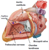
How does the right ventricle get its sponge-like appearance?

A

The right ventricle is covered by a number of irregular bridges (trabeculae carneae)

20
Q

How does the right ventricle help to prevent the tricuspid valve opening during systole?

A
  • The right ventricle contains three papillary muscles, which project into the chamber via their heart tendons (chordae tendineae)
  • They attach to the edges of the tricuspid valve leaflets & contract to prevent the back-flow of blood
21
Q

What is found at the apex of the right ventricular outflow tract?

A

Pulmonary valve – leads to the pulmonary artery

22
Q

Which vascular structures open into the left atrium?

A

Four pulmonary veins

23
Q

What is found in the inferior wall of the left atrium?

A

Mitral valve – opens into the left ventricle

24
Q

How can the walls of the left and right atriums be distinguished?

A

Left atrium wall is thicker than that of the right atrium

25
How can the structure of the left and right ventricles be distinguished?
**-** Left ventricle **cavity** is cone shaped and longer - **Wall thickness** is 9 to 11 mm (3x right ventricle) - **Trabeculae carneae** are finer and more numerous - Larger **papillary muscles** (only 2) **- Chordae tendineae** are thicker and less numerous
26
Describe the opening into the aorta from the left ventricle
- Surrounding the aortic valve opening is a fibrous ring to which is attached the three cusps of the valve - Above the right and left aortic valve cusps in the aortic wall are the origins of the **right** and **left coronary arteries**
27
What is the aortic vestibule?
**Aortic vestibule** is a smooth-walled part of the left ventricular cavity located just inferior to the aortic valve
28
Describe location and function of the cardiac veins
- **Function:** return blood from the myocardial capillaries to the right atrium predominantly via the coronary sinus - **Location**: lie in the epicardial fat, usually superficial to their arterial counterparts
29
Describe the anatomical course of the left coronary artery
- The left coronary artery passes between the left atrium and the pulmonary trunk to reach the AV groove - There, it divides into the **left anterior descending artery** (LAD) and the **circumflex artery**
30
Describe the anatomical course of the left anterior descending artery
LAD travels within the anterior interventricular groove toward the cardiac apex: - It gives off **septal branches** that supply the anterior 2/3 of the IV septum - It gives off **diagonal branches** that supply the anterior surface of the left ventricle
31
Describe the anatomical course of the circumflex artery
- Continues within the left AV groove and passes around the left border of the heart to reach the posterior surface - Gives off **large obtuse marginal branches** that supply the lateral and posterior wall of the left ventricle
32
Describe the anatomical course and function of the right coronary artery
- **Course**: travels in the right AV groove, passing posteriorly between the right atrium and ventricle - **Function**: supplies blood to the right ventricle via acute marginal branches
33
In most people (85%), the distal RCA gives rise to a large branch, the posterior descending artery. Describe the anatomical course of this artery
- Travels from the inferoposterior aspect of the heart to the apex - It supplies blood to the inferior and posterior walls of the ventricles and the posterior 1/3 of the IV septum
34
Where does the AV nodal artery arise from?
Just before giving off the posterior descending branch, the RCA usually gives off the **AV nodal artery**
35
Blood supply to the SAN is through the SA Nodal artery. Describe the variation in its origins.
- In 70% of normal hearts, blood supply is **derived from the RCA** - In 25% of normal hearts blood supply is **derived from the circumflex** artery - In 5% of normal hearts, blood supply is **derived from the RCA** and circumflex artery
36
The heart lies in the middle mediastinum. What is the mediastinum?
The **mediastinum** is the intervening region in the thoracic cavity between the right and left pleural cavities which are occupied by the lungs
37
Identify the structures visible from the anterior surface of the heart
38
Identify the structures visible from the posterior surface of the heart
39
What are the oblique and transverse pericardial sinuses?
- The **transverse sinus** is a passage between the aorta and pulmonary artery posteriorly and the superior vena cava anteriorly - The **oblique sinus** is the pericardial cul-de-sac behind the heart which opens into the pericardial space
40
Identify and name the major arteries comprising the vascular system in the head and neck region
- Brachiocephalic trunk - Common Carotid artery - Internal Carotid artery - External Carotid artery - Superficial Temporal artery - Facial artery - Maxillary artery
41
Identify and name the major arteries comprising the vascular system in the thorax
Thoracic (Descending) Aorta & its intercostal branches
42
Identify and name the major arteries comprising the vascular system in the abdomen
- Abdominal Aorta & its paired branches - Celiac artery - Superior Mesenteric artery - Inferior Mesenteric artery - Suprarenal arteries - Renal arteries - Ovarian/Testicular arteries & its unpaired branches
43
Identify and name the major arteries comprising the vascular system in the pelvis
- Common Iliac artery - External Iliac artery - Internal Iliac artery
44
Identify and name the major arteries comprising the vascular system in the upper limb
- Subclavian artery - Axillary artery - Brachial artery - Radial artery - Ulnar artery
45
Identify and name the major arteries comprising the vascular system in the lower limb
- Femoral artery - Popliteal artery - Anterior and Posterior Tibial arteries
46
Identify and name the major veins in the vascular system in the head and neck region
- External jugular vein - Internal jugular vein - Right and left brachiocephalic vein
47
Identify and name the major veins in the vascular system in the thorax
Superior vena cava
48
Identify and name the major veins in the vascular system in the abdomen
- Inferior Vena cava - Right and left renal veins - Left and right testicular vein
49
Identify and name the major veins in the vascular system in the pelvis
- Common Iliac vein - External Iliac vein - Internal Iliac vein
50
Identify and name the major veins in the vascular system in the upper limb
- Subclavian vein - Cephalic vein - Basilic vein - Median cubital vein
51
Identify and name the major veins in the vascular system in the lower limb
- Femoral vein - Long Saphenous vein - Short Saphenous vein - Popliteal veins