Reversible Cell Injury & Cellular Accumulations Flashcards
What is reversible cell injury?
Injured cell can regain homeostasis & return to a morphologically & functionally normal state
Cellular swelling is the SINGLE FEATURE that can be recognized under a light microscope
Swollen cells have a paler cytoplasm
Fatty change (lipid accumulation) & glycogen accumulation are also considered to be reversible hepatocyte injuries
What is the mechanism for cell swelling (reversible cell injury)?

Hypoxic Injury/ Mitochondrial Damage
Depletion of cellular O2 → decreased oxidative phosphorylation → less ATP produced → failure of ATP dependent Na+/K+ ATPase pumps → influx of Na+/Ca2+/ H2O into cell → Cell loses K+ & Mg2+
Influx of H2O causes cell swelling
Direct Membrane Injury that results in Cell Swelling
Free radical damage (lipid peroxidation)
Covalent binding by toxins
Interference with ion channels (ionophore toxicity)
Insertion of transmembrane complexes
Bacterial cytotoxins
MAC complex from complement system - Immune Molecules
Gross Appearance of Reversible Cell Injury

Increased pallor (paler color)
Increased weight
Left is abnormal
Cellular Swelling under Light Microscopy

Paler looking
Cells are enlarged w/ many small, colorless vacuoles (pinched off segments of ER, Golgi & swollen mitochondria
Paler cytoplasm
More cytoplasm present
Some cells might have increased eosinophilia of the cytoplasm
As cell swelling progresses to irreversible cell injury, specifically necrosis, eosinophilia becomes more apparent
Darker pink, glassy appearance of cytoplasm
Balooning Degenration in the Oral Mucosal Epithelium
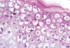
Reversible cell injury
Progression to colorless cytoplasm:
Pale pink → Clear
Arrows point to viral inclusions (pink)
Progression from
Reversible Cell Injury to Irreversible Cell Injury
Progression to irreversible cell injury when reversible is no longer reversible
Irreversible cell injury leads to cell death via:
Apoptosis
“Programmed cell death”
Not usually associated with membrane rupture that occurs with oncosis
Necrosis
Death by swelling and subsequent cell membrane rupture
Intracellular Accumulations
(What are they?)
Injured cells can accumulate endogenous by-products or exogenous substances b/c of:
- Metabolic abnormalities
- Genetic mutations
- Lacking enzyme
- Defect in protein folding
- Exposure to indigestible exogenous substances
Substances can accumulate:
- Cytoplasm
- Within organelles
- In the nucleus
- May be produced within the cell or elsewhere
4 Pathways to Abnormal Intracellular Accumulations
Inadequate removal of a normal substance
- Defect in packaging & transport mechanisms
Genetic or acquired defects that result in an accumulation of an abnormal endogenous substance
- Folding, packaging, transport, secretion defects
Failure to degrade metabolites due to inherited enzyme deficiency
- Storage diseases
Accumulation of an abnormal exogenous substance
- Cell doesn’t have the ability to degrade, transport or remove substance
- Ex. Carbon or silica particles
- Lungs affected (pulmonary macrophages)
Types of Intracellular Accumulations
Lipids
Lipidosis- Steatosis- Fatty change (synonomous)
Glycogen
Proteins
Autophagic Vacuoles
Crystalline Protein Inclusions
Viral Inclusions
Lead
Lipid Intracellular Accumulations

Accumulation of lipids within parenchymal cells (functional tissue of an organ)
- Most common in liver
- Liver is main organ in lipid metabolism
- Kidney is 2nd most common
All classes of lipids can accumulate:
- Triglycerides
- Cholesterol/cholesterol ethers
- Phospholipids
Causes of Intracellular Lipid Accumulations (3)

Increased mobilization of free fatty acids
- Metabolic machinery can’t keep up
Abnormal metabolism in the liver
- Ex. Hepatic toxicosis
Impaired release of lipoproteins
- Ex. Cat with a genetic lipoprotein lipase deficiency
- Ex. hepatic lipidosis in toy breed puppies
Fatty Liver, Hepatic Lipidosis
Gross Appearance

Steatosis
Uniform pale yellow-tan color
Liver is enlarged, with rounded edges
- Normal margins of a liver lobe should come to a point
Liver bulges on incision
May feel greasy
May float in formalin
Fatty Liver, Hepatic Lipidosis
(Lipid Intracellular Accumulations)
Light Microscopy

Severely affected liver
All hepatocytes contain unstained, sharply defined cytoplasmic lipid vacuoles
Nucleus is displaced to the periphery of the cell

Intracellular Lipid Accumulations
Cholesterol and Cholesterol Esters
(Atherosclerosis)

Atherosclerosis
- Accumulation of cholesterol esters w/in SM of arterial vessel and w/in macrophages
- Filled with lipid vacuoles containing cholesterol & cholesterol esters
- Cells have a foamy appearance
- Only significant in pigs, chickens and rabbits
Intracellular Lipid Accumulations
Cholesterol & Cholesterol Esters
Xanthomas

Xanthomas
- Accumulation of cholesterol-laden macrophages in tissues (mainly skin)
- Arise from metabolism disorders
- Hyperlipidemia in cats
- Diabetes mellitus
- high blood glucose levels & glucose spills into urine
- High fat diets
Intracellular Glycogen Accumulations
Stored mainly in hepatocytes & muscle during homeostasis
Stores can be depleted with starvation (anorexia)
Glycogen accumulation occurs in:
- Metabolic abnormalities of skeletal muscle
- Glycogen storage diseases (glycogenesis)
- In the liver
Intracellular Glycogen Accumulations in the Liver

Glucocorticoid (Steroid) Hepatopathy - chronic excess of corticosteroids (endogenous or exogenous)
- Hyperadrenocorticism (Cushing’s)
- Functional pituitary or adrenal tumors
- Excess exogenous corticosteroids (iatrogenic)
- Prolonged or high-dose corticosteroid accumulation
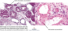
Image: Glucocorticoid-induced Hepatopathy, Liver, Dog
- Extensive accumulation of glycogen in hepatocytes
- Results in a enlarged, pale-brown to beige liver
- Arrows point to swollen hepatocytes with huge amounts of cytoplasmic vacuolation from glycogen accumulation
Lipid Vacuoles vs. Glycogen Vacuoles
(Intracellular Accumulations in the Liver

Lipid Vacuoles:
- More discrete
- Round
- Displace nucleus of hepatocyte
Glycogen Vacuoles:
- Blend better with cytoplasmic constituents
- Lacey and eosinophilic
- Irregular shape
- No displacement of nucleus
Both found in the liver
If you put a section of fatty liver in formalin…
It will float
Intracellular Protein Accumulations
as
Reabsorption Droplets in Proximal Renal Tubules
Reabsorption droplets in proximal renal tubules
- Seen in renal disease where there is protein loss in the urine
- Protein-losing nephropathy (low protein in blood & high in urine)
Disease that affect glomerular filtration →
- More protein leaking into renal tubules →
- heavy reabsorption of proteins by proximal tubular epithelial cells →
- protein droplet accumulation in cells (they can only absorb so much)
- heavy reabsorption of proteins by proximal tubular epithelial cells →
Intracellular Protein Accumulations
Mott Cells & Russell Bodies

Proteins that accumulate may be normally produced by the cell but are present in excessive amounts
- Ex. Plasma cell producing too much immunoglobulin
- “Mott Cell”
- Accumulation of excessive immunoglobulin expands ER and produces large, homogenous eosinophilic inclusions = Russell bodies
- Russell bodies = ER swollen with protein bodies
Intracellular Accumulations - Autophagic Vacuoles

Autophagy
- Process where cell eats its own contents
- Survival mechanism during periods of nutrient deprivation
Involved in many physiologic and pathologic processes
- H&E appear as small, clear, & colorless vacuoles in the sarcoplasm of myofibrils
- Look like holes in skeletal muscle

Intracellular Accumulations
Crystalline Protein Inclusions

Rhomboid crystalline protein inclusions
- Aka crystalloids
- Common in hepatocytes & tubular epithelial cells of older dogs
- Marker of cell aging
- Appear as rectangular, pink, intranuclear inclusions
Intracellular Accumulations
Viral Inclusions
Presence of viral inclusions depends on chronicity of disease
Expect to see inflammation & remodeling
- Early in disease = inclusions usually present
- If animal survives, more chronic or reparative lesions are seen, then inclusions probably wont be there
- Viral inclusions can be intranuclear, intracytoplasmic or both
- Generally, DNA viruses have intranuclear inclusions
- RNA viruses have intracytoplasmic
- Exceptions: Pox virus, Canine Distemper (heat-shock protein)
Intracellular Accumulations
Viral Inclusions
Canine herpesvirus & Herpesviral nephritis

Canine herpesvirus:
- Intranuclear red inclusions
- Chromatin from nucleus is marginated (pushed out)
Herpesviral nephritis:
- Turkey egg kidney
- Splotchy
- Red foci correlate to areas of necrosis
Intracellular Accumulations
Viral Inclusions
Canine parvovirus & Canine parvovirus enteritis

Canine parvovirus:
- Pink, intranuclear inclusions
Canine parvovirus enteritis
- Necrotizing enteritis (dead mucosa)
Intracellular Accumulations
Viral Inclusions
Canine Adenovirus & Adenoviral Hepatitis

Canine adenovirus
- Pink, intranuclear inclusions
- Larger inclusions than canine herpes virus
Adenoviral hepatitis
Intracellular Accumulations
Viral Inclusions
Canine Distemper Virus

Canine Distemper Virus (CDV)
- Pink intranuclear & intracytoplasmic inclusions
Bronchointerstitial pneumonia
- Lung was never deflated
- Something preventing exhalation
Intracellular Accumulations
Viral Inclusions
Rabies Virus in a Cow

Rabies virus in a cow
- Negri bodies
- Red to brown intracytoplasmic inclusions
Intracellular Accumulations
Lead

Lead Poisoning
- Can produce intranuclear inclusions in renal tubular epithelial cells
- Mixture of lead + protein
- Acid-fast positive inclusions (red)
