Restrictive lung disease Flashcards
Define what restrictive lung disease is
- This is where there is a decrease in the total volume of air that the lungs are able to hold.
- Causes may be intrinsic (caused by disease of the lungs itself) or extrinsic (caused by disease outside of the lung - often related to disease affecting the expansion of the chest wall)
Describe the pathology & causes of intrinsic causes of restrictive lung disease
There is impaired alveolar gas exchange to O2 whilst CO2 gas exchange remains unimpaired. Due to inflammation &/or scar tissue, & aveolar inflammation caused by the interstitial lung diseases (ILD’s) or due to alveolar inflammation caused by oedema, haemorrhage (vasculitis) or infection
Describe the pathophysiology & causes of extrinsic causes of restrictive lung disease
Causes:
- Thoracic/extra-thoracic = obesity, kypho-scoliosis, ascites, diaphrgmatic palsy
- Neuromuscular disorders = MND or myasthenia gravis
- Pleural diseases = diffuse pleural thickening, mesothelioma or large pleural effusions
These causes result in impaired alveolar ventilation (hypoventilation) which results in a rise in PaCO2 & a decrease in PaO2
How is the general diagnosis of restrictive lung disease made ?
A combination of clinical exammination, PFT’s & radiological assessment
How is the diagnosis of specific ILD’s made ?
- This requires careful history taking regarding -Occupational, hobbies, smoking & drug history
- Often then supported by blood tests & radiological CT patterns of disease, MDT’s & occasionally lung biopsy
What are the PFT’s which show restrictive lung disease ?
- Decreased FVC with a FEV1/FVC > 70
- Decreased DLCO (<80% of predicted)
Recall what DLCO is
This measures the ability of the lungs to transfer gas from inhaled air to the red blood cells in pulmonary capillaries
List the causes of a decreased DLCO
- Anaemia
- Emphysema
- ILD’s
- Pulmonary oedema
- PE
What is DLCO used to monitor ?
Treatment response in ILD’s (it is more senstive than FVC)
What are the different Tx’s for the causes of extrinsic causes of ILD?
- Lifestyle, weight loss Mx for obesity
- Treatment of neuromuscular disease e.g. myridostigamine in MG
- Intercostal or ascitic drainge for ascites or pleural effusion
- Corrective spinal surgery for scoliosis
- Decortication for chronic empyema
- Diaphragmatic plication for diaphragmatic paralysis etc
Define what the lung parenchyma is
This is the portion of lung involved in gas transfer i.e. the alveoli, alveolar ducts & resp bronchioles
Define what interstitial lung disease is
- This is the generic term used to describe a number of conditions that primarily affect the lung parenchyma in a diffuse manner,
- They are characterised by chronic inflammation &/or progressive interstitial fibrosis & share a number of clinical pathological features
What are the 3 different classifications of ILD’s ?
- Those with a known cause
- Those associated with systemic disorders
- Idiopathic pulmonary fibrosis (unknown cause)
List the known causes of ILD’s
- Occupational/environmental e.g. asbestosis, beryllosis, silicosis, cotton workers lung (byssinosis), coal workers pneumoconiosis
- Drugs e.g. nitrofuratonin, bleomycin, amiodarone, sulfasalazine & busulfan
- Hypersensitivity reactions e.g. EAA
- Infections e.g. TB, fungi, viral
- GORD
List the systemic disorders associated with causing ILD
- Sarcoidosis
- RA
- SLE, systemic sclerosis, mixed connective tissue disease & sjorgens syndrome
What are the signs/symptoms of ILD’s ?
Symptoms:
- Progressive SOB
- Dry cough
- Malaise
- Weight loss
- Arthralgia
Signs:
- Bilateral fine-end inspiratory crackles
- Finger clubbing
- Restrictive spirometry pattern & decreased DLCO
- Abnormal CXR or CT
- Cyanosis
How should everyone with suspected ILD be investigated/ diagnosed ?
- Detailed history + clinical exammination
- Blood tests to help identify underlying cause + ABG
- Spirometry & DLCO
- CXR
- HR-CT
MDT then review this info to made diagnosis or deicde on further tests
What blood tests are done with investigating someone for ILD?
- FBC, ESR, CRP
- Immunoglobulins (connective tissue & vasculitis screen - ANCA’s with MPO/PR3 Abs)
- ANA
- Rheumatoid factor
If a confident diagnosis cannot be made by the MDT based on the initial tests then what is done to definitivley diagnose ILD?
Bronchoalveolar lavage or transbronchial biopsy &/or surgical lung biopsy
Describe what extrinsic allergica alveolitis (EAA) is
- It is a type of ILD, EAA is also known as hypersensitivity pneumonitis & is a condition where in sensitised individuals, inhalation of allergens (fungal spores or avian proteins) provoke a hypersensitivity reaction
- In the acute phase (type III hypersensitvity) alveoli are inflated with acute inflam cells. With chronic exposure (type IV) hypersensitivity granuloma formation & oblierative bronchiolitis occur
List the causes of EAA
- Bird-fanciers lung - avain proteins
- Farmers lung - spores of saccharopolyspora
- Mushroom workers lung - thermophilic actinomycetes
- Malt workers lung - aspergillus clavatus
- Bagassosis or sugar workers lung - thermoactinomyces sacchari
- Drugs - gold, bleomcyin, sulfasalazine
What are the acute features of EAA & when do they occur ?
4-8hrs after exposure to allergens:
- SOB
- Dry cough
- Fever
- Myalgia
What are the features of chronic EAA?
Features of ILD outlined previously
What are the Ix findings of ILD suggestive of EAA?
- CXR - upper zone fibrosis (honeycomb lung)
- Bronchoalveolar lavage - ly,phocytosis & mast cells
- Bloods - neutrophilia, positive serum precipitins
What is the management of EAA?
Acute Mx = Remove allergen & give O2 then:
- Give oral prednisolone
- In cases of progressive fibrosis give anti-fibrotic therapy (Pirfenidone or Nintedanib)
Chronic:
- Avoid future exposure to allergens or wear facemask or +ve pressure helment
- Long-term steroids if breathless or low DLCO
- May ve entitled to compensation (UK industrial injuries act)
Define what sarcoidosis is
- It is a multi-system disorder of unknown aetiology characterised by non-caseating granuloma formation at various sites in the body with a predliction for the lungs & thoracic cavity.
- It is possibly due to an imbalance of the immune system with type 4 hypersensitivity
Who is most commonly affected by sarcoidosis ?
Young adults & people of african decent
What are the common systems affects by sarcoidosis ?
- Lungs
- Lymph nodes
- Joints
- Liver
- Skin
- eyes
Describe the typical progression of the disease if someone develops sarcoidosis
- Upto 50% are asymptomatic & are diagnosed on the basis of routine CXR
- Those who are symptomatic develop acute disease which can then go into remission but in roughly 10-30% they will develop a chronic, progressive pattern of disease
What are the acute clinical features of sarcoidosis ?
- Eryhthema nodosum
- Bilateral hilar lymphadenopathy
- Polyarthralgia
- Uveitis
- Parotitis (inflam of parotid glands)
- Fever (swinging)
What clinical feature is shown in the pic ?

Erythema nodosum
What clinical feature is shown in the pic ?
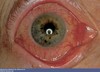
Uveitis
What are the constitutional symptoms which may be present in someone with sarcoidosis ?
- Fever & night sweats
- Malaise
- Fatigue
- Weight loss
What are the features of chronic sarcoidosis ?
- Lung infiltrates - dry cough, dyspnoea, bilateral hilar lymphadenopathy
- Skin infiltrates - erythema nodosum, lupus pernio (chronic raised purpulish lesions affecting cheeks & nose)
- Peripheral lymphadenopathy
- Eye - anterior uveitis
- Hypercalcaemia
- Other organs - hepatitis, heart disease (cardiomyopathies, arrhythmias), neurological (bells palsy, lymphocytic meningitis) splenomegaly, renal (stones)
How is sarcoidosis diagnosed?
There is no one diagnostic test, but generally needs histological evidence of granulomatous inflammation, exclusion of alternative causes & evidence of systemic disease
- Bloods done - hypercalcaemia & increased ESR & serum ACE
- Negative mantoux test
- CXR which shows BHL +/- interstitial infiltrates
- CT scan which shows peripheral nodular infiltrates
- Tissue biopsy done (e.g. transbronchial, skin, lymph nodes) which hows non-caesating granulomas
- PFT’s show restrictive disease pattern
How is sarcoidosis staged ?
On CXR:
- Stage 0 = normal
- Stage 1 = BHL
- Stage 2 = BHL + interstitial infiltrates
- Stage 3 = Diffuse interstitial infiltrates only
- Stage 4 = Diffuse fibrosis
What is the management of sarcoidosis ?
1st line = oral steroids (prednisolone) if they are symptomatic stage ≥ 2 or 3 OR if hypercalcaemia, eye, heart or neuro involvement then treat regardless of stage or symptoms
Do patients with sarcoidosis who have asymptomatic stage 2 or 3 disease with only midly abnormal lung function require treatment ?
No
What are the 2nd and 3rd line treatment options for sarcoidosis ?
- 2nd line = immunosuppressants e.g. methrotrexate or asathioprine etc
- 3rd line = anti-TNF
Define what idiopathic pulmonary fibrosis (IPF) is
It is a ILD characterised by progressive fibrosis of the interstitium of the lungs. Whilst there are many causes of ILD, the term IPF is reserved for when no underlying cause exists
Who is IPF typically seen in ?
Patients aged 50-70 & more so in men
What are the clinical features of IPF?
That of ILD:
- Progressive dyspnoea
- Bilateral fine end-inspiratory crackles/crepitations
- Dry cough
- Finger clubbing
- Cyanosis
How is IPF diagnosed ?
- As previous flashcard for ILD, do all those Ix’s & then diagnose IPF only on the basis of MDT consensus
- Then 2nd line as per ILD is a biopsy if needed
What is the investigation of choice for investigating someone suspected of having IPF?
HR-CT
Describe the pathology of IPF
- Hetergenous fibrosis in alveolar walla with fibroblastic foci & destruction of archiecture causing honeycombing
- Inflammation is minimal
What CXR features are suggestive of IPF?
Bilateral interstitial shadowing - ground-glass appearance later progressing to honeycombing
What is the management of IPF?
- Pulmonary rehabiliation + supportive tx (oxygen) +/- pirfenidone or nintedanib
- If no contraindications & young will eventually require lung transplantation
What are the criteria which must be met for pirfenidone or nintedanib use in IPF?
- Person has a FEV between 50-80% predicted
- Company provides discount for the drug
- Tx is stopped if there is evidence of disease progression (decline of ≥ 10% in FVC within any 12-month period)
What is the prognosis of IPF?
Poor - llife expectancy is around 3-4 years
Define what pneumoconiosis is
This refers to a sub-group of ILD’s caused by the inhalation & retention of dusts in the lungs
What are the main types of pneuoconiosis
- Coal workers pneumoconiosis (CWP) + Progressive massive pneumoconiosis
- Silicosis
- Asbestosis
What is the cause of CWP + PMP?
It is caused by long-term exposure to coal dust particles
What are the clinical features of CWP?
- Usually asymptomatic, but CXR shows many round opacties esp in the upper zone
- Restrictive LFT’s (spirometry) seen
- Commonly have co-exisiting chronic bronchitis
What is the management of CWP + PWP ?
- Avoid exposure to coal dust
- Treat co-exisiting chronic bronchitis
- May be eligable for compensation via the industrial injuries act
Describe what PWP is
This is where there is progression of CWP resulting in progressive dysponea, fibrosis & eventually cor pulmonale
How is pneumoconiosis investigated ?
As per ILD guidelines
What is the cause of silicosis ?
Inhalation of silicon dioxide (silica) particles
What are the characteristic CXR features of silicosis ?
Diffuse miliary or nodular pattern in upper & mid zones and ‘egg-shell’ calcification of the hilar lymph nodes
What occupation are at risk of silicosis ?
- Mining
- Slate workers
- Foundries
- Potteries
What is the management of silicosis ?
Avoid exposure & claim compensation under industrial injuries act
What are the clinical features of silicosis ?
Primarily progressive dyspnoea
What causes asbestosis ?
Inhalation of asbestos fibres
What jobs increase risk of asbestose exposure ?
Building trades
What are the clinical features of asbestosis ?
Similar to all the other ILD’s:
- Progressive dysponea
- Fine end-inspiratory crackles
- Finger clubbing
What is the CXR features of asbestosis ?
- Pleural plaques
- Eventually diffuse pleural thickening and lower lobe fibrosis
What does asbestosis increase the risk of ?
Mesothelioma & bronchical adenocarcinoma
What is the management of asbestosis ?
- Symptomatic Tx
- Often eligable for compensation via the UK industrial injuries act
Essentially what is the treatment of all the pneumoconioses?
Avoidance & compensation
What is caplans syndrome ?
The association between RA, pneumoconiosis & pulmonary rheumatoid nodes
