Renal Pathology 2 Flashcards

1
Q
A

Normal adult kidney

Capsule has been removed & pattern of fetal lobulations still persists (sometimes does). Hilum has some adipose tissue. There is a simple renal cyst (smooth surface, small, clear fluid) - not uncommon in adults.

2
Q
A

Normal Kidney

Cut surface of the kidney with good cortical-medullary demarcation and pelvic adipose tissue

Light outer cortex & darker medulla with renal pyramids into which the collecting ducts coalesce and rains into calyces and central pelvis

3
Q
A

Normal kidney

4
Q
A

Acute pyelonephritis

5
Q
A

Acute Pyelonephritis

6
Q
A

Acute Pyelonephritis

7
Q
A

Acute Pyelonephritis

8
Q
A

Acute Pyelonephritis

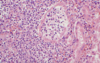
Ascending bacterial infection leading to acute pyelonephritis. Numerous PMNs seen filling renal tubules across center/right of this pic. These leukocytes may form into a cast within the tubule. Casts appearing in the urine orginate in the distal renal tubules and collecting ducts.

9
Q
A

Acute Pyelonephritis

10
Q
A

Acute Pyelonephritis

11
Q
A

Acute Pyelonephritis

12
Q
A

Renal Papillary Necrosis

13
Q
A

Sloughed renal papillary in Renal Papillary Necrosis

14
Q
A

Sloughed renal papilla in renal papillary necrosis

15
Q
A

Distended calyx & ureter in hydronephrosis

16
Q
A

Atrophy of renal parenchyma = non-functional in hydronephrosis

17
Q
A

Hydronephrosis

18
Q
A

Hydronephrosis

19
Q
A

Hydronephrosis

20
Q
A

Nephrolithiasis

21
Q
A

Staghorn Calculus

Urolithiasis

22
Q
A

Staghorn Calculus

Struvite stone, usually forms in alkaline urine

23
Q
A

Uric acid calculi

Form in acidic urine, radiolucent

24
Q
A

Urolithiasis

Calcium oxalate stones

25
Urolithiasis
26
Urolithiasis
27
Urolithiasis
28
Nephrosclerosis Granular surface due to tiny infarcts throughout renal cortex
29
Hyaline arteriosclerosis in nephrosclerosis
30
Hyaline arteriosclerosis in nephrosclerosis
31
Nephrosclerosis Small red dot petechiae hemorrhage (Flea bitten kidney)
32
Nephrosclerosis (fibrinoid necrosis)
33
Nephrosclerosis Onion-skinning hyperplastic arteriolitis
34
Nephrosclerosis
35
Autosomal dominant PKD
36
Autosomal Dominant PKD
37
AD PKD
38
AD PKD
39
AD PKD
40
AD PKD
41
AR PKD
42
AR PKD
43
AR PKD
44
AR PKD
45
AR PKD
46
AR PKD
47
AR PKD
48
Acquired Dialysis-associated cystic kidney disease
49
Acquired Dialysis-associated cystic kidney disease
50
Renal Cell Carcinoma Bright yellow/orange because tumor cells have lipid in cytoplasm
51
Renal Cell Carcinoma
52
Renal Cell Carcinoma Propensity to spread to renal vein & hematogenously
53
Renal Cell Carcinoma Adipocytes with empty cytoplasm
54
Renal Cell Carcinoma
55
Transitional Cell Carcinoma | (In renal pelvis, not yellow)
56
Transitional Cell Carcinoma
57
Transitional Cell Carcinoma of Ureter
58
Transitional Cell Carcinoma of Bladder
59
Transitional Cell Carcinoma
60
Transitional Cell Carcinoma
61
Transitional Cell Carcinoma
62
Transitional Cell Carcinoma
63
Wilm's Tumor (Nephroblastoma) Usually large by the time diagnosis occurs
64
Wilm's Tumor (Nephroblastoma) Left side has cells that are mostly nucleus with less cytoplasm
65
Wilm's Tumor (Nephroblastoma)
66
Wilm's Tumor (Nephroblastoma) Left: primitive; Right: glandular structure
67
Wilm's Tumor (Nephroblastoma)
68
What are the clinical features of acute pyelonephritis?
Fever, malaise, flank pain, urinary frequency, dysuria
69
What is found in the lab work of someone with acute pyelonephritis?
Pyuria, bacteruria, sometimes hematuria, WBC casts
70
What are 2 common agents that cause acute pyelonephritis via hematogenous spread?
Staphylococcus E. coli
71
What are some common organisms that cause acute pyelonephritis via ascending infection?
E. coli Proteus Enterobacter Klebsiella Enterococcus Pseudomonas
72
What are some risk factors/causes (besides bacteria) of acute pyelonephritis?
1. Short urethra (females) 2. Stasis of urine (any cause) 3. Structural abnormalities of urinary tract (causes obstruction) 4. VUR 5. Catheterization of bladder 6. Diabetes mellitus
73
What are some complications of acute pyelonephritis?
Perinephric abscess Pyonephrosis Renal papillary necrosis Sepsis
74
What are some causes of papillary necrosis?
Acute pyelonephritis with obstruction Excessive intake of analgesics, esp. phenacitin DM Sickle cell disease
75
What are some clinical hallmarks of papillary necrosis?
Inability to concentrate urine Gross hematuria Proteinuria Colicky flank pain Sloughing of renal papilla
76
What is hydronephrosis?
Dilation of renal pelvis and calyces associated with progressive atrophy of kidneys due to obstruction of urine outflow
77
What are some causes of hydronephrosis?
Transitional cell carcinoma of renal pelvis/ureter Carcinoma of cervix Blood clot Ureteral stone Retroperitoneal fibrosis Pregnancy/endometriosis BPH Uretheral stricture
78
What is a main clinical finding of hydronephrosis?
Postrenal azotemia
79
What are the clinical findings of urolithiasis?
Flank pain radiating toward groin Hematuria
80
What are the 4 main types of urinary tract calculi?
Calcium oxalate & calcium phosphate (70%) Magnesium ammonion phosphate (struvite/staghorn calculus (15-20%) Uric acid (5-10%) Cystine (1-2%)
81
What is the difference betwen benign and accelerated nephrosclerosis?
Benign: normal/small kidney with finely granular cortical surface. Hyaline arteriolosclerosis, tubular atrophy, interstitial fibrosis, glomerular sclerosis Accelerated: (malignant HTN), vascular injury to arterioles & small arteries (vasculopathy)
82
What are the clinical findings in nephrosclerosis?
Acute renal failure Hematuria, proteinuria Rapid increased in BP (\>200/120) Encephalopathy
83
What are the gross and microscopic findings in nephrosclerosis?
Gross: swollen kidneys with petechial hemorrage (flea bitten kidney) Microscopic: fibrinoid necrosis of arterioles, hyperplastic arteriolitis (onion skinning)
84
What is the incidence of AD PKD? What is the mutation and its frequency?
1/400-1/1,000 in US PKD1 (85%) - polycystin 1 (chromosome 16p) PKD2 (15%) - polycystin 2 (chromosome 4q)
85
What is the chronologic sequence of events of AD PKD?
Bilateral cysts develop by 3rd decade (many also have liver/pancreas cysts) Chronic renal failure by 40-60 Intracranial berry aneurysms (15-20% of patients)
86
What is the incidence of AR PKD?
1:10,000 - 1:50,000
87
Who does AR PKD affect?
Neonates, infants, and young children 75% of children die in perinatal period
88
What is AR PKD associated with?
Congenital hepatic fibrosis -\> portal hypertension
89
How is AR PKD different from AD?
AR PKD has numerous elongated cysts arranged at right angles to cortex w/ cylindrical enlargement of collecting tubules
90
How common is Acquired Dialysis-associated cystic kidney disease?
Found in ~35% of patients treated with long-term dialysis
91
What is Acquired Dialysis-associated cystic kidney disease?
Cystic disease superimposed on other types of kidney pathology Probably due to obstruction of tubules by interstitial fibrosis or crystals
92
What are the gross and microscopic findings of Acquired Dialysis-associated cystic kidney disease?
Gross: Numerous 0.5-0.2 cm cysts containing clear fluid Microscopic: Cysts lined by hyperplastic or flattened tubular epithelium, sometimes contain Ca2+ oxalate crystals
93
What are some complications of Acquired Dialysis-associated cystic kidney disease?
Hemorrhage into cysts or retroperitoneum Renal cell carcinoma (7%)
94
What is the most common primary malignancy of the kidney?
Renal cell carcinoma (80% of renal cancers)
95
What syndromes are renal cell carcinoma associated with?
von-Hippel Lindau Syndrome Paraneoplastic sydnrome: EPO, PTH, etc.
96
What are the clinical findings of renal cell carcinoma?
Classic triad: Hematuria, CV angle pain, flank mass (but only seen in 10%)
97
What are the histological findings of renal cell carcinoma?
Epithelioid cells arranged in nests, often with clear cytoplasm (clear cell carcinoma) due to intracellular lipid/glycogen
98
Where does renal cell carcinoma have a tendency to invade and metastasize?
Tendency to invade renal vein and metastasize to bone, lung, CNS (late metastasis)
99
What are some of the genetic associations of renal cell carcinoma?
Translocation, deletion on chromosome 3 --\> sporadic clear cell Loss of VHL, inactivated/hypermethylated VHL --\> hereditary clear cell
100
Urothelial (transitional cell) carcinomas represent \_\_% of primary renal tumors?
5-10%
101
Where do Urothelial (transitional cell) carcinomas occur?
anywhere in the urinary tract, including renal pelvis Patients may have multiple tumors in different locations
102
Describe a Urothelial (transitional cell) carcinoma histologically & grossly
Composed of tumor cells resembling normal transitional epithelium (urothelium) Gross: firm, white, sometimes papillary white cauliflower mass
103
What is the most common childhood tumor of the kidney?
Wilm's tumor most common from 1-7 y.o.
104
What are the clinical findings of Wilm's tumor?
Palpable mass in abdomen or flank HTN (due to renin secretion)
105
Describe the gross and microscopic findings of Wilm's tumor?
Large, solid, circumscribed tumor Resembles renal blastema (primitive mesodermal tissue) but may have "differentiation" in form of primitive tubular structures, glomeruli, or other mesenchymal tissue
106
What is the treatment for Wilm's tumor?
Combined surgery, chemo, radiation
107
What is the overall long-term survival of Wilm's tumor?
\>90%