Regulation Flashcards

1
Q

Adipose hormones (Adipokines) act as sensors

Describe the role of Adiponectin

A
  • Affects metabolism of faIy acids and carbohydrates
    • ↑ rate of FA β-oxidation in muscle
    • ↓ FA synthesis and gluconeogenesis in liver
    • ↑ glucose uptake in muscle
    • ↑ insulin sensitivity
  • Effects of adiponectin are indirect as adiponectin acts through AMP dependent kinase (AMPK)
    • AMPK is a fuel sensor regulating metabolic activity
2
Q

Absense of adiponectin causes desensitization to what?

A

Insulin

Poor glucose tolerance; ingestion of dietary carbohydrates results in long-lasting rise in blood glucose, similar to type 2 diabetes.

3
Q

Compare and contrast Leptin and Adiponectin

A
  • Leptin
    • Controled at the level of CNS
    • Regulation of food intake and appetite
    • Levels are higher in obesity
    • Leptin resistance prevents anti-obesity effects
  • Adiponectin
    • Acts through tissue metabolism
      • (less so through CNS)
    • Levels are lower in obesity
    • Hypoadiponectinemia + type 2 diabetes.
4
Q

Ghrelin

A
  • Produced when hungry and about to eat a meal
  • Peptide hormone produced in cells lining the stomach
  • Acts on hypothalamus
  • Stimulates appetite via plasma
  • Levels peak just before a meal and drops after a meal
  • Exceptionally high levels in Prader-Willi syndrome
5
Q

PYY

A
  • Peptide hormone produced by endocrine cells in the lining of the small intestines.
  • Acts on hypothalamus to signal satiety
  • Appetite suppresant, plasma levels peak after a meal
6
Q

Orexigenic vs Anorexigenic neurons

A

Leptin acts on anorexigenic cells to release α-MSH, upregulating satiety and metabolism.

Leptin also acts on orexigenic cells to inhibit NPY, which is the “eat more” signal.

Ghrelin stimulates NPY.

PYY inhibits NPY.

7
Q

What are the 2 mechanisms for degrading proteins?

A

Lysosomal degradation

  • Nonselective
    • material taken up by endocytosis
    • intracellular constitutents enclosed in vesicles
  • Highly selective
    • Chaperone-mediated autophagy

Proteasome degradation

  • Ubiquitination
8
Q

Chapernone-mediated autophagy (CMA)

A

Critical during times of starvation in the liver and kidney.

Proteins are selected for degradation based on the KFERQ protein sequence. This sequence is exposed during gradual unfolding and turnover. 30% of liver and kidney proteins have this sequence.

9
Q

What is Lamp2a?

A

Cytosolic chaperone protein recognizes KFERQ proteins and targets them to LAMP-2A at surface of lysosomes.

  • LAMP-2A monomers aggregate to form multimeric complexes
  • Protein is moved into the lysosome through the LAMP-2A complex (with assistance from lysosomal chaperone)
  • LAMP-2A levels control CMA activity:
    • Upregulation of Lamp-2A increases CMA Downregulation of Lamp-2A decreases CMA
10
Q

Proteasome degradation

A
  • Housekeeping
    • Eliminates misfolded or damaged proteins
  • Regulatory
    • rapid system of degradation for proteins involved in regulatory function, like during the cell cycle
  • A protein tagged with a chain of at least 4 ubiquitin molecules binds the 19S cap
11
Q

The 1st step in the catabolism of most AAs in the liver

A
12
Q

2nd step in the catabolism of most AAs in the liver

A

Oxidative deamination catalyzed by glutamate dehydrogenase

13
Q

What is transdeamination?

A

Steps 1 and 2 of Amino Acid catabolism

14
Q

Two nitrogen groups enter the urea cycle.

How do they do so?

A

1 enters as carbamoyl phosphate

1 enters as aspartate

*Liver mitochondria

15
Q

Enzyme that forms Carbamoyl Phosphate

A

Carbamoyl phosphate synthetase:

couples HCO3 with NH4 to form carbamoyl phosphate

16
Q

Where does the Urea Cycle take place?

A
  • Mitochondrial
    • Glutamate dehydrogenase
    • Aspartate aminotransferase
    • Carbamoyl phosphate synthetase
    • 1 st step of Urea cycle
  • Cytosolic
    • Aminotransferases
17
Q

What happens to NH4 from AA breakdown in extrahepatic tissue where there is no urea cycle?

A

Extrahepatic tissue transports NH4 through the bloodstream in the form of Glutamine. Once in the liver, NH4+ is removed from glutamine and is processed in the urea cycle.

  • Glutamine Synthetase extrahepatically.
  • followed by Glutaminase in the liver.

For muscle, Glucose-alanine cycle.

18
Q

How is the Urea Cycle regulated?

A
  • Long-term regulation
    • upregulation of urea cycle enzymes
      • prolonged starvation
      • high protein diets
  • Short-term regulation
    • Allosteric activation of carbamoyl phsophate synthetase by N-acetylglutamate (a glutamate derivative whose concentration is proportional to glutamate).
19
Q

Glucose-alanine cylce

A
  • Amino groups are transferred to alanine.
  • Alanine transports amino groups to liver for the urea cycle.
  • Carbon skeleton is used for gluconeogenesis.
  • Glucose is essentially forming pyruvate, which picks up the nitrogen via transamination.
20
Q

Ketogenic vs. Glucogenic amino acids

A

Glucogenic AA’s can all result in net synthesis of glucose:

  • Pyruvate
  • α-ketoglutarate
  • Succinyl-CoA
  • Fumarate
  • Oxaloacetate

Ketogenic AA’s → acetyl CoA ⇒ ketone bodies or Fatty Acids.

21
Q

Formyl tratrahydrofolate

A

derived from Folic Acid, important donor of single carbons in biological systems.

22
Q

De novo synthesis of purines

A

Production of IMP

Ribose Pentose Phosphate Pathway makes PRPP, which is the precursor of IMP (Inosine monophosphate).

PRPP makes purines and pyramidines, and some amino acids.

First committed step to purine synthesis is glutamine-PRPP aminotransferase, to make Phosphoribosylamine.

23
Q

Amino acid synthesis

carbon-skeleton precursors

A

Glycolysis, CAC, and PPP

24
Q

In amino acid synthesis,

Where are amino groups derived from?

A

Glutamate and Glutamine

25
Q

Regulation of Purine Synthesis

A

AMP (adenine monophosphate) and GMP (guanine monophosphate) don’t really get stockpiled, but IMP does.

When IMP builds up, it blocks its own formation.

AMP and GMP independently regulate their own production.

AMP also regulates PRPP synthetase (the very first step).

PRPP is a feedforward allosteric activator. A lot of PRPP accelerates flux through the pathway, whereas IMP causes feedback inhibition.

26
Q

Regulation of pyrimidine synthesis in animals

A

Carbamoyl Phosphate Synthetase II is in the cytosol and has nothing to do with the Urea Cycle.

Carbamoyl phosphate + Aspartate makes N-Carbamoylaspartate, which eventually reacts with PRPP, ending up as uradine triphosphate and then cytidine triphosphate.

27
Q

Purine Synthesis

A
28
Q

If PRPP builds up, what happens?

A

Generation of more Purines and more Pyrimidines

29
Q

How are deoxynucleotides made?

A

Ribonucleotide Reductase

Disregulation in the levels of individual deoxynucleosides can increase the chance of mutations; a lack can be lethal.

30
Q

Ribonucleotide reductase enzyme mechanism

A

The ribonucleotide reductase enzyme utilizes a free radical, which is transfered from outside to inside the active side, where the free raidical is used for enzyme catalysis.

31
Q

Synthesis of Thymine

A

Reduction depends on tetrahydrofolate, suscpetible to anti-folate drugs.

32
Q

Purine catabolism

A

Relies on Xanthine Oxidase to convert purines to Uric Acid.

AMP → IMP → Xanthine → Uric Acid.

Guanine → Xanthine → Uric Acid.

33
Q

Gout

A

Impaired uric acid excretion or overproduction of uric acid, which is non-soluble and builds up in joints. Purine rich foods should be avoided.

34
Q

Thymidine monophosphate (TMP or Thymidylate) snythesis

A

Thymidylate synthetase is an enzyme that catalyzes the conversion of deoxyuridine monophosphate (dUMP) to deoxythymidine monophosphate (dTMP). The cycle depends on folate, a target for chemotherapy.

35
Q

Purine salvage pathways

A

Recycling the nitrogen base instead of making it from scratch

Enzymes are HGPRT and APRT

If something goes wrong with the enzymes, PRPP accumulates, activating the production of IMP, whose degradation causes an increase in Uric Acid and therefore gout.

36
Q

Pyrimidine Catabolism

A

Source of NH4+ and Urea

Source of malonyl-CoA (FA synthesis)

Source of Succinyl-CoA (CAC intermediate)

37
Q

How are TAGs broken down?

A

Triacylglycerols (TAGs) are broken down by lipases

Lipases are located in dfferent locations to serve distinct functions.

  • Intestinal lumen (pancreatic lipase)
    • Absorption of FA from diet into intestine
  • Capillary walls (lipoprotein lipase)
    • Absorption from chylomicrons and VLDLs into tissue
  • Intracellular (hormone-sensitive lipase)
    • Break down cellular fat stores in adipose tissue
38
Q

What is Bile?

A

derivatives of cholesterol that act as detergents to solubilize dietary fats

39
Q

Characteristics of Lipoproteins

A
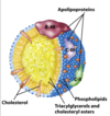
  • TAG + cholesterol, cholesteryl esters, and specific proteins.
    • Phospholipid head group on outside.
    • Apolipoproteins coat the surface.
  • Apolipoproteins determine the fate of the particle.
    • high affinity ligands for lipoprotein receptors
40
Q

Exogenous lipoprotein metabolism

A
  • Dietary TAGs absorbed in the intestines are packaged with dietary cholesterol into Chylomicrons
    • transport of dietary fat to tissue through lymphatic system
  • Once in tissue, lipoprotein lipases (LPL) in the capillaries hydrolyze TAGs into FAs and glycerol
    • Muscle FAs are oxidized for energy
    • Adipose FAs are re-esterified as TAGs for storage
  • Remnants of Chylomicrons (depleted of most TAGs but containing cholesterol and apolipoproteins) are taken up in the liver
41
Q

Different Apolipoproteins

A
  • ApoB48
    • Intestine (Chylomicrons)
  • ApoB100
    • Liver (VLDLs)
  • ApoE
    • IDL but not LDL
  • ApoCII
    • acquired from HDL
42
Q

Roles of Apolipoproteins

A
  • Chylomicrons that drain into the blood interact with HDL
    • HDL gives Chylomicrons ApoCII
    • ApoCII interacts with Lipoprotein Lipase on capillaries
    • Once Chylomicrons become small and dense enough, they exchange ApoCII and gain ApoE from HDL
  • As VLDL particles are metabolized, they become more dense, becoming IDL particles
  • 50% of IDLs exchange ApoCII for ApoE
    • ApoE is high affinity substrate for internalization
    • So, these IDLs are taken up by the liver
  • 50% of IDL become LDL, losing ApoCII
    • LDLs stay in the plasma
      • LDLs bind to LDL-R (receptors)
43
Q

Lipoproteins in order by size

A
44
Q

Chylomicrons vs VLDL

A

Chylomicrons and VLDLs are coated with ApoB lipoproteins.

ApoB is not exchangeable.

45
Q

HDL

A
  • HDL serves as a reservoir for the exchangeable of apolipoproteins.
  • HDL particles extract excess cholesterol from cell-surface membranes as a cholesterol scavanger
  • Transports the cholesterol to the liver where it is converted to bile salt
46
Q

What is HMG-CoA reductase?

A

HMG-CoA reductase is the key regulatory step in cholesterol biosynthesis.

47
Q

Process of LDL endocytosis

A
  • Tissue receptors specifically bind ApoB-100
  • LDL is internalized in vesicles which fuse with lysosomes
  • Lysosomes hydrolyze cholesteryl esters
  • Cholesterol and FA are released into cytosol
  • LDL receptor is recycled to surface
  • Free cholesterol goes to ER
48
Q

LDL absorption and subsequent regulation

A

↑ [cholesterol] in Endoplasmic Reticulum

↓ HMG-CoA reductase

↓ the rate of cholesterol biosynthesis

↓ synthesis of LDL receptors

↑ ACAT

49
Q

What is ACAT?

A

ACAT is a liver enzyme that converts cholesterol to cholesteryl esters.

It replaces the hydrophilic hydroxyl group with a hydrophobic lipid, so that it can be packaged up inside a VLDL.

50
Q

Serum Albumin

A

Free Fatty Acids are hydrophobic; they’re mobilized through the blood on Serum Albumin.

51
Q

Role of Hormone sensitive lipase in TAG metabolism

A
52
Q

How and where are fatty acids are activated and transported for β oxidation?

A

Activation of FA occurs on the cytosolic side of the outer mitochondrial membrane. Activation requires ATP, and the product is Fatty Acyl-CoA.

Fatty Acyl CoA runs through a Carnitine shuttle into the matrix.

β oxidation occurs in the mitochondria.

53
Q

How is fatty acyl CoA transported into mitochondria?

A

Carnitine Shuttle

  1. Fatiy acyl group of activated FA is transferred to carnitine
  2. Acyl carnitine enters the mito matrix through a carnitine transporter
  3. Carnitine acyltransferase in the mito matrix transfers a fatty acyl group to a CoA
  4. Carnitine returns to the cytosol through the transporter
54
Q

β oxidation

A

Oxidation of acyl-CoA (fatty acid aattached to a Coenzyme A)

  • Each round of β oxidation yields
    • 1 FADH2
    • 1 NADH
    • 1 acetyl-CoA for oxidation by citric acid cycle, which yields:
      • 3 NADH
      • 1 FADH2
      • 1 GTP
55
Q

β oxidation in odd-chain fatty acids

A

Odd-chain fatty acid β oxidation produces a bunch of Acetyl-CoAs plus one additional Propionyl-CoA, which is a CAC intermediate and feeds directly into it, increasing the amount of intermediates.

56
Q

Regulation of fatty acid oxidation

A
  • Regulation by Malonyl CoA of carnitine shuttle
  • Regulation by energy state of the cell
  • High levels of NADH/NAD+ inhibit β-oxidation enzymes
  • Hormonal regulation of Hormone sensitive lipase
    • ↑ Glucagon and epinephrine
    • ↑ cAMP
    • ↑ cAMP-dependent phosphorylation of perilipin and hormone sensitive lipase
    • ↑ activity of hormone sensitive lipase
    • ↑ free fatty acids
    • ↑ β-oxidaEon
  • Transcriptional regulation of FA oxidation enzymes
  • PPAR family of nuclear receptors with fatty acid ligands
    • Act as transcriptional factors
    • Triggered with increased energy demand
57
Q

Perilipin in TAG mobilization

A

The protein coating that protects a lipid droplet is called perilipin, and when perilipin is phosphorylated, activated hormone sensitive lipase can gain access to the lipid and break it down to individual fatty acids.

58
Q

Medium Chain acyl-Coa dehydrogenase deficiency

A

Inability to break down some medium-sized fatty acids.

Symptoms: hypoketonic hypoglycemia (low ketone bodies, low blood sugar).

59
Q

FA synthesis vs. degradation

A

Degradation is an oxidative process (which needs NAD+), whereas synthesis is a reductive process (utilizing NADPH).

60
Q

Starting fatty acid synthesis

A

Acetyl-CoA in mitochondria is transported to the cytoplasm for FA synthesis by the Tricarboxylate transport system, which involves the citrate transporter.

Mitochondrial acetyl CoA cannot exit to the cytosol for FA biosynthesis.

  1. Oxaloacetate + Acetyl-CoA makes Citrate.
  2. Citrate can be transported out of the mitochondrial matrix and into the cytosol.
  3. There, the citrate is converted back to Oxaloacetate and Acetyl-CoA.
  4. Oxaloacetate can return to the matrix.
61
Q

What is the regulatory step for fatty acid synthesis?

A

The 1st step of fatty acid synthesis is rate-controlling.

It’s catalyzed by acetyl-CoA carboxylase (ACC).

Bicarbonate Carboxyl group + acetyl CoA → Malonyl-CoA

62
Q

Palmitate formation by Fatty Acid Synthase

A
  • FAS is “charged” with acetyl (from acetyl CoA) and malonyl (from malonyl CoA)
    • CoAs are released
  • The fatty acyl chain grows by 2 carbon units donated by malonyl-CoA
    • Loss of CO 2 at each step
  • Reductions at each step (2 NADPH)
    • [NADPH is from the PPP]
63
Q

Energy requirements of FA synthesis

A

Acetyl-CoA

Used in the iniEal loading of acetyl CoA (1 per palmitate)

Used in the production of every Malonyl-CoA (7 per palmitate)

ATP

Used in the production of Malonyl-CoA (7 per palmitate)

NADPH

Reducing power is required by 2 active sites of FAS (14 per palmitate)

Overal reaction for 1 molecule of palmitate:

8 Acetyl-CoA + 7 ATP + 14 NADPH + 14 H+

palmitate + 8 CoA + 7 ADP + 7 Pi + 14 NADP + 6 H2O

Additionally, ATP is used for movement of acetyl-CoA into the cytosol

64
Q

Synthesis of unsaturated FAs

A

elongation and desaturation

Mammals cannot introduce double-bonds beyond the 9th carbon.

65
Q

Regulation of FA synthesis and degradation through acetyl-CoA carboxylase (ACC)

A

Allosteric regulation

  • upregulation by Citrate (signalling high acetyl CoA and ATP)
    • citrate also inhibits PFK-1 and downregulates glycolysis
  • downregulation by Palmitoyl CoA (signal of excess of FA)

Covalent modification

  • phosphorylation/dephosphorylation triggered by hormones

Acetyl-CoA carboxylase also inhibits FA oxidation through Malonyl-CoA

  • Malonyl-CoA inhibits carnitine acyltransferase (the carnitine shuttle)
  • When FA synthesis is underway, there’s no transport of fatty acyl groups into the mitochondria for β oxidation.
66
Q

Overview of fatty acid metabolism

A
67
Q

Insulin and glucagon’s effect on Acetyl-CoA carboxylase

A

Glucagon - phosphorylation

Insulin - dephosphorilation

68
Q

Making triacylglycerides (TGAs)

A
69
Q

Cholesterol Biosynthesis

A

2 Acetyl-CoA ⇨ HMG-CoA ⇨ Mevalonate

HMG-CoA Reductase - a membrane protein of the smooth ER - is the key regulatory step of cholesterol production.

Subsequent steps as follows.

70
Q

Regulation of Cholesterol Synthesis

A

HMG-CoA reductase: rate determining step of cholesterol synthesis

Sterol regulatory element-binding proteins (SREBPs)

SCAP (SREBP cleavage-activating protein) is a sterol sensor.

  • High* [cholesterol] ➠ transcriptionally active SREBP bound to SCAP.
  • Low* [cholesterol] ➠ change in SCAP frees SREBP’s active domain.
71
Q

HMG-CoA reductase regulation overview

A
72
Q

Hypercholesterolemia and Statins

A

Statins ≋ competitive inhibitors of HMG-CoA reductase

Reduction of endogenous cholesterol synthesis

(inhibition of HMG-CoA reductase)

Increase in LDL receptors

⬇︎ levels of blood LDL-cholesterol

73
Q

Summary of FA metabolism

A
74
Q

There are three different ketone bodies, what are they?

When are they formed?

A
  • Acetone
    • exhaled
  • Acetoacetate
    • transported to extrahepatic tissues
  • β-hydroxybutyrate
    • transported to extrahepatic tissues

Ketone bodies are formed during starvation when FAs are being heavily used - increased lipolysis + inadequate glucose uptake.

When Acetyl-CoA can’t enter CAC because of low OAA (depleted for gluconeogenesis), Acetyl-CoA accumulates and forms ketone bodies.

75
Q

Leptin

A

Leptin is released by adipose in response to rising adipose mass.

Acts on hypothalamus of the brain to suppress appetite

  • Promotes ⬇︎ in fuel intake
    • Signals brain to release α-MSH - neuronal signal to eat less
    • Inhibits the release of NPY - neuronal signal to eat more
  • Promotes ⬆︎ in expenditure of energy
    • ⬆︎ blood pressure and heart rate
76
Q

Glucose 6-phosphate, Pyruvate, and Acetyl-CoA

Key junctions in metabolism

A
77
Q

Primary oxidative fuel in the liver

A

FAs are oxidative fuel for the liver

FA synthesis with ample fuel supply

Synthesized FAs are released in VLDLs

During starvation, FAs are converted to ketone bodies.

78
Q

dietary amino acids and the liver

A

The liver absorbs majority of dietary amino acids

Protein synthesis or amino acid catabolism depending on fuel needs

Urea cycle to remove nitrogen

79
Q

Metabolic cycles in the muscle

A

Regenerating NAD+

Disposing of NH3

80
Q

Metabolic activity of cardiac muscle

A

The heart can use lactate produced by active muscle directly

Functions highly aerobically (high density of mitochondria)

Very limited glycogen reserves

Fatty Acids are the main fuel source

Also glucose, ketone bodies, and lactate

81
Q

What organs can do gluconeogenesis?

A

Liver and Kidney

82
Q

Fuel metabolism by different tissues

A
83
Q

Hormonal effects on Fuel Metabolism

A
84
Q

AMP-dependent kinase

A

Activated by high AMP:ATP ratio (not net concentrations)

Phosphorylates enzymes:

  • turns off ATP consuming anabolic pathways
  • turns on ATP generating catabolic pathways

Example targets: FA synthase, acetyl-CoA carboxylase, hormone sensitive lipase, HMG-CoA reductase, glycogen synthase, GLUT4

85
Q

AMPK target enzymes

A
  • FA synthase
  • acetyl-CoA carboxylase
  • hormone sensitive lipase
  • HMG-CoA reductase
  • glycogen synthase
86
Q

What are PPARs?

A

Peroxisome proliferator-activated receptors

A nuclear receptor involved in transcriptional regulation of fat metabolism: essentially, lipid sensors.

They bind hydrophobic ligands, like FAs.

PPARs bind to another nuclear receptor RXR prior to binding DNA and then act as transcription factors.

87
Q

What and where are the three types of PPARs?

A
  • PPARα: (alpha)
    • Starvation response
  • PPARγ: (gamma)
    • Master regulator of adipogenesis
    • instructs a cell to become an adipocyte
  • PPARδ: (delta)
    • Senses dietary lipid.
88
Q

NPY

A

neuronal signal to eat more

89
Q

α-MSH

A

neuronal signal to eat less

90
Q

What is parabiosis?

A

Parabiosis experiments to study a continuous, low-volume blood exchange between living animals.

91
Q

Where are Ketone Bodies made?

A

In the liver.

92
Q

Sources of glucose during starvation

A
93
Q

Pharmacological Treatments for Type 2 diabetes

A